



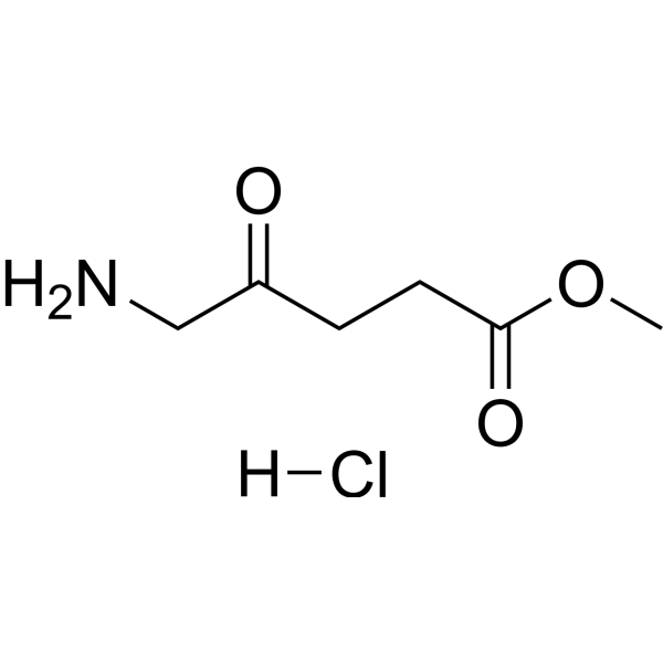
Methyl aminolevulinate hydrochloride
Based on 1 publication(s) in Google Scholar
Methyl aminolevulinate hydrochloride is a sensitizer used in photodynamic therapy (PDT). Methyl aminolevulinate hydrochloride penetrates the skin and induces the production of photoactive porphyrins including protoporphyrin IX in cells; upon exposure to appropriate light, it generates ROS, which triggers cellular oxidation and cell death. Methyl aminolevulinate hydrochloride acts as a photo-damage reversing agent through epidermal reconstruction, cytokine-mediated activation of dermal fibroblasts, elastin breakdown, new collagen formation, and compression of dilated capillaries. Methyl aminolevulinate hydrochloride reduces the expression of the proliferation marker Ki-67 and the early skin carcinogenesis marker TP53. Methyl aminolevulinate hydrochloride delays the onset of ultraviolet-induced skin tumors and reduces tumor burden in hairless mice. Methyl aminolevulinate hydrochloride is applicable to research related to actinic keratosis and basal cell carcinoma.
For research use only. We do not sell to patients.
- Purity : 98.98%
- CAS No.: 79416-27-6
- Formula: C6H12ClNO3
- Molecular Weight:181.62
-
Storage:
4°C, sealed storage, away from moisture
* In solvent : -80°C, 6 months; -20°C, 1 month (sealed storage, away from moisture)

To place orders, for customer services and technical support, please contact: MedChemExpress USA
Tel: 609-228-6898 E-mail: [email protected] [email protected]


-
 Biological Activity
-
Chemical Information
-
Solvent & Solubility
- Protocols
- Purity & Documentation
- References
-
 Help & FAQs


-
HY-N0305
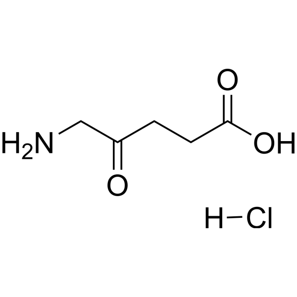
5-Aminolevulinic acid hydrochlorideHeme Precursor -

HY-A0169AR
Methyl aminolevulinate hydrochloride (Standard)Sensitizer -
HY-W010981
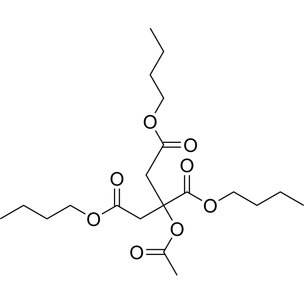
Acetyl tributyl citratePharmaceutical Excipient -

HY-W010981R
Acetyl tributyl citrate (Standard)Standard -

HY-78608
4-Boc-(trans)-Cyclohexanediamine-2-ethyl acetatePROTAC Linker
Publications Citing Use of MedChemExpress (MCE) Methyl aminolevulinate hydrochloride
More

Biological Activity
Description
In Vitro
Methyl aminolevulinate (0.0001-1.4 mmol/L) hydrochloride induces concentration-dependent formation of photoactive porphyrins in cultures of mouse adenocarcinoma LM2 cells, with the highest yield of 48 ng/105 cells observed at the concentration of 1.4 mmol/L; additionally, 90% of porphyrins remain intracellular at all tested concentrations[1].
Methyl aminolevulinate (1.2-1.8 mmol/L, 3 h) hydrochloride shows no intrinsic cytotoxicity against mouse adenocarcinoma LM2 cells at the concentration of 1.8 mmol/L, but induces 98% cell death when cells are treated with 1.2 mmol/L for 3 h followed by irradiation with 3 J/cm2[1].
MedChemExpress (MCE) has not independently confirmed the accuracy of these methods. They are for reference only. Further protocols information, click here.
-
Cell Line:murine adenocarcinoma LM2 cell
-
Concentration:1.8 mmol/L (intrinsic cytotoxicity); 1.2 mmol/L (photodynamic cytotoxicity)
-
Incubation Time:3 h (photodynamic cytotoxicity)
-
Result:Showed no cytotoxicity to LM2 cells at 1.8 mmol/L when used alone.
Induced 98% cell death when used at 1.2 mmol/L for 3 h followed by irradiation with 3 J/cm2 light.
In Vivo
MedChemExpress (MCE) has not independently confirmed the accuracy of these methods. They are for reference only.
-
Animal Model:Hairless mice (UV radiation-induced tumor model)[1]
-
Dosage:8-20%
-
Administration:topical; weekly; 26 weeks (UV-exposed mice); up to 36 weeks (non-UV-exposed mice)
-
Result:Delayed the appearance of UV-induced small tumors (AK or in situ SCC) by 2-3 weeks.
Delayed larger lesions (invasive SCC) by 5-7 weeks.
Caused an approximate 2- to 2.5-fold decrease in the number of UV-induced tumors per mouse after 26 weeks.
Significantly increased the tumor-free time on the methyl aminolevulinate-PDT side compared with the vehicle-PDT side in contralaterally treated mice.
Prevented tumor formation in non-UV-exposed mice treated for up to 36 weeks.
Induced higher levels of photoactive porphyrins (PAPs) in UV-induced tumors compared with normal skin of hairless mice.
Induced PAP formation only at the sites where it was applied.
Caused faster clearance of PAP than that formed by topical aminolevulinic acid.


Chemical Information
-
CAS No. 79416-27-6
-
Appearance Solid
-
Molecular Weight 181.62
-
Formula C6H12ClNO3
-
Color White to off-white
-
SMILES
O=C(OC)CCC(CN)=O.[H]Cl
-
Shipping
Room temperature in continental US; may vary elsewhere.
-
Storage
4°C, sealed storage, away from moisture
* In solvent : -80°C, 6 months; -20°C, 1 month (sealed storage, away from moisture)


Publications (1)
-
Journal Impact Factor
-
Most Recent
-
Sci Data
High-throughput drug screening identifies novel therapeutics for Low Grade Serous Ovarian Carcinoma. [Abstract]2024 Sep 19;11(1):1024. PMID: 39300112


Solvent & Solubility
In Vitro:
H2O : 100 mg/mL (550.60 mM; Need ultrasonic)
DMSO : 100 mg/mL (550.60 mM; Need ultrasonic; Hygroscopic DMSO has a significant impact on the solubility of product, please use newly opened DMSO)
Please refer to the solubility information to select the appropriate solvent. Once prepared, please aliquot and store the solution to prevent product inactivation from repeated freeze-thaw cycles.
Storage method and period of stock solution: -80°C, 6 months; -20°C, 1 month (sealed storage, away from moisture). When stored at -80°C, please use it within 6 months. When stored at -20°C, please use it within 1 month.
* Note: If you choose water as the stock solution, please dilute it to the working solution, then filter and sterilize it with a 0.22 μm filter before use.
Please refer to the solubility information to select the appropriate solvent. Once prepared, please aliquot and store the solution to prevent product inactivation from repeated freeze-thaw cycles.
Storage method and period of stock solution: -80°C, 6 months; -20°C, 1 month (sealed storage, away from moisture). When stored at -80°C, please use it within 6 months. When stored at -20°C, please use it within 1 month.
* Note: If you choose water as the stock solution, please dilute it to the working solution, then filter and sterilize it with a 0.22 μm filter before use.
Concentration (start) × Volume (start) = Concentration (final) × Volume (final)
In Vivo:
Select the appropriate dissolution method based on your experimental animal and administration route.
- For the following dissolution methods, please ensure to first prepare a clear stock solution using an In Vitro approach and then sequentially add co-solvents:
- To ensure reliable experimental results, the clarified stock solution can be appropriately stored based on storage conditions. As for the working solution for In Vivo experiments, it is recommended to prepare freshly and use it on the same day.
- The percentages shown for the solvents indicate their volumetric ratio in the final prepared solution. If precipitation or phase separation occurs during preparation, heat and/or sonication can be used to aid dissolution.
Add each solvent one by one: 10% DMSO 40% PEG300 5% Tween-80 45% Saline
Solubility: ≥ 2.5 mg/mL (13.77 mM); Clear solution
This protocol yields a clear solution of ≥ 2.5 mg/mL (saturation unknown).
Taking 1 mL working solution as an example, add 100 μL DMSO stock solution (25.0 mg/mL) to 400 μL PEG300, and mix evenly; then add 50 μL Tween-80 and mix evenly; then add 450 μL Saline to adjust the volume to 1 mL.
Preparation of Saline: Dissolve 0.9 g sodium chloride in ddH₂O and dilute to 100 mL to obtain a clear Saline solution.
Add each solvent one by one: 10% DMSO 90% (20% SBE-β-CD in Saline)
Solubility: ≥ 2.5 mg/mL (13.77 mM); Clear solution
This protocol yields a clear solution of ≥ 2.5 mg/mL (saturation unknown).
Taking 1 mL working solution as an example, add 100 μL DMSO stock solution (25.0 mg/mL) to 900 μL 20% SBE-β-CD in Saline, and mix evenly.
Preparation of 20% SBE-β-CD in Saline (4°C, storage for one week): 2 g SBE-β-CD powder is dissolved in 10 mL Saline, completely dissolve until clear.
In Vivo Dissolution Calculator
Please enter the basic information of animal experiments:
-
-
-
-
Recommended: Prepare an additional quantity of animals to account for potential losses during experiments.
Working solution concentration: 0.22 mg/mL
This product has good water solubility, please refer to the measured solubility data in water/PBS/Saline for details.

Protocols
-
RNA extraction experimental
By lysing cells, releasing RNA, and removing impurities such as proteins and DNA, high-purity RNA products are finally obtained. The commonly used traditional method is the guanidine isothiocyanate/phenol/chloroform method (Trizol), which is suitable for a variety of animal materials including animal tissues, microorganisms, cultured cells, etc., and most plant materials.
-
Ki-67 Immunostaining Proliferation Assay
Ki-67 immunostaining measures the growth fraction of a cell population by detecting Ki-67, a nuclear antigen present in proliferating cells and absent in quiescent G0 cells. The readout is the percentage of Ki-67-positive nuclei among total counted cells, commonly called the Ki-67 labeling index or proliferation index.
-
Cell Cytotoxicity Assay
Cytotoxicity assays are usually based on the assessment of cell membrane damage, which can also be indirectly detected by measuring cell viability. Detection methods include MTT assay, CKK-8 assay, LDH assay and ATP assay, etc.
-
Mammalian live/dead viability and cytotoxicity staining
Live/dead viability and cytotoxicity staining assays are based on the simultaneous detection of intracellular esterase activity in metabolically active (viable) cells and membrane integrity loss in non-viable cells. In commonly used dual-staining approaches, membrane-permeant fluorogenic substrates are converted by intracellular esterases into fluorescent products in live cells, while impermeant DNA-binding dyes selectively enter cells with compromised plasma membranes and label nucleic acids in dead or dying cells, enabling discrimination between viable and non-viable populations by fluorescence microscopy or flow cytometry.
-
PCNA Immunodetection Proliferation Assay
PCNA immunodetection measures proliferative activity by detecting proliferating cell nuclear antigen, a nuclear protein associated with DNA polymerase δ function and DNA replication. The assay readout is the proportion of PCNA-positive nuclei among total counted cells, but PCNA labeling is not identical to BrdU labeling because PCNA can mark late G1/early S-associated replication competence and may persist beyond active DNA synthesis depending on fixation and extraction conditions.
-
ROS/oxidative-stress fluorescent staining
ROS/oxidative-stress fluorescent staining uses cell-permeant fluorogenic probes that become fluorescent after oxidation inside cells or tissues; commonly used examples include DCFH-DA/DCFDA for broad cellular oxidant detection, DHE for superoxide-related signal detection, MitoSOX for mitochondrial superoxide-related signal detection, and CellROX probes for oxidative-stress-associated fluorescence readouts. The assay detects probe oxidation rather than a single ROS species unless the probe and analysis method have been chemically validated for that species. DCFH-DA enters cells, is deacetylated by intracellular esterases to DCFH, and produces fluorescent DCF after oxidation, so the readout is used as an operational measure of total cellular oxidative stress rather than a species-specific ROS measurement. DHE and MitoSOX can report superoxide-related oxidation, but red fluorescence alone can include non-specific ethidium-like oxidation products; HPLC or optimized spectral approaches are
-
Apoptosis Solutions
Apoptosis is a regulated, generally non-lytic cell-death pathway that removes unwanted, damaged, infected, or abnormal cells through coordinated morphological changes, caspase activation, DNA fragmentation, and membrane remodeling. The intrinsic apoptosis pathway is controlled mainly by mitochondrial outer membrane permeabilization, BCL-2 family proteins, cytochrome c release, apoptosome formation, caspase-9 activation, and downstream executioner caspase-3/7 activation. The extrinsic apoptosis pathway is initiated by death receptors such as Fas, TNFR, and TRAIL receptors, which recruit adaptor proteins and activate caspase-8 before engaging executioner caspases or mitochondrial amplification through BID cleavage. Apoptosis is linked to many phenotypes, including cancer cell killing, tissue homeostasis, immune regulation, neurodegeneration, infection response, and treatment-induced cytotoxicity; unresolved questions include how apoptosis interacts with necroptosis, pyroptosis, ferroptos
-
Fibers: Elastic Fiber Staining
Elastic fiber staining is a histochemical technique used to selectively visualize elastin-rich structures such as elastic fibers in connective tissues (e. g. , blood vessels, lung, dermis) based on the affinity of specific dyes or oxidation products for elastin-associated amino acid residues and cross-linked elastic matrix components. Classical methods such as Verhoeff-Van Gieson (VVG), resorcin-fuchsin (Weigert-type stains), or aldehyde fuchsin rely on differential binding of dye complexes to elastic fibers, allowing them to be distinguished from collagen and other extracellular matrix components by contrast staining (typically black or deep purple elastic fibers against red collagen counterstain). These methods are widely used in histopathology to evaluate elastic fiber integrity, fragmentation, or remodeling in vascular diseases, pulmonary pathology, and connective tissue disorders.
-
Research Protocol for Inflammation-related Diseases
The NLRP3 inflammasome is a cytosolic innate immune signaling platform that integrates priming signals and danger-signal activation to promote caspase-1 activation, maturation of IL-1β and IL-18, and gasdermin D-mediated pyroptotic cell death. The core experimental logic is to determine whether inflammatory disease phenotypes are driven by increased NLRP3 expression, ASC-containing inflammasome assembly, caspase-1 cleavage, GSDMD cleavage, and extracellular release of IL-1β/IL-18 rather than by nonspecific cell injury alone. The pathway is strongly linked to inflammation-related disease phenotypes because monosodium urate crystals activate NALP3/NLRP3 inflammasome signaling in gout-like crystal inflammation, cholesterol crystals activate NLRP3 inflammasomes in atherogenesis models, and DSS-induced intestinal inflammation has been reported to involve NLRP3 inflammasome activity. However, experimental colitis studies also show context-dependent protective effects of NLRP3 inflammasome co
-
Cell Viability Determination by MTT Colorimetric Assay
The following protocol uses the MTT colorimetric assay as a classic literature-established method for assessing cell viability/metabolic activity in cultured mammalian cells. MTT[3-(4,5-dimethylthiazol-2-yl)-2,5-diphenyltetrazolium bromide] is reduced by metabolically active cells to a colored formazan product; the amount of formazan is quantified spectrophotometrically and provides an indirect measure of metabolically active viable cells. Importantly, MTT reduction reflects cellular oxidoreductase/metabolic activity rather than an absolute direct count of living cells, so changes in cellular metabolism can alter the signal independently of cell number.


Purity & Documentation
-
Data Sheet (275 KB)
-
SDS (393 KB)
- English - EN (393 KB)
- Français - FR (393 KB)
- Deutsch - DE (393 KB)
- Norwegian - NO (393 KB)
- Español - ES (393 KB)
- Swedish - SV (393 KB)
- Italian - IT (393 KB)
- Korean - KR (393 KB)
- Portuguese - PT (393 KB)
-
Handling Instructions (2659 KB)
References
[1]. Siddiqui MA, et al. Topical methyl aminolevulinate. Am J Clin Dermatol. 2004;5(2):127-37; discussion 138-40. [Content Brief]
[2]. Szeimies RM, et al. Photoaging and Cosmetic Result with Artificial Daylight Photo-dynamic Therapy Using Methyl Aminolevulinate. Acta Derm Venereol. 2025;105:adv43245. Published 2025 Apr 22. [Content Brief]


Complete Stock Solution Preparation Table
Please refer to the solubility information to select the appropriate solvent. Once prepared, please aliquot and store the solution to prevent product inactivation from repeated freeze-thaw cycles.
Storage method and period of stock solution: -80°C, 6 months; -20°C, 1 month (sealed storage, away from moisture). When stored at -80°C, please use it within 6 months. When stored at -20°C, please use it within 1 month.
| Optional Solvent | Concentration Solvent Mass | 1 mg | 5 mg | 10 mg | 25 mg |
|---|---|---|---|---|---|
| H2O / DMSO | 1 mM | 5.5060 mL | 27.5300 mL | 55.0600 mL | 137.6500 mL |
| 5 mM | 1.1012 mL | 5.5060 mL | 11.0120 mL | 27.5300 mL | |
| 10 mM | 0.5506 mL | 2.7530 mL | 5.5060 mL | 13.7650 mL | |
| 15 mM | 0.3671 mL | 1.8353 mL | 3.6707 mL | 9.1767 mL | |
| 20 mM | 0.2753 mL | 1.3765 mL | 2.7530 mL | 6.8825 mL | |
| 25 mM | 0.2202 mL | 1.1012 mL | 2.2024 mL | 5.5060 mL | |
| 30 mM | 0.1835 mL | 0.9177 mL | 1.8353 mL | 4.5883 mL | |
| 40 mM | 0.1377 mL | 0.6883 mL | 1.3765 mL | 3.4413 mL | |
| 50 mM | 0.1101 mL | 0.5506 mL | 1.1012 mL | 2.7530 mL | |
| 60 mM | 0.0918 mL | 0.4588 mL | 0.9177 mL | 2.2942 mL | |
| 80 mM | 0.0688 mL | 0.3441 mL | 0.6883 mL | 1.7206 mL | |
| 100 mM | 0.0551 mL | 0.2753 mL | 0.5506 mL | 1.3765 mL |
* Note: If you choose water as the stock solution, please dilute it to the working solution, then filter and sterilize it with a 0.22 μm filter before use.


Methyl aminolevulinate hydrochloride Related Classifications
HY-A0169A Related Classifications
 Powered by Bioz