



IL-36 gamma/IL-1F9 Protein, Human
Based on 1 Customer Validation
IL-36 gamma (IL-1F9), a subform of IL-36 family, belongs to IL-1 superfamily. IL-36 gamma mediates inflammatory response. L-36 beta binds to IL-36R and recruits the co-receptor IL-1RacP, and thereby activating NF-κB and MAPK signaling pathways, but the activation requires N-terminal cleavage by neutrophil granule-derived proteases. IL-36 gamma/IL-1F9 Protein, Human is a recombinant human IL-36 gamma without any tag, which is produced in E. coli.
- Species: Human
- Source: E. coli
-
Storage:Stored at -20°C for 2 years from date of receipt. After reconstitution, it is stable at 4°C for 1 week or -20°C for longer (with carrier protein). It is recommended to freeze aliquots at -20°C or -80°C for extended storage.


To place orders, for customer services and technical support, please contact: MedChemExpress USA
Tel: 609-228-6898 E-mail: [email protected] [email protected]


-
 Biological Activity
-
Technical Parameters
-
Product Properties
- Documentation
- References
- Help & FAQs


-

HY-P70702
IL-36 alpha/IL-1F6 Protein, Human -
HY-P74803AF
Animal-Free IL-36 beta/IL-1F8 Protein, Human (His) -
HY-P70568
IL-13 Protein, Human (HEK293, His) -
HY-P72544
IL-36 gamma/IL-1F9 Protein, Mouse -
HY-P7037
IL-2 Protein, Human -
HY-P7050
M-CSF Protein, Human -
HY-P70445
IL-4 Protein, Human -
HY-P70591
IL-18 Protein, Human -
HY-P72545
IL-36 beta/IL-1F8 Protein, Human -
HY-P71857
IL-36 alpha/IL-1F6 Protein, Human (158a.a)


Biological Activity
Description
IL-36 gamma (IL-1F9), a subform of IL-36 family, belongs to IL-1 superfamily. IL-36 gamma mediates inflammatory response. L-36 beta binds to IL-36R and recruits the co-receptor IL-1RacP, and thereby activating NF-κB and MAPK signaling pathways, but the activation requires N-terminal cleavage by neutrophil granule-derived proteases[1][2]. IL-36 gamma/IL-1F9 Protein, Human is a recombinant human IL-36 gamma without any tag, which is produced in E. coli.
Background
IL-36 gamma (IL-1F9), a subform of IL-36 family, belongs to IL-1 superfamily. IL-36 gamma is expressed in peripheral blood lymphocytes, keratinocytes, bronchial epithelial cells and THP-1 cells[3].
The sequence of amino acids in IL-36 gamma differs in different species. Human IL-36 gamma shares <55% aa sequence identity with mouse.
IL-36 gamma has β-trefoil structure. L-36 gamma binds to IL-36R and recruits the co-receptor IL-1RAcP. So that heterodimeric signaling complex brings Toll/IL-1R (TIR) domains of the 2 receptor chains in close proximity, and thereby activating NF-κB and MAPK signaling pathways[1]. But the activation requires N-terminal cleavage at Val1518 by neutrophil granule-derived proteases, such as cathepsin G, elastase and proteinase-3[1][2]. IL-36 gamma is associated with chronic inflammation and cancers. IL-36 gamma is up-regulated in skin psoriatic lesions, inaganglionic and ganglionic areas of the colon in patients with Hirschsprung's disease, and inflamed mucosa of patients with inflammatory bowel disease (IBD)[1][4].
IL-36 gamma is a pro-inflammatory factor. IL-36 gamma mediates inflammatory response through the activation of NF-κB and MAPK signaling pathway[2].
In Vitro
IL-36 gamma (human, 50 ng/mL for 4 h or 100 ng/mL for 24 h) promotes the expression of pro-inflammatory cytokines and antibacterial peptides in NHOKs (oral keratinocytes)[5].
IL-36 gamma (human, 0.1 ng/mL, 3 days) promotes colony formation, migration and invasion of AGS cells[6].
Verified Bioactivity
The ability to induce IL-8 secretion in A431 human epithelial carcinoma cells has an ED50 value of 5-20 ng/mL.
MCE Validation Data
-
Purity - SDS-PAGE
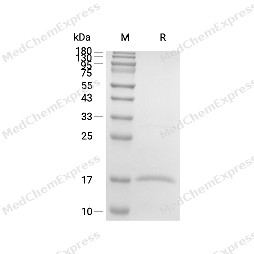
Purity - SDS-PAGE
-
Bioactivity - Cell-Based Assay
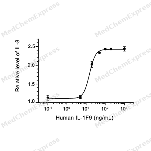
Bioactivity - Cell-Based Assay


Technical Parameters
-
Species Human
-
Source E. coli
-
Tag Tag Free
-
Accession
Q9NZH8-1 (S18-D169)
-
Protein Length
Full Length of Isoform-1
-
Synonyms
IL36G; Interleukin 1-Related Protein 2; Prev. IL1F9; Interleukin-36 Gamma; IL-1RP2; IL1RP2; IL-1F9; Interleukin 1 Family, Member 9; IL-1H1; IL-1 Related Protein 2; IL1H1; IL-1-Related Protein 2; IL1E; IL-1-Epsilon; Interleukin-1 Homolog 1; IL-1 Epsilon; I
-
AA Sequence
SMCKPITGTINDLNQQVWTLQGQNLVAVPRSDSVTPVTVAVITCKYPEALEQGRGDPIYLGIQNPEMCLYCEKVGEQPTLQLKEQKIMDLYGQPEPVKPFLFYRAKTGRTSTLESVAFPDWFIASSKRDQPIILTSELGKSYNTAFELNIND
-
Predicted Molecular Mass
17 kDa
-
Molecular Weight
Approximately 14-16 kDa, based on SDS-PAGE under reducing conditions.
-
Purity
≥ 95%, as determined by reducing SDS-PAGE.


Product Properties
Lyophilized powder
Lyophilized from a 0.22 μm filtered solution of 20 mM Tris-HCl, 100 mM NaCl, 0.1 mM EDTA, pH 8.0.
Note: For SPR assay, please replace the buffer. Primary amine components (e.g., Tris, imidazole) can affect protein-coupled chips.
<1 EU/μg, determined by LAL method.
It is not recommended to reconstitute to a concentration less than 100 μg/mL in ddH2O. For long term storage it is recommended to add a carrier protein (0.1% BSA, 5% HSA, 10% FBS or 5% Trehalose).
Stored at -20°C for 2 years from date of receipt. After reconstitution, it is stable at 4°C for 1 week or -20°C for longer (with carrier protein). It is recommended to freeze aliquots at -20°C or -80°C for extended storage.
Room temperature in continental US; may vary elsewhere.


Documentation
-
Data Sheet (264 KB)
-
SDS (252 KB)
- English - EN (252 KB)
- Français - FR (252 KB)
- Deutsch - DE (252 KB)
- Norwegian - NO (252 KB)
- Español - ES (252 KB)
- Swedish - SV (252 KB)
- Italian - IT (252 KB)
- Korean - KR (252 KB)
- Portuguese - PT (252 KB)
-
Handling Instructions (2659 KB)


References
[1]. Bassoy EY, et al. Regulation and function of interleukin-36 cytokines. Immunol Rev. 2018 Jan;281(1):169-178. [Content Brief]
[2]. Zhou L,et al. Interleukin-36: Structure, Signaling and Function. Adv Exp Med Biol. 2021;21:191-210. [Content Brief]
[3]. Gresnigt MS, et al. Biology of IL-36 cytokines and their role in disease. Semin Immunol. 2013 Dec 15;25(6):458-65. [Content Brief]
[4]. Neurath MF. IL-36 in chronic inflammation and cancer. Cytokine Growth Factor Rev. 2020 Oct;55:70-79. [Content Brief]
[5]. Xu P, et al. The mechanism on Prevotella melaninogenica promoting the inflammatory progression of oral lichen planus. Clin Exp Immunol. 2022 Aug 19;209(2):215-224. [Content Brief]
[6]. Le N, Luk I, et al. IL-36G promotes cancer-cell intrinsic hallmarks in human gastric cancer cells. Cytokine. 2022 Jul;155:155887. [Content Brief]

Calculators
Concentration (start) × Volume (start) = Concentration (final) × Volume (final)


 Powered by Bioz

