
17 Results for "

nuclear stain

" in MedChemExpress (MCE) Product Catalog:
Products (17)

17 Results for "nuclear stain" in MCE Product Catalog:

160
160 Publications Verification
Cat. No.: HY-D0815
CAS No.: 25535-16-4
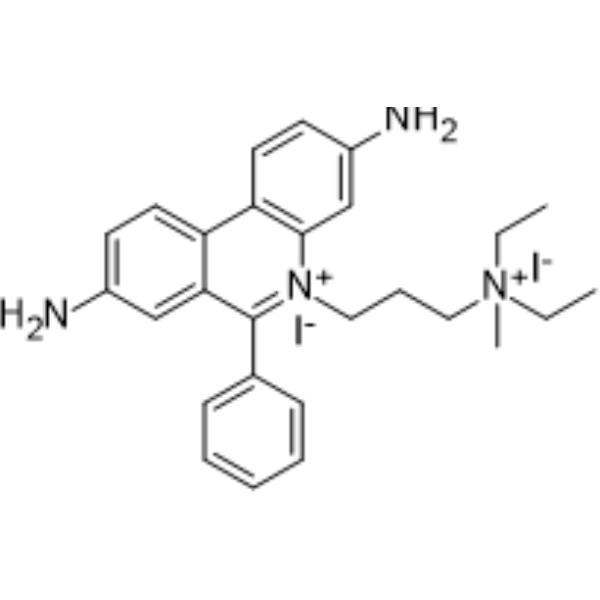
Propidium Iodide (PI) is a nuclear staining agent that stains DNA. Propidium Iodide is an analogue of ethidine bromide that emits red fluorescence upon embedding in double-stranded DNA. Propidium Iodide cannot pass through living cell membranes, but it can pass through damaged cell membranes to stain the nucleus. Propidium Iodide has a fluorescence wavelength of 493/617 nm and a wavelength of 536/635 nm after Mosaic with DNA. Propidium Iodide is commonly used in the detection of apoptosis (apoptosis) or necrosis (necrosis), and is often used in flow cytometry analysis.
loading...
    loading...
11
11 Cited Publications
Cat. No.: HY-N0116
CAS No.: 517-28-2
Synonyms: Natural Black 1; Haematoxylin
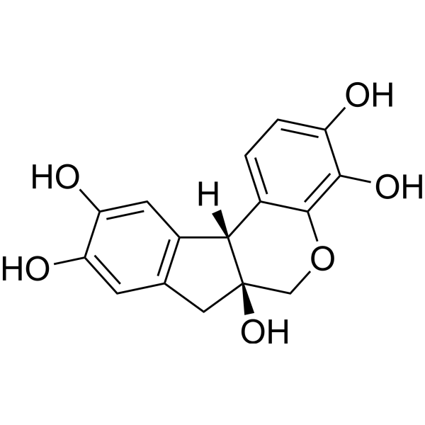
Hematoxylin (Natural Black 1), a naturally occurring flavonoid compound derived from Caesalpinia sappan Linn.. Hematoxylin is a nuclear stain in histology and is also a potent Aβ42 fibrillogenesis inhibitor with an IC50 of 1.6 μM.
loading...
    loading...
2
2 Cited Publications
Cat. No.: HY-D0944
CAS No.: 51811-82-6
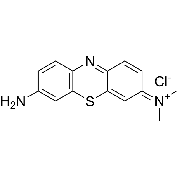
Giemsa stain is a composite dye composed of methylene azure, methylene blue, eosin and other components. Giemsa stain stains chromatin, nuclear membranes, specific cytoplasmic components and microorganisms. Giemsa stain is used in research on cytological and parasitological staining .
loading...
    loading...
1
1 Cited Publications
Cat. No.: HY-126367
CAS No.: 5141-20-8
Purity:  ≥75.0%
Synonyms: Acid Green 5
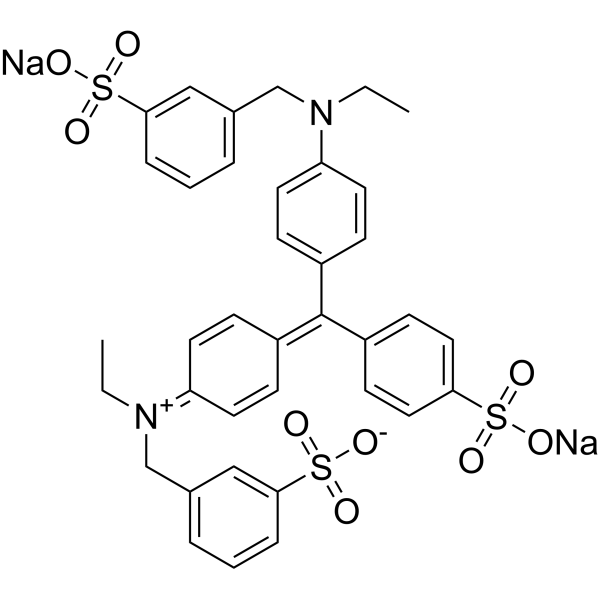
Light green SF yellowish (Acid Green 5) is a water-soluble triarylmethane dye. Light green SF yellowish serves as a histological stain that selectively labels mitochondria, collagen, and cartilage, while being an essential component of Papanicolaou staining. Light green SF yellowish is commonly used as a cytoplasmic counterstain for nuclear stains, and is applied in Masson's trichrome staining for collagen fibers, Pap staining, and cytological polychromatic staining in histopathology. Light green SF yellowish also induces growth inhibition and local fibrosarcomas in rats and exerts mild pulmonary tumorigenicity in mice .
loading...
    loading...
1
1 Cited Publications
Cat. No.: HY-D1423
CAS No.: 68654-25-1
Target:  

Fluorescent Dye

Research Areas:  

Others

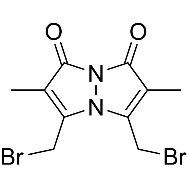
Dibromobimane is a Fluorescent dye and probe that can be used for the detection of intracellular reduced glutathione and the analysis of spatial proximity of closely adjacent cysteine thiol groups. Dibromobimane activates fluorescence through binding to thiol groups; its excitation/emission wavelengths are Ex/Em = 393/477 nm for glutathione detection, and Ex/Em = 385/477 nm for cysteine thiol cross-linking. Dibromobimane uses two equivalent bromomethyl groups to cross-link cysteine thiolates with a distance ranging from 3-6 Å. Dibromobimane can be applied to 96-well microplate-based high-throughput assays for labeling intracellular glutathione, and its fluorescence signal is normalized against nuclear staining results to eliminate the impact of cell density .
loading...
    loading...
Cat. No.: HY-DY1006
CAS No.: 25535-16-4
Propidium Iodide (PI) (solution) is a nuclear staining agent that stains DNA. Propidium Iodide is an analogue of ethidine bromide that emits red fluorescence upon embedding in double-stranded DNA. Propidium Iodide cannot pass through living cell membranes, but it can pass through damaged cell membranes to stain the nucleus. Propidium Iodide has a fluorescence wavelength of 493/617 nm and a wavelength of 536/635 nm after Mosaic with DNA. Propidium Iodide is commonly used in the detection of apoptosis (apoptosis) or necrosis (necrosis) , and is often used in flow cytometry analysis.
Solvent and Concentration: Sterile water: 1 mg/mL
The 1 mL volume is defined as the base specification. All larger sizes correspond to incremental volumes of this base.
loading...
    loading...
Cat. No.: HY-W088068
CAS No.: 68988-92-1
Wright's stain is a composite cell stain that mainly binds to intracellular nucleic acids, proteins and other components through thiazine dyes (such as methylene blue) and eosin. Wright's stain is pH-dependent (optimal pH 6.4-6.7) and achieves cell morphology resolution by differentially staining the cytoplasm and nucleus. Under alkaline conditions, thiazine dyes bind to nucleic acids to form purple, and acidic eosin binds to cytoplasmic proteins to form red, which can form contrasting cell morphological features. Wright's stain can clearly display the fine structures of blood cells and bone marrow cells (such as nuclear chromatin and granules) and quickly evaluate cell morphological abnormalities .
loading...
    loading...
Cat. No.: HY-117070
CAS No.: 157199-63-8
Purity:  99.00%
TO-PRO-3 iodide is a cell-impermeant nucleic acid stain. TO-PRO-3 iodide is useful as a nuclear counterstain and dead cell indicator (Ex/Em = 642/661 nm).
loading...
    loading...
Cat. No.: HY-D0946
CAS No.: 37247-10-2
Azure II is a dye that can be used for nuclear stain .
loading...
    loading...
Cat. No.: HY-D1771
CAS No.: 25137-58-0
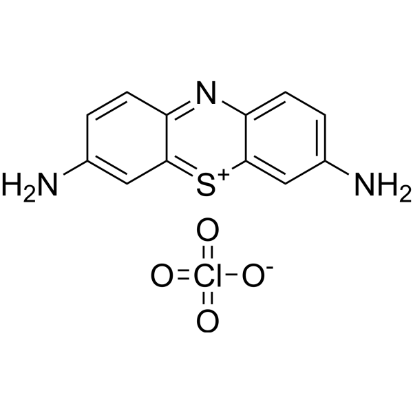
Thionin perchlorate is a general nuclear stain for chromatin and mucin.
loading...
    loading...
Cat. No.: HY-W713888
CAS No.: 66687-07-8
Aniline blue diammonium is a component of commonly used polychrome stains. Aniline blue diammonium is used to stain collagen fibers in tissue sections using Masson′s trichrome protocol for staining multiple components. Collagen is stained blue by this method. The dye is suitable for selective staining of callose in plant specimens and staining histones for assessing nuclear maturity. Aniline blue diammonium is used in Gomori′s one-step trichrome stain and Mallory′s connective tissue stain for tissue including kidney and intestine.
loading...
    loading...
Cat. No.: HY-K0611

MCE Modified Van Gieson Staining Kit employs Celestine Blue and Mayer’s hematoxylin for nuclear staining, providing clearer and more stable nuclear visualization while facilitating longer preservation of stained sections. Ponceau S is used for collagen fiber staining, offering stable coloration and strong resistance to fading. This method enables effective differentiation between collagen fibers and muscle fibers, and can assist in distinguishing collagen fiber–derived tumors from myogenic tumors to a certain extent. It is also suitable for observing tissue or organ injury, repair processes, and the degree of fibrosis.

loading...
    loading...
Cat. No.: HY-DY2027
Target:  

Fluorescent Dye

Research Areas:  

Others

Nissl Staining Solution (Thionin Method) is a neural tissue staining solution with thionin as the core component, which stains Nissl bodies purplish blue and nuclear chromatin blue. Nissl Staining Solution (Thionin Method) relies on positively charged thionin molecules to specifically bind to the negatively charged nucleic acid components in the rough endoplasmic reticulum inside Nissl bodies, and achieves differential staining by regulating pH through a buffer system. Nissl Staining Solution (Thionin Method) has pH-dependent staining selectivity; it exhibits the highest staining specificity for neuronal Nissl bodies under pH 3.65 conditions, and can efficiently label glial processes under pH 4.6 conditions .
loading...
    loading...
Cat. No.: HY-K0609

MCE Modified Masson Staining Kit employs Celestine Blue hematoxylin for light nuclear staining, offering shorter differentiation time compared to conventional methods. It features low toxicity, environmental friendliness, simple operation, and stable performance. The staining results show clear coloration and high contrast. The stained sections can be stored for long periods with minimal fading, facilitating long-term preservation and image analysis. This kit is widely used in studies of connective tissue, muscle tissue, and collagen fibers, and is suitable for histological observation and related pathological analyses.

loading...
    loading...
Cat. No.: HY-137889
CAS No.: 347337-33-1
Target:  

CMV

Research Areas:  

Infection

MLS8969 is a selective human cytomegalovirus (HCMV) entry inhibitor with an EC50 of 0.12 μM (HFFs). MLS8969 reduces viral protein levels and inhibits nuclear staining of HCMV pp65. MLS8969 can be used in studies related to human cytomegalovirus infection .
loading...
    loading...
Cat. No.: HY-D3417
CAS No.: 157199-57-0
Target:  

Fluorescent Dye

Research Areas:  

Others

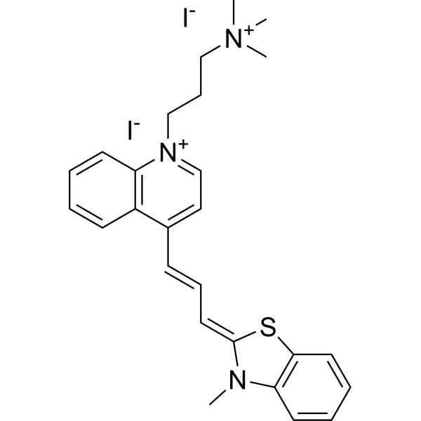
BO-TAP-1 is a cell-impermeable cyan fluorescent carbocyanine monomeric nucleic acid dye that can be used for microarray nucleic acid staining, multicolor fluorescent labeling of cell nuclei and chromosomes, cell detection and counting, and flow cytometry. BO-TAP-1 is non-fluorescent in the absence of nucleic acids, but its fluorescence is significantly enhanced upon binding to double-stranded DNA. The maximum excitation and emission wavelengths of the BO-TAP-1–DNA complex are 454 nm and 487 nm, respectively. BO-TAP-1 can be used in research related to nucleic acid detection and cell analysis.
loading...
    loading...
Cat. No.: HY-DY2025
Target:  

Fluorescent Dye

Research Areas:  

Neurological Disease

Luxol Fast Blue Staining Solution is a copper phthalocyanine-based histological dye that specifically binds to lipid-rich structures such as myelin sheaths and mitochondria, staining them blue-green. Luxol Fast Blue Staining Solution is suitable for displaying myelin sheaths in neural tissues, staining mitochondria in tissues such as kidney/liver, and staining sections for phase contrast microscopy. It is often used in combination with PAS, Phloxine or Cresyl Violet to enhance structural contrast .
loading...
    loading...
  • 1

