



Light green SF yellowish
Based on 1 publication(s) in Google Scholar
Light green SF yellowish (Acid Green 5) is a water-soluble triarylmethane dye. Light green SF yellowish serves as a histological stain that selectively labels mitochondria, collagen, and cartilage, while being an essential component of Papanicolaou staining. Light green SF yellowish is commonly used as a cytoplasmic counterstain for nuclear stains, and is applied in Masson's trichrome staining for collagen fibers, Pap staining, and cytological polychromatic staining in histopathology. Light green SF yellowish also induces growth inhibition and local fibrosarcomas in rats and exerts mild pulmonary tumorigenicity in mice.
For research use only. We do not sell to patients.
- Purity : 75.0%
- CAS No.: 5141-20-8
- Formula: C37H34N2Na2O9S3
- Molecular Weight:792.85
-
Storage:
4°C, protect from light
* In solvent : -80°C, 6 months; -20°C, 1 month (protect from light)

To place orders, for customer services and technical support, please contact: MedChemExpress USA
Tel: 609-228-6898 E-mail: [email protected] [email protected]


-
 Biological Activity
-
Chemical Information
-
Solvent & Solubility
- Protocols
- Purity & Documentation
- References
-
 Help & FAQs


-
HY-D0718
Nile RedHydrophobic Fluorescent Stain -
HY-B1102
Evans BlueL-glutamate Uptake Inhibitor -

HY-DY1008
Nile Red (solution)Hydrophobic Fluorescent Stain -
HY-111391
Resazurin sodiumBacterial Inhibitor -
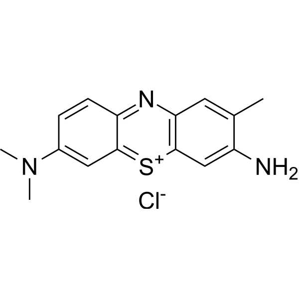
HY-D0220
Toluidine BlueBasic Dye -
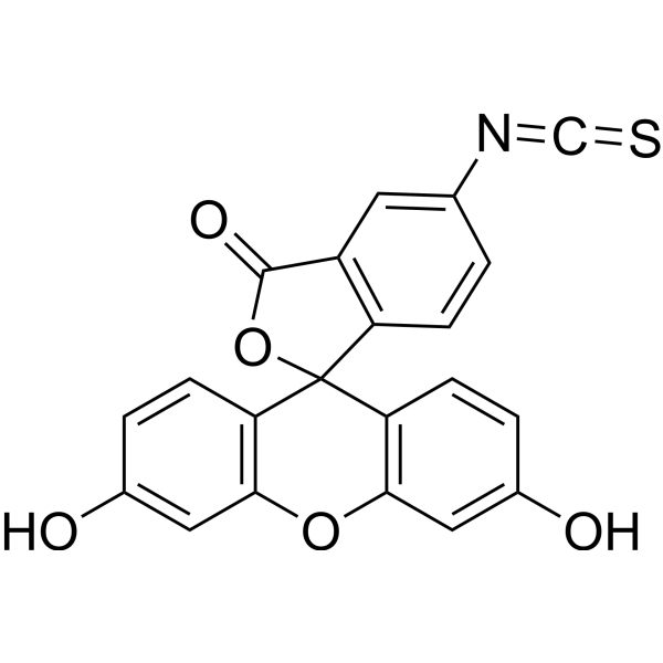
HY-66019
FITCFluorochrome -
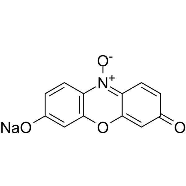
HY-111391A
Resazurin sodium, indicatorBiochemical Assay Reagent -
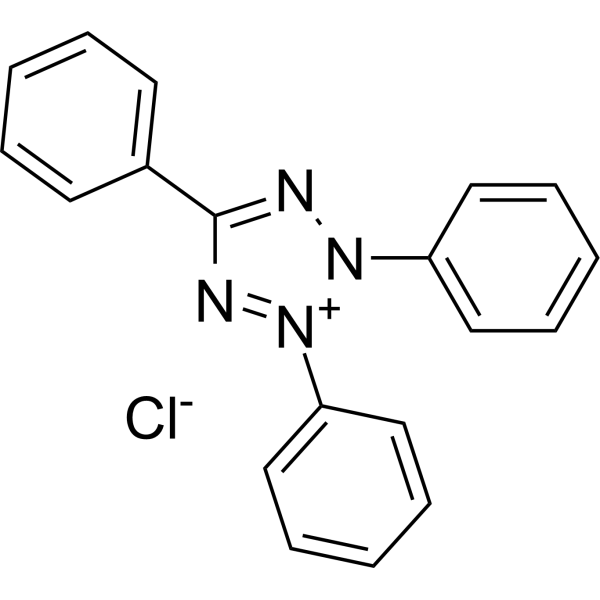
HY-D0714
Tetrazolium RedBiochemical Assay Reagent -
HY-D0014
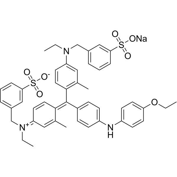
Brilliant blue G-250Dye/P2×7R Antagonist -
HY-B1571
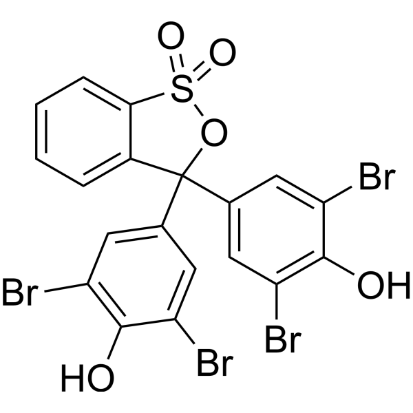
Bromophenol bluepH Indicator -
HY-Y0695
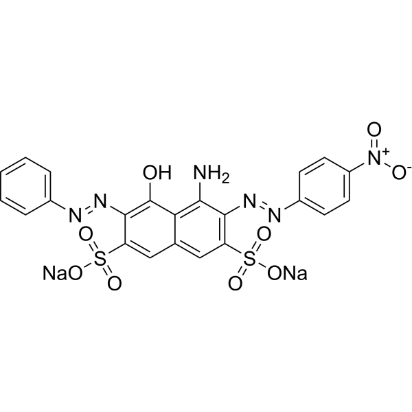
Amido Black 10BAzo Dye -

HY-111391R
Resazurin sodium (Standard)Bacterial Inhibitor
Publications Citing Use of MedChemExpress (MCE) Light green SF yellowish
More

Biological Activity
Description
In Vivo
Light green SF yellowish (1.0-2.0% dietary concentration; dietary; continuously; 2 years) increases survival at 1.0% dietary concentration, decreases survival at 2.0% dietary concentration, and produces a statistically significant increase in total lung tumour incidence without increasing malignant lung tumour or other organ tumour incidence when fed to C3HeB/FeJ mice for 2 years[3].
Light green SF yellowish (0.5-5.0% dietary concentrations; dietary; continuously; 2 years) induces growth inhibition in Osborne-Mendel rats at high dietary levels, with no significant differences in tumour incidence or reagent-related lesions[3].
MedChemExpress (MCE) has not independently confirmed the accuracy of these methods. They are for reference only.
-
Animal Model:Osborne-Mendel (weanling, 50 per group, evenly divided by sex)[3]
-
Dosage:0.5%, 1.0%, 2.0%, 5.0% dietary concentrations
-
Administration:dietary; continuously; 2 years
-
Result:Induced growth inhibition in male rats at 2.0% and 5.0% dietary levels, and in female rats at 5.0% dietary level.
Showed no statistically significant differences in tumour incidence (including malignant tumours) between treated groups and controls.
Exhibited no gross or microscopic lesions attributable to the reagent.


Chemical Information
-
CAS No. 5141-20-8
-
Appearance Solid
-
Molecular Weight 792.85
-
Formula C37H34N2Na2O9S3
-
Color Purple to purplish red
-
SMILES
O=S(C1=CC(CN(CC)C2=CC=C(/C(C3=CC=C(S(=O)(O[Na])=O)C=C3)=C4C=C/C(C=C/4)=[N+](CC)\CC5=CC=CC(S(=O)([O-])=O)=C5)C=C2)=CC=C1)(O[Na])=O
-
Synonyms
Acid Green 5
-
Shipping
Room temperature in continental US; may vary elsewhere.
-
Storage
4°C, protect from light
* In solvent : -80°C, 6 months; -20°C, 1 month (protect from light)


Publications (1)
-
Journal Impact Factor
-
Most Recent
-
Sci Rep
Catabolic mediators from TLR2-mediated proteoglycan aggrecan peptide-stimulated chondrocytes are reduced by Lactobacillus-conditioned media. [Abstract]2024 Aug 5;14(1):18043. PMID: 39103466


Solvent & Solubility
In Vitro:
DMSO : 50 mg/mL (63.06 mM; ultrasonic and warming and heat to 60°C; Hygroscopic DMSO has a significant impact on the solubility of product, please use newly opened DMSO)
H2O : 5 mg/mL (6.31 mM; ultrasonic and warming and heat to 60°C)
Please refer to the solubility information to select the appropriate solvent. Once prepared, please aliquot and store the solution to prevent product inactivation from repeated freeze-thaw cycles.
Storage method and period of stock solution: -80°C, 6 months; -20°C, 1 month (protect from light). When stored at -80°C, please use it within 6 months. When stored at -20°C, please use it within 1 month.
* Note: If you choose water as the stock solution, please dilute it to the working solution, then filter and sterilize it with a 0.22 μm filter before use.
Please refer to the solubility information to select the appropriate solvent. Once prepared, please aliquot and store the solution to prevent product inactivation from repeated freeze-thaw cycles.
Storage method and period of stock solution: -80°C, 6 months; -20°C, 1 month (protect from light). When stored at -80°C, please use it within 6 months. When stored at -20°C, please use it within 1 month.
* Note: If you choose water as the stock solution, please dilute it to the working solution, then filter and sterilize it with a 0.22 μm filter before use.
Concentration (start) × Volume (start) = Concentration (final) × Volume (final)

Protocols
-
Carcinogenicity Bioassay
A carcinogenicity bioassay detects whether long-term exposure to a test substance increases benign or malignant tumor incidence, changes tumor spectrum, or shortens tumor latency in experimental animals; the classical rodent design exposes rats and/or mice to multiple dose levels for most of their lifespan, followed by complete necropsy and histopathologic diagnosis of neoplastic and non-neoplastic lesions. The readout is tumor incidence by organ, sex, species, dose group, and survival status; interpretation requires concurrent controls, dose-response assessment, survival-adjusted tumor statistics, and pathology review because mortality, spontaneous tumor background, and body-weight effects can influence apparent tumor rates.
-
Connective Tissue: Masson's Trichrome/Collagen Trichrome Staining
Masson’s Trichrome (collagen/trichrome staining) is a histological technique that differentially stains tissue compartments using sequential acidic dyes to distinguish collagen from muscle and cytoplasmic components based on dye affinity and tissue permeability differences, enabling visualization of fibrosis and connective tissue architecture in histological sections. The classical formulation typically uses Weigert's iron hematoxylin for nuclear staining, Biebrich scarlet-acid fuchsin for cytoplasm and muscle, and aniline blue (or light green variants) for collagen, producing a characteristic blue/green collagen signal contrasted against red cytoplasm and dark nuclei. The staining principle relies on selective displacement of smaller dye molecules by larger anionic dyes in collagen-rich regions under controlled acidified conditions, which enhances collagen-specific dye retention. This property makes the method widely used for fibrosis assessment in organs such as heart, liver, lung, a
-
Soft Agar Colony Formation Assay
Soft agar colony formation assay measures anchorage-independent growth, in which transformed or tumorigenic cells proliferate as colonies in a semisolid agar matrix while many non-transformed adherent cells fail to proliferate without attachment; classic studies showed that growth in semisolid medium correlates with tumorigenicity in nude mice, and later protocol papers describe the method as a stringent in vitro assay for malignant transformation. The readout is the number, size, morphology, or signal intensity of colonies formed within agar after incubation; published formats include manual colony counting after staining, 96-well or 384-well quantitative formats, DNA-binding dye detection, MTT/tetrazolium-based detection, digital image analysis, and PCR-based marker detection from soft agar cultures.


Purity & Documentation
-
Data Sheet (283 KB)
-
SDS (418 KB)
- English - EN (418 KB)
- Français - FR (418 KB)
- Deutsch - DE (418 KB)
- Norwegian - NO (418 KB)
- Español - ES (418 KB)
- Swedish - SV (418 KB)
- Italian - IT (418 KB)
- Korean - KR (418 KB)
- Portuguese - PT (418 KB)
-
Handling Instructions (2659 KB)
References
[1]. Mittal A, et al. Decoloration treatment of a hazardous triarylmethane dye, Light Green SF (Yellowish) by waste material adsorbents. J Colloid Interface Sci. 2010;342(2):518-527. [Content Brief]
[2]. Harman JW. The Selective Staining of Mitochondria. Stain Technol. 1950;25(2):69-72.
[3]. Hansen WH, et al. Chronic toxicity of three food colourings: Guinea Green B, Light Green SF Yellowish and Fast Green FCF in rats, dogs and mice. Food Cosmet Toxicol. 1966 Aug;4(4):389-410. [Content Brief]
[4]. Sengprasert P, et al. Catabolic mediators from TLR2-mediated proteoglycan aggrecan peptide-stimulated chondrocytes are reduced by Lactobacillus-conditioned media. Sci Rep. 2024;14(1):18043. Published 2024 Aug 5. [Content Brief]


Complete Stock Solution Preparation Table
Please refer to the solubility information to select the appropriate solvent. Once prepared, please aliquot and store the solution to prevent product inactivation from repeated freeze-thaw cycles.
Storage method and period of stock solution: -80°C, 6 months; -20°C, 1 month (protect from light). When stored at -80°C, please use it within 6 months. When stored at -20°C, please use it within 1 month.
| Optional Solvent | Concentration Solvent Mass | 1 mg | 5 mg | 10 mg | 25 mg |
|---|---|---|---|---|---|
| H2O / DMSO | 1 mM | 1.2613 mL | 6.3064 mL | 12.6127 mL | 31.5318 mL |
| 5 mM | 0.2523 mL | 1.2613 mL | 2.5225 mL | 6.3064 mL | |
| DMSO | 10 mM | 0.1261 mL | 0.6306 mL | 1.2613 mL | 3.1532 mL |
| 15 mM | 0.0841 mL | 0.4204 mL | 0.8408 mL | 2.1021 mL | |
| 20 mM | 0.0631 mL | 0.3153 mL | 0.6306 mL | 1.5766 mL | |
| 25 mM | 0.0505 mL | 0.2523 mL | 0.5045 mL | 1.2613 mL | |
| 30 mM | 0.0420 mL | 0.2102 mL | 0.4204 mL | 1.0511 mL | |
| 40 mM | 0.0315 mL | 0.1577 mL | 0.3153 mL | 0.7883 mL | |
| 50 mM | 0.0252 mL | 0.1261 mL | 0.2523 mL | 0.6306 mL | |
| 60 mM | 0.0210 mL | 0.1051 mL | 0.2102 mL | 0.5255 mL |
* Note: If you choose water as the stock solution, please dilute it to the working solution, then filter and sterilize it with a 0.22 μm filter before use.


Light green SF yellowish Related Classifications
HY-126367 Related Classifications
 Powered by Bioz