


Crizotinib
Based on 89 publication(s) in Google Scholar
Crizotinib (PF-02341066) is an orally bioavailable, ATP-competitive ALK and c-Met inhibitor with IC50s of 20 and 8 nM, respectively. Crizotinib inhibits tyrosine phosphorylation of NPM-ALK and tyrosine phosphorylation of c-Met with IC50s of 24 and 11 nM in cell-based assays, respectively. Crizotinib is also a ROS1 inhibitor. Crizotinib has effective tumor growth inhibition.
For research use only. We do not sell to patients.
- Purity: 99.74%
- CAS No.: 877399-52-5
- Formula: C21H22Cl2FN5O
- Molecular Weight:450.34
-
Storage:Powder -20°C, 3 years , 4°C, 2 years ; In solvent -80°C, 1 year , -20°C, 6 months

To place orders, for customer services and technical support, please contact: MedChemExpress USA
Tel: 609-228-6898 E-mail: [email protected] [email protected]


-
 Biological Activity
-
Chemical Information
-
Solvent & Solubility
- Protocol
- Purity & Documentation
- References
-
 Help & FAQs
-


Kinase Inhibitor Library
HY-L009
-
Protein Tyrosine Kinase Compound Library
HY-L016
-
FDA-Approved Drug Library
HY-L022
-
Anti-Cancer Compound Library
HY-L025
-
Drug Repurposing Compound Library
HY-L035
-
NMPA-Approved Drug Library
HY-L053
-
Orally Active Compound Library
HY-L061
-
Glutamine Metabolism Compound Library
HY-L064
-
FDA Approved & Pharmacopeial Drug Library
HY-L066
-
Anti-Lung Cancer Compound Library
HY-L075
-
Drug-Induced Liver Injury (DILI) Compound Library
HY-L076
-
Anti-Pancreatic Cancer Compound Library
HY-L077
-
Anti-Blood Cancer Compound Library
HY-L079
-
Targeted Therapy Drug Library
HY-L080
-
Angiogenesis-Related Compound Library
HY-L088
-
Anti-Liver Cancer Compound Library
HY-L101
-
Rare Diseases Drug Library
HY-L102
-
Anti-Colorectal Cancer Compound Library
HY-L103
-
EMA-Approved Drug Library
HY-L116
-
FDA-Approved Anticancer Drug Library
HY-L122
-
Human Metabolite Library
HY-L123
-
Anti-Prostate Cancer Compound Library
HY-L124
-
Target Protein Ligand Library
HY-L129
-
Non-steroidal Anti-Inflammatory Compound Library
HY-L130
-
Cancer Stem Cells Compound Library
HY-L135
-
Heterocyclic Compound Library
HY-L138
-
Off-patent Drug Library
HY-L141
-
Mitochondrial Protection Compound Library
HY-L144
-
Membrane Protein-targeted Compound Library
HY-L149
-
Membrane Receptor-targeted Compound Library
HY-L150
-
Mitochondrial Toxicity Compound Library
HY-L155
-
Cytokine Inhibitors Library
HY-L161
-
Serine/Threonine Kinase Inhibitor Library
HY-L164
-
Anti-Hematopathy Compound Library
HY-L171
-
Anti-Ovarian Cancer Compound Library
HY-L173
-
Multi-Target Compound Library
HY-L176
-
Bioactive Compound Library Max
HY-L181
-
Kinase Inhibitor Library Mini
HY-L009M
-
MCE Bioactive Compound Library
HY-L001V
-
Drug Repurposing Compound Library Plus
HY-L035P
-
FDA-Approved Drug Library Plus
HY-L022P
-
FDA-Approved Drug Library Mini
HY-L022M
-
Bioactive Compound Library
HY-L001
-
Anti-Gastric Cancer Compound Library
HY-L184
-
Anti-Brain Cancer Compound Library
HY-L188
-
Protein Kinase Compound Library
HY-L196
-
High-Throughput Bioactive Compound Library
HY-L205
-
Anti-Cancer Approved Drug Library
HY-L213
-
Mass Spectrometry Human Metabolite Library
HY-L215
-
Cardiotoxic Compound Library
HY-L225
-
Posttranslational Modification Library
HY-L226
-
Nephrotoxicity Compound Library
HY-L229
-
FDA Kinase Inhibitor Library
HY-L230
-
RNA Binding Bioactive Compound Library
HY-L248


Publications Citing Use of MedChemExpress (MCE) Crizotinib
More- Signal Transduct Target Ther. 2024 Mar 9;9(1):65. [Abstract]
- Cancer Cell. 2026 May 11;44(5):983-994.e5. [Abstract]
- J Hematol Oncol. 2018 Aug 29;11(1):109. [Abstract]
- Cancer Discov. 2024 Sep 13:OF1-OF20. [Abstract]
- Cancer Discov. 2023 Mar 1;13(3):598-615. [Abstract]
- Cancer Discov. 2018 Mar;8(3):354-369. [Abstract]
- Nat Biomed Eng. 2018 Aug;2(8):578-588. [Abstract]
- Blood. 2022 Feb 3;139(5):717-731. [Abstract]
- Cancer Res. 2015 Nov 1;75(21):4548-59. [Abstract]
- Nat Commun. 2025 Jul 17;16(1):6587. [Abstract]
- Sci Transl Med. 2021 Sep;13(609):eabb3738. [Abstract]
- Sci Transl Med. 2018 Jul 18;10(450):eaaq1093. [Abstract]
- Acta Pharm Sin B. 2026 Mar 2.
- Biomark Res. 2024 Jan 25;12(1):13. [Abstract]
- J Exp Clin Cancer Res. 2025 Jul 19;44(1):215. [Abstract]
- J Exp Clin Cancer Res. 2022 Mar 29;41(1):113. [Abstract]
- MedComm (2020). 2025 Jul 21;6(8):e70286. [Abstract]
- Cell Rep Med. 2025 Apr 2:102053. [Abstract]
- Cell Rep Med. 2024 Mar 19;5(3):101472. [Abstract]
- Cell Rep Med. 2023 Feb 21;4(2):100911. [Abstract]
- Cancer Lett. 2026 May 29:656:218624. [Abstract]
- Cancer Lett. 2018 May 28:422:19-28. [Abstract]
- EBioMedicine. 2023 Jan:87:104410. [Abstract]
- J Pharm Anal. 2021 Dec;11(6):799-807. [Abstract]
- J Transl Med. 2023 Aug 5;21(1):530. [Abstract]
- Oncogene. 2024 Sep;43(40):2995-3002. [Abstract]
- Oncogene. 2018 Mar;37(11):1417-1429. [Abstract]
- Leukemia. 2025 Aug 14. [Abstract]
- Int J Biol Macromol. 2025 Jun 4;318(Pt 1):144963. [Abstract]
- Clin Transl Med. 2025 May;15(5):e70338. [Abstract]
- Blood Adv. 2025 Jul 2:bloodadvances.2024015322. [Abstract]
- Blood Adv. 2023 Aug 8;7(15):4049-4063. [Abstract]
- J Med Chem. 2024 Oct 24;67(20):18098-18123. [Abstract]
- J Med Chem. 2021 Oct 14;64(19):14344-14357. [Abstract]
- J Med Chem. 2021 Mar 11;64(5):2725-2738. [Abstract]
- Sci Signal. 2015 Dec 8;8(406):ra125. [Abstract]
- Sci Signal. 2014 Oct 28;7(349):ra102. [Abstract]
- Mol Cancer Ther. 2025 Jul 2;24(7):1005-1019. [Abstract]
- Talanta. 2019 Aug 15:201:217-225. [Abstract]
- Eur J Med Chem. 2017 Oct 20:139:674-697. [Abstract]
- Anal Chim Acta. 2026 May 12;1412:345649.
- Cell Rep Methods. 2023 Oct 23;3(10):100599. [Abstract]
- Pharmaceuticals (Basel). 2026 Feb 27;19(3):381. [Abstract]
- Int J Mol Sci. 2022 Sep 17;23(18):10895. [Abstract]
- Stem Cell Reports. 2017 Dec 12;9(6):1948-1960. [Abstract]
- ACS Omega. 2023 Jun 14;8(25):22603-22612. [Abstract]
- Int J Antimicrob Agents. 2025 Jun;65(6):107470. [Abstract]
- Cancer Sci. 2025 Jul 23. [Abstract]
- Transl Oncol. 2022 Jan;15(1):101272. [Abstract]
- Transl Oncol. 2021 Jan;14(1):100887. [Abstract]
- Cancers (Basel). 2024
- Spectrochim Acta A Mol Biomol Spectrosc. 2021 Oct 5:259:119884. [Abstract]
- Cancers (Basel). 2017 Oct 30;9(11). pii: E149. [Abstract]
- Spectrochim Acta A Mol Biomol Spectrosc. 2014 Oct 15;131:347-54. [Abstract]
- J Pharm Investig. 2025 Dec 29.
- Mol Oncol. 2017 Aug;11(8):996-1006. [Abstract]
- Bioengineering (Basel). 2025 Oct 19;12(10):1121. [Abstract]
- Proteomes. 2023 Jun 2;11(2):20. [Abstract]
- Biochim Biophys Acta Mol Cell Res. 2020 Jul;1867(7):118712. [Abstract]
- Saudi Pharm J. 2015 Jan;23(1):75-84. [Abstract]
- Dis Model Mech. 2016 Sep 1;9(9):941-52. [Abstract]
- Arch Biochem Biophys. 2024 Mar:753:109905. [Abstract]
- Exp Cell Res. 2020 Aug 1;393(1):112054. [Abstract]
- Cancer Med. 2020 Jun;9(12):4350-4359. [Abstract]
- Invest New Drugs. 2023 Apr;41(2):254-266. [Abstract]
- Front Oncol. 2020 May 12;10:696. [Abstract]
- J Cancer Res Clin Oncol. 2021 Jan;147(1):167-175. [Abstract]
- PLoS One. 2025 Jan 21;20(1):e0308747. [Abstract]
- PLoS One. 2021 Jun 8;16(6):e0252907. [Abstract]
- PLoS One. 2019 Feb 11;14(2):e0212048. [Abstract]
- Fundam Clin Pharmacol. 2021 Oct;35(5):919-929. [Abstract]
- Biol Methods Protoc. 2025 Feb 13;10(1):bpaf012. [Abstract]
- Eur J Drug Metab Pharmacokinet. 2021 Sep;46(5):625-635. [Abstract]
- Biomed Chromatogr. 2024 Oct;38(10):e5986. [Abstract]
- Biomed Chromatogr. 2019 Oct;33(10):e4611. [Abstract]
- J Solution Chem. 2014 Jul;43(7):1282-1295.
- Braz J Pharm Sci. 2015 Jun;51(2):2175-9790.
- bioRxiv. 2025 Dec 5.
- Patent. US20250269018A1.
- bioRxiv. 2024 Nov 6:2024.11.04.621884. [Abstract]
- Eberhard Karls Universität Tübingen. 2023 Sep 18.
- Research Square Print. 26 Oct, 2022
- Research Square Print. September 14th, 2022.
- Research Square Preprint. 2022 Feb.
- Evid Based Complement Alternat Med. 2019 Nov 7;2019:4253846. [Abstract]
- Oncotarget. 2019 Aug 13;10(48):4937-4950. [Abstract]
- Technical University of Munich. 24.01.2018.
- Oncotarget. 2016 May 17;7(20):29011-22. [Abstract]
- Oncotarget. 2014 May 15;5(9):2688-702. [Abstract]
 Customer Validation & Images

-
WB
-
WB
-
WB
-
WB
-
WB


Biological Activity
IC50: 20 nM (ALK), 8 nM (c-Met)[3]
|
Cell Line
|
Type | Value | Description | References |
|---|---|---|---|---|
| A549 | IC50 |
0.008 μM
Compound: 63, Crizotinib, PF-02341066
|
Inhibition of human recombinant c-MET kinase expressed in A549 cells assessed as inhibition of HGF-induced autophosphorylation by ELISA method
Inhibition of human recombinant c-MET kinase expressed in A549 cells assessed as inhibition of HGF-induced autophosphorylation by ELISA method
|
[PMID: 21812414] |
| A549 | IC50 |
>1 nM
Compound: Crizotinib
|
Antiproliferative activity against human A549 cells after 72 hrs by MTT assay
Antiproliferative activity against human A549 cells after 72 hrs by MTT assay
|
[PMID: 24785465] |
| A549 | IC50 |
4.084 μM
Compound: Crizotinib
|
Cytotoxicity against human A549 cells after 48 hrs by MTT assay
Cytotoxicity against human A549 cells after 48 hrs by MTT assay
|
[PMID: 24900830] |
| A549 | IC50 |
>10 μM
Compound: Crizotinib
|
Cytotoxicity against human A549 cells assessed as reduction in cell proliferation after 72 hrs by MTT assay
Cytotoxicity against human A549 cells assessed as reduction in cell proliferation after 72 hrs by MTT assay
|
[PMID: 27474925] |
| A549 | IC50 |
>1 μM
Compound: Crizotinib
|
Antiproliferative activity against human A549 cells harboring EGFR after 72 hrs by MTT assay
Antiproliferative activity against human A549 cells harboring EGFR after 72 hrs by MTT assay
|
[PMID: 29174809] |
| A549 | IC50 |
0.1343 μM
Compound: Crizotinib
|
Antiproliferative activity against human A549 cells after 72 hrs by Alamarblue assay relative to control
Antiproliferative activity against human A549 cells after 72 hrs by Alamarblue assay relative to control
|
[PMID: 29202410] |
| A549 | IC50 |
>1 μM
Compound: Crizotinib
|
Antiproliferative activity against human A549 cells after 72 hrs by MTT assay
Antiproliferative activity against human A549 cells after 72 hrs by MTT assay
|
[PMID: 30223120] |
| A549 | IC50 |
1.17 μM
Compound: Crizotinib
|
Cytotoxicity against human A549 cells harboring ALK G1202R mutation incubated for 72 hrs by MTT assay
Cytotoxicity against human A549 cells harboring ALK G1202R mutation incubated for 72 hrs by MTT assay
|
[PMID: 30927566] |
| A549 | IC50 |
2.31 μM
Compound: Crizotinib
|
Cytotoxicity against human A549 cells incubated for 72 hrs by MTT assay
Cytotoxicity against human A549 cells incubated for 72 hrs by MTT assay
|
[PMID: 30927566] |
| A549 | IC50 |
1.06 μM
Compound: Crizotinib
|
Antiproliferative activity against human A549 cells assessed as reduction in cell viability incubated for 72 hrs by MTT assay
Antiproliferative activity against human A549 cells assessed as reduction in cell viability incubated for 72 hrs by MTT assay
|
[PMID: 31260890] |
| A549 | IC50 |
5.7 μM
Compound: PF-02341066
|
Cytotoxicity against EGFR-positive human A549 cells incubated for 72 hrs by MTT assay
Cytotoxicity against EGFR-positive human A549 cells incubated for 72 hrs by MTT assay
|
[PMID: 33069075] |
| A549 | IC50 |
0.41 μM
Compound: Crizotinib
|
Antiproliferative activity against human A549 cells assessed as inhibition of cell proliferation measured after 72 hrs by CCK8 assay
Antiproliferative activity against human A549 cells assessed as inhibition of cell proliferation measured after 72 hrs by CCK8 assay
|
[PMID: 34237620] |
| ASPC1 | GI50 |
2.38 μM
Compound: 2
|
Antiproliferative activity against human ASPC1 cells harbouring KRAS G12D mutant and RON delta 165 assessed as cell growth inhibition measured after 72 hrs by MTS assay
Antiproliferative activity against human ASPC1 cells harbouring KRAS G12D mutant and RON delta 165 assessed as cell growth inhibition measured after 72 hrs by MTS assay
|
[PMID: 37736180] |
| BaF3 | IC50 |
0.19 μM
Compound: 2, PF-2341066
|
Cytotoxicity against mouse BAF3 cells expressing Tel-ALK after 48 hrs by CellTiter-Glo assay
Cytotoxicity against mouse BAF3 cells expressing Tel-ALK after 48 hrs by CellTiter-Glo assay
|
[PMID: 21572589] |
| BaF3 | IC50 |
0.28 μM
Compound: 2, PF-2341066
|
Cytotoxicity against mouse BAF3 cells expressing EML4-ALK after 48 hrs by MTS assay
Cytotoxicity against mouse BAF3 cells expressing EML4-ALK after 48 hrs by MTS assay
|
[PMID: 21572589] |
| BaF3 | IC50 |
0.62 μM
Compound: 2, PF-2341066
|
Cytotoxicity against mouse BAF3 cells expressing ALK F1174L mutant coexpressing EML4 after 48 hrs by MTS assay
Cytotoxicity against mouse BAF3 cells expressing ALK F1174L mutant coexpressing EML4 after 48 hrs by MTS assay
|
[PMID: 21572589] |
| BaF3 | IC50 |
2.2 μM
Compound: 2, PF-2341066
|
Cytotoxicity against mouse BAF3 cells expressing ALK L1196M mutant coexpressing EML4 after 48 hrs by MTS assay
Cytotoxicity against mouse BAF3 cells expressing ALK L1196M mutant coexpressing EML4 after 48 hrs by MTS assay
|
[PMID: 21572589] |
| BaF3 | IC50 |
150.8 nM
Compound: 1, PF-02341066
|
Inhibition of NPM-fused ALK (unknown origin) expressed in mouse BAF3 cells after 2 to 3 days by luciferase reporter gene assay
Inhibition of NPM-fused ALK (unknown origin) expressed in mouse BAF3 cells after 2 to 3 days by luciferase reporter gene assay
|
[PMID: 23742252] |
| BaF3 | IC50 |
1643 nM
Compound: 1, PF-02341066
|
Inhibition of TEL-fused insulin receptor (unknown origin) expressed in mouse BAF3 cells after 2 to 3 days by luciferase reporter gene assay
Inhibition of TEL-fused insulin receptor (unknown origin) expressed in mouse BAF3 cells after 2 to 3 days by luciferase reporter gene assay
|
[PMID: 23742252] |
| BaF3 | IC50 |
3479 nM
Compound: 1, PF-02341066
|
Cytotoxicity against mouse BAF3 cells after 2 to 3 days by luciferase reporter gene assay
Cytotoxicity against mouse BAF3 cells after 2 to 3 days by luciferase reporter gene assay
|
[PMID: 23742252] |
| BaF3 | IC50 |
0.051 μM
Compound: Crizotinib
|
Inhibition of NPM/ALK (unknown origin) transfected in mouse BAF3 cells assessed as cell growth inhibition after 72 hrs by [3H]-thymidine incorporation assay
Inhibition of NPM/ALK (unknown origin) transfected in mouse BAF3 cells assessed as cell growth inhibition after 72 hrs by [3H]-thymidine incorporation assay
|
[PMID: 24468632] |
| BaF3 | IC50 |
0.26 μM
Compound: Crizotinib
|
Inhibition of NPM/ALK L1196M mutant (unknown origin) transfected in mouse BAF3 cells assessed as cell growth inhibition after 72 hrs by [3H]-thymidine incorporation assay
Inhibition of NPM/ALK L1196M mutant (unknown origin) transfected in mouse BAF3 cells assessed as cell growth inhibition after 72 hrs by [3H]-thymidine incorporation assay
|
[PMID: 24468632] |
| BaF3 | IC50 |
0.98 μM
Compound: Crizotinib
|
Cytotoxicity against mouse BAF3 cells assessed as growth inhibition after 72 hrs by [3H]-thymidine incorporation assay
Cytotoxicity against mouse BAF3 cells assessed as growth inhibition after 72 hrs by [3H]-thymidine incorporation assay
|
[PMID: 24468632] |
| BaF3 | IC50 |
>2000 nM
Compound: PF-02341066
|
Antiproliferative activity against mouse BaF3 cells harboring CD74-ROS1 G2032R mutant assessed as reduction in cell viability incubated for 72 hrs by CellTiter-Glo assay
Antiproliferative activity against mouse BaF3 cells harboring CD74-ROS1 G2032R mutant assessed as reduction in cell viability incubated for 72 hrs by CellTiter-Glo assay
|
[PMID: 25733882] |
| BaF3 | IC50 |
18 nM
Compound: PF-02341066
|
Antiproliferative activity against mouse BaF3 cells harboring CD74-ROS1 assessed as reduction in cell viability incubated for 72 hrs by CellTiter-Glo assay
Antiproliferative activity against mouse BaF3 cells harboring CD74-ROS1 assessed as reduction in cell viability incubated for 72 hrs by CellTiter-Glo assay
|
[PMID: 25733882] |
| BaF3 | IC50 |
259 nM
Compound: PF-02341066
|
Antiproliferative activity against mouse BaF3 cells harboring CD74-ROS1 L2026M mutant assessed as reduction in cell viability incubated for 72 hrs by CellTiter-Glo assay
Antiproliferative activity against mouse BaF3 cells harboring CD74-ROS1 L2026M mutant assessed as reduction in cell viability incubated for 72 hrs by CellTiter-Glo assay
|
[PMID: 25733882] |
| BaF3 | IC50 |
127.4 nM
Compound: 1
|
Antiproliferative activity against mouse BAF3/TPR-Met cells after 72 hrs
Antiproliferative activity against mouse BAF3/TPR-Met cells after 72 hrs
|
[PMID: 26005523] |
| BaF3 | IC50 |
144 nM
Compound: 4
|
Inhibition of EML4-ALK C1156Y mutant (unknown origin) expressed in mouse Ba/F3 cells assessed as cell viability after 72 hrs by MTS assay
Inhibition of EML4-ALK C1156Y mutant (unknown origin) expressed in mouse Ba/F3 cells assessed as cell viability after 72 hrs by MTS assay
|
[PMID: 26568289] |
| BaF3 | IC50 |
328 nM
Compound: 4
|
Inhibition of EML4-ALK G1202R mutant (unknown origin) expressed in mouse Ba/F3 cells assessed as cell viability after 72 hrs by MTS assay
Inhibition of EML4-ALK G1202R mutant (unknown origin) expressed in mouse Ba/F3 cells assessed as cell viability after 72 hrs by MTS assay
|
[PMID: 26568289] |
| BaF3 | IC50 |
512 nM
Compound: 4
|
Inhibition of EML4-ALK G1269A mutant (unknown origin) expressed in mouse Ba/F3 cells assessed as cell viability after 72 hrs by MTS assay
Inhibition of EML4-ALK G1269A mutant (unknown origin) expressed in mouse Ba/F3 cells assessed as cell viability after 72 hrs by MTS assay
|
[PMID: 26568289] |
| BaF3 | IC50 |
549 nM
Compound: 4
|
Inhibition of EML4-ALK L1196M mutant (unknown origin) expressed in mouse Ba/F3 cells assessed as cell viability after 72 hrs by MTS assay
Inhibition of EML4-ALK L1196M mutant (unknown origin) expressed in mouse Ba/F3 cells assessed as cell viability after 72 hrs by MTS assay
|
[PMID: 26568289] |
| BaF3 | IC50 |
56 nM
Compound: 4
|
Inhibition of wild type EML4-ALK (unknown origin) expressed in mouse Ba/F3 cells assessed as cell viability after 72 hrs by MTS assay
Inhibition of wild type EML4-ALK (unknown origin) expressed in mouse Ba/F3 cells assessed as cell viability after 72 hrs by MTS assay
|
[PMID: 26568289] |
| BaF3 | IC50 |
645 nM
Compound: 4
|
Inhibition of EML4-ALK L1152R mutant (unknown origin) expressed in mouse Ba/F3 cells assessed as cell viability after 72 hrs by MTS assay
Inhibition of EML4-ALK L1152R mutant (unknown origin) expressed in mouse Ba/F3 cells assessed as cell viability after 72 hrs by MTS assay
|
[PMID: 26568289] |
| BaF3 | IC50 |
65 nM
Compound: 4
|
Inhibition of EML4-ALK S1206Y mutant (unknown origin) expressed in mouse Ba/F3 cells assessed as cell viability after 72 hrs by MTS assay
Inhibition of EML4-ALK S1206Y mutant (unknown origin) expressed in mouse Ba/F3 cells assessed as cell viability after 72 hrs by MTS assay
|
[PMID: 26568289] |
| BaF3 | IC50 |
81 nM
Compound: 4
|
Inhibition of EML4-ALK F1174L mutant (unknown origin) expressed in mouse Ba/F3 cells assessed as cell viability after 72 hrs by MTS assay
Inhibition of EML4-ALK F1174L mutant (unknown origin) expressed in mouse Ba/F3 cells assessed as cell viability after 72 hrs by MTS assay
|
[PMID: 26568289] |
| BaF3 | IC50 |
857 nM
Compound: 4
|
Inhibition of EML4-ALK 1151Tins mutant (unknown origin) expressed in mouse Ba/F3 cells assessed as cell viability after 72 hrs by MTS assay
Inhibition of EML4-ALK 1151Tins mutant (unknown origin) expressed in mouse Ba/F3 cells assessed as cell viability after 72 hrs by MTS assay
|
[PMID: 26568289] |
| BaF3 | IC50 |
927 nM
Compound: 4
|
Cytotoxicity against mouse BA/F3 cells assessed as cell viability after 72 hrs by MTS assay
Cytotoxicity against mouse BA/F3 cells assessed as cell viability after 72 hrs by MTS assay
|
[PMID: 26568289] |
| BaF3 | IC50 |
1237 nM
Compound: Crizotinib
|
Cytotoxicity against mouse BaF3 cells assessed as inhibition of cell growth in presence of IL-3
Cytotoxicity against mouse BaF3 cells assessed as inhibition of cell growth in presence of IL-3
|
[PMID: 27780853] |
| BaF3 | GI50 |
1.1 μM
Compound: 9
|
Antiproliferative activity against mouse BAF3 cells after 72 hrs by CellTiter-Glo assay
Antiproliferative activity against mouse BAF3 cells after 72 hrs by CellTiter-Glo assay
|
[PMID: 28850922] |
| BaF3 | IC50 |
340 nM
Compound: 1
|
Antiproliferative activity against mouse BAF3 cells harboring EML4-ALK L1196M mutant after 72 hrs by SRB or CCK8 assay
Antiproliferative activity against mouse BAF3 cells harboring EML4-ALK L1196M mutant after 72 hrs by SRB or CCK8 assay
|
[PMID: 29288940] |
| BaF3 | IC50 |
48.6 nM
Compound: 1
|
Antiproliferative activity against mouse BAF3 cells harboring CD74-ROS1 after 72 hrs by SRB or CCK8 assay
Antiproliferative activity against mouse BAF3 cells harboring CD74-ROS1 after 72 hrs by SRB or CCK8 assay
|
[PMID: 29288940] |
| BaF3 | IC50 |
564 nM
Compound: 1
|
Antiproliferative activity against mouse BAF3 cells harboring EML4-ALK G1202R mutant after 72 hrs by SRB or CCK8 assay
Antiproliferative activity against mouse BAF3 cells harboring EML4-ALK G1202R mutant after 72 hrs by SRB or CCK8 assay
|
[PMID: 29288940] |
| BaF3 | IC50 |
57.5 nM
Compound: 1
|
Antiproliferative activity against mouse BAF3 cells harboring EML4-ALK after 72 hrs by SRB or CCK8 assay
Antiproliferative activity against mouse BAF3 cells harboring EML4-ALK after 72 hrs by SRB or CCK8 assay
|
[PMID: 29288940] |
| BaF3 | IC50 |
594 nM
Compound: 1
|
Antiproliferative activity against mouse BAF3 cells harboring CD74-ROS1 G2032R mutant after 72 hrs by SRB or CCK8 assay
Antiproliferative activity against mouse BAF3 cells harboring CD74-ROS1 G2032R mutant after 72 hrs by SRB or CCK8 assay
|
[PMID: 29288940] |
| BaF3 | IC50 |
644 nM
Compound: 1
|
Antiproliferative activity against IL3-stimulated mouse BAF3 cells after 72 hrs by SRB or CCK8 assay
Antiproliferative activity against IL3-stimulated mouse BAF3 cells after 72 hrs by SRB or CCK8 assay
|
[PMID: 29288940] |
| BaF3 | IC50 |
>1 μM
Compound: Crizotinib
|
Antiproliferative activity against mouse BAF3 cells harboring G1202R mutation after 72 hrs by MTT assay
Antiproliferative activity against mouse BAF3 cells harboring G1202R mutation after 72 hrs by MTT assay
|
[PMID: 30223120] |
| BaF3 | IC50 |
>5 μM
Compound: Crizotinib
|
Cytotoxicity against mouse BA/F3 cells harboring ALK G1202R mutation incubated for 72 hrs by MTT assay
Cytotoxicity against mouse BA/F3 cells harboring ALK G1202R mutation incubated for 72 hrs by MTT assay
|
[PMID: 30927566] |
| BaF3 | IC50 |
>5 μM
Compound: Crizotinib
|
Cytotoxicity against mouse BA/F3 cells incubated for 72 hrs by MTT assay
Cytotoxicity against mouse BA/F3 cells incubated for 72 hrs by MTT assay
|
[PMID: 30927566] |
| BaF3 | IC50 |
4.336 nM
Compound: Crizotinib
|
Antiproliferative activity against mouse BAF3 cells expressing wild type CD74/ROS1 assessed as reduction in cell viability incubated for 72 hrs by CCK8 assay
Antiproliferative activity against mouse BAF3 cells expressing wild type CD74/ROS1 assessed as reduction in cell viability incubated for 72 hrs by CCK8 assay
|
[PMID: 31260890] |
| BaF3 | IC50 |
58.5 nM
Compound: Crizotinib
|
Antiproliferative activity against mouse BAF3 cells expressing CD74/ROS1 L2026M mutant assessed as reduction in cell viability incubated for 72 hrs by CCK8 assay
Antiproliferative activity against mouse BAF3 cells expressing CD74/ROS1 L2026M mutant assessed as reduction in cell viability incubated for 72 hrs by CCK8 assay
|
[PMID: 31260890] |
| BaF3 | IC50 |
643.5 nM
Compound: Crizotinib
|
Antiproliferative activity against mouse BAF3 cells expressing CD74/ROS1 G2032R mutant assessed as reduction in cell viability incubated for 72 hrs by CCK8 assay
Antiproliferative activity against mouse BAF3 cells expressing CD74/ROS1 G2032R mutant assessed as reduction in cell viability incubated for 72 hrs by CCK8 assay
|
[PMID: 31260890] |
| BaF3 | IC50 |
111 nM
Compound: Crizotinib
|
Antiproliferative activity against mouse BaF3 cells harbouring ALK wild type assessed as reduction in cell viability by Celltitre-Glo luminescent assay
Antiproliferative activity against mouse BaF3 cells harbouring ALK wild type assessed as reduction in cell viability by Celltitre-Glo luminescent assay
|
[PMID: 35421578] |
| BaF3 | IC50 |
284 nM
Compound: Crizotinib
|
Antiproliferative activity against mouse BaF3 cells harbouring ALK C1156Y mutant assessed as reduction in cell viability by Celltitre-Glo luminescent assay
Antiproliferative activity against mouse BaF3 cells harbouring ALK C1156Y mutant assessed as reduction in cell viability by Celltitre-Glo luminescent assay
|
[PMID: 35421578] |
| BaF3 | IC50 |
704 nM
Compound: Crizotinib
|
Antiproliferative activity against mouse BaF3 cells harbouring ALK L1196M mutant assessed as reduction in cell viability by Celltitre-Glo luminescent assay
Antiproliferative activity against mouse BaF3 cells harbouring ALK L1196M mutant assessed as reduction in cell viability by Celltitre-Glo luminescent assay
|
[PMID: 35421578] |
| CAPAN-1 | GI50 |
5.12 μM
Compound: 2
|
Antiproliferative activity against human CAPAN-1 cells harbouring KRAS G12V mutant and RON delta 155 assessed as cell growth inhibition measured after 72 hrs by MTS assay
Antiproliferative activity against human CAPAN-1 cells harbouring KRAS G12V mutant and RON delta 155 assessed as cell growth inhibition measured after 72 hrs by MTS assay
|
[PMID: 37736180] |
| CHO | GI50 |
3.2 μM
Compound: 9
|
Antiproliferative activity against CHO cells after 72 hrs by CellTiter-Glo assay
Antiproliferative activity against CHO cells after 72 hrs by CellTiter-Glo assay
|
[PMID: 28850922] |
| COLO 205 | IC50 |
2.449 μM
Compound: Crizotinib
|
Antiproliferative activity against human COLO205 cells after 72 hrs by Alamarblue assay
Antiproliferative activity against human COLO205 cells after 72 hrs by Alamarblue assay
|
[PMID: 29202410] |
| DEL | GI50 |
309 nM
Compound: Crizotinib
|
Growth inhibition of human DEL cells harboring NPM-ALK incubated for 72 hrs by CyQuant cell proliferation assay
Growth inhibition of human DEL cells harboring NPM-ALK incubated for 72 hrs by CyQuant cell proliferation assay
|
[PMID: 27780853] |
| EBC-1 | IC50 |
0.053 μM
Compound: 1
|
Antiproliferative activity against human EBC1 cells expressing elevated levels of constitutively active c-Met after 72 hrs by SRB assay
Antiproliferative activity against human EBC1 cells expressing elevated levels of constitutively active c-Met after 72 hrs by SRB assay
|
[PMID: 22863529] |
| EBC-1 | IC50 |
0.023 μM
Compound: Crizotinib
|
Antiproliferative activity against human EBC1 cells after 72 hrs
Antiproliferative activity against human EBC1 cells after 72 hrs
|
[PMID: 23993328] |
| EBC-1 | IC50 |
6.9 nM
Compound: Crizotinib, PF2341066
|
Antiproliferative activity against cMET-amplified human EBC1 cells after 72 hrs
Antiproliferative activity against cMET-amplified human EBC1 cells after 72 hrs
|
[PMID: 24900831] |
| EBC-1 | IC50 |
0.021 μM
Compound: Crizotinib
|
Antiproliferative activity against human EBC1 cells after 72 hrs
Antiproliferative activity against human EBC1 cells after 72 hrs
|
[PMID: 25537530] |
| EBC-1 | IC50 |
21.8 nM
Compound: 1
|
Antiproliferative activity against human EBC1 cells after 72 hrs
Antiproliferative activity against human EBC1 cells after 72 hrs
|
[PMID: 26005523] |
| EBC-1 | IC50 |
19 nM
Compound: Crizotinib
|
Antiproliferative activity against human EBC1 cells after 72 hrs by SRB assay
Antiproliferative activity against human EBC1 cells after 72 hrs by SRB assay
|
[PMID: 26698536] |
| EBC-1 | IC50 |
0.044 μM
Compound: Crizotinib
|
Cytotoxicity against human EBC1 cells assessed as inhibition of cell proliferation after 72 hrs by sulforhodamine B assay
Cytotoxicity against human EBC1 cells assessed as inhibition of cell proliferation after 72 hrs by sulforhodamine B assay
|
[PMID: 27017548] |
| EBC-1 | IC50 |
0.013 μM
Compound: Crizotinib
|
Antiproliferative activity against human EBC1 cells after 72 hrs by Alamarblue assay
Antiproliferative activity against human EBC1 cells after 72 hrs by Alamarblue assay
|
[PMID: 29202410] |
| EBC-1 | IC50 |
39 nM
Compound: Crizotinib
|
Antiproliferative activity against human EBC1 cells after 72 hrs by Cell Titer-Glo assay
Antiproliferative activity against human EBC1 cells after 72 hrs by Cell Titer-Glo assay
|
[PMID: 29602036] |
| EBC-1 | IC50 |
6.9 nM
Compound: Crizotinib
|
Antiproliferative activity against human EBC-1 cells
Antiproliferative activity against human EBC-1 cells
|
[PMID: 38733884] |
| GBM | EC50 |
3200 nM
Compound: 5
|
Synergistic antiproliferative activity against patient-derived GBM cells assessed as cell viability after 48 hrs in presence of temozolomide by 5-Ethynyl-2'-deoxyuridine incorporation assay
Synergistic antiproliferative activity against patient-derived GBM cells assessed as cell viability after 48 hrs in presence of temozolomide by 5-Ethynyl-2'-deoxyuridine incorporation assay
|
[PMID: 31378572] |
| GBM | EC50 |
4.3 μM
Compound: 5
|
Cytotoxicity against patient-derived GBM cells assessed as LDH release after 96 hrs by spectrophotometric method
Cytotoxicity against patient-derived GBM cells assessed as LDH release after 96 hrs by spectrophotometric method
|
[PMID: 31378572] |
| GBM | EC50 |
5600 nM
Compound: 5
|
Antiproliferative activity against patient-derived GBM cells assessed as cell viability after 48 hrs by 5-Ethynyl-2'-deoxyuridine incorporation assay
Antiproliferative activity against patient-derived GBM cells assessed as cell viability after 48 hrs by 5-Ethynyl-2'-deoxyuridine incorporation assay
|
[PMID: 31378572] |
| GBM | IC50 |
480 nM
Compound: 5
|
Synergistic antiproliferative activity against patient-derived GBM cells assessed as reduction in cell viability after 48 hrs in presence of temozolomide by 5-Ethynyl-2'-deoxyuridine incorporation assay
Synergistic antiproliferative activity against patient-derived GBM cells assessed as reduction in cell viability after 48 hrs in presence of temozolomide by 5-Ethynyl-2'-deoxyuridine incorporation assay
|
[PMID: 31378572] |
| GBM | IC50 |
540 nM
Compound: 5
|
Antiproliferative activity against patient-derived GBM cells assessed as reduction in cell viability after 48 hrs by 5-Ethynyl-2'-deoxyuridine incorporation assay
Antiproliferative activity against patient-derived GBM cells assessed as reduction in cell viability after 48 hrs by 5-Ethynyl-2'-deoxyuridine incorporation assay
|
[PMID: 31378572] |
| GL261 | IC50 |
3.43 μM
Compound: Cri
|
Anti-proliferative activity against mouse GL261 cells assessed as cell viability incubated for 2 hrs by CCK8 assay
Anti-proliferative activity against mouse GL261 cells assessed as cell viability incubated for 2 hrs by CCK8 assay
|
[PMID: 37861443] |
| HCC78 | IC50 |
21 nM
Compound: PF-02341066
|
Antiproliferative activity against human HCC78 cells harboring SLC34A2-ROS1 fusion protein assessed as reduction in cell viability incubated for 72 hrs by CellTiter-Glo assay
Antiproliferative activity against human HCC78 cells harboring SLC34A2-ROS1 fusion protein assessed as reduction in cell viability incubated for 72 hrs by CellTiter-Glo assay
|
[PMID: 25733882] |
| HCC78 | IC50 |
47 nM
Compound: PF-02341066
|
Antiproliferative activity against human HCC78 cells assessed as reduction in cell viability incubated for 72 hrs by CellTiter-Glo assay
Antiproliferative activity against human HCC78 cells assessed as reduction in cell viability incubated for 72 hrs by CellTiter-Glo assay
|
[PMID: 25733882] |
| HCC78 | IC50 |
0.34 μM
Compound: Crizotinib
|
Cytotoxicity against human HCC78 cells harboring SLC34A2-ROS1 assessed as reduction in cell proliferation after 72 hrs by MTT assay
Cytotoxicity against human HCC78 cells harboring SLC34A2-ROS1 assessed as reduction in cell proliferation after 72 hrs by MTT assay
|
[PMID: 27474925] |
| HCC78 | IC50 |
0.17 μM
Compound: Crizotinib
|
Antiproliferative activity against human HCC78 cells harboring SLC34A2-ROS1 after 72 hrs by MTT assay
Antiproliferative activity against human HCC78 cells harboring SLC34A2-ROS1 after 72 hrs by MTT assay
|
[PMID: 29174809] |
| HCC78 | IC50 |
2 μM
Compound: Crizotinib
|
Antiproliferative activity against human HCC78 cells after 48 hrs by MTT assay
Antiproliferative activity against human HCC78 cells after 48 hrs by MTT assay
|
[PMID: 29174814] |
| HCC78 | IC50 |
889 nM
Compound: 1
|
Antiproliferative activity against human HCC78 cells harboring SLC34A2-ROS1 after 72 hrs by SRB or CCK8 assay
Antiproliferative activity against human HCC78 cells harboring SLC34A2-ROS1 after 72 hrs by SRB or CCK8 assay
|
[PMID: 29288940] |
| HCC78 | IC50 |
0.17 μM
Compound: Crizotinib
|
Antiproliferative activity against human HCC78 cells harboring SLC34A2-ROS1 after 72 hrs by MTT assay
Antiproliferative activity against human HCC78 cells harboring SLC34A2-ROS1 after 72 hrs by MTT assay
|
[PMID: 30223120] |
| HCC78 | IC50 |
0.23 μM
Compound: Crizotinib
|
Cytotoxicity against human HCC78 cells incubated for 72 hrs by MTT assay
Cytotoxicity against human HCC78 cells incubated for 72 hrs by MTT assay
|
[PMID: 30927566] |
| HCC78 | IC50 |
19.19 μM
Compound: Crizotinib
|
Antiproliferative activity against ROS1-addicted human HCC78 cells assessed as reduction in cell viability incubated for 72 hrs by MTT assay
Antiproliferative activity against ROS1-addicted human HCC78 cells assessed as reduction in cell viability incubated for 72 hrs by MTT assay
|
[PMID: 31260890] |
| HCC78 | IC50 |
0.17 μM
Compound: PF-02341066
|
Cytotoxicity against human HCC78 cells harboring SLC34A2-ROS1 incubated for 72 hrs by MTT assay
Cytotoxicity against human HCC78 cells harboring SLC34A2-ROS1 incubated for 72 hrs by MTT assay
|
[PMID: 33069075] |
| HCC827 | IC50 |
>10 μM
Compound: Crizotinib
|
Cytotoxicity against human HCC827 cells after 48 hrs by MTT assay
Cytotoxicity against human HCC827 cells after 48 hrs by MTT assay
|
[PMID: 24900830] |
| HCC827 | IC50 |
7.25 μM
Compound: Crizotinib
|
Cytotoxicity against human HCC827 cells assessed as growth inhibition after 72 hrs by MTT assay
Cytotoxicity against human HCC827 cells assessed as growth inhibition after 72 hrs by MTT assay
|
[PMID: 27396929] |
| HCT-116 | IC50 |
14.82 μM
Compound: Crizotinib
|
Antiproliferative activity against human HCT116 cells after 72 hrs
Antiproliferative activity against human HCT116 cells after 72 hrs
|
[PMID: 23993328] |
| HCT-116 | IC50 |
0.2536 μM
Compound: Crizotinib
|
Antiproliferative activity against human HCT116 cells after 72 hrs by Alamarblue assay relative to control
Antiproliferative activity against human HCT116 cells after 72 hrs by Alamarblue assay relative to control
|
[PMID: 29202410] |
| HEK293 | IC50 |
2887 nM
Compound: 63, Crizotinib, PF-02341066
|
Inhibition of IR in human HEK293 cells assessed as growth factor-induced autophosphorylation by sandwich ELISA method
Inhibition of IR in human HEK293 cells assessed as growth factor-induced autophosphorylation by sandwich ELISA method
|
[PMID: 21812414] |
| HEK293 | IC50 |
294 nM
Compound: 63, Crizotinib, PF-02341066
|
Inhibition of AXL in human HEK293 cells assessed as growth factor-induced autophosphorylation by sandwich ELISA method
Inhibition of AXL in human HEK293 cells assessed as growth factor-induced autophosphorylation by sandwich ELISA method
|
[PMID: 21812414] |
| HEK293 | IC50 |
37 nM
Compound: Crizotinib
|
Inhibition of recombinant human ALK expressed in HEK293 cells assessed as reduction in ALK autophosphorylation at Tyr1604 residue incubated for 60 mins by sandwich ELISA
Inhibition of recombinant human ALK expressed in HEK293 cells assessed as reduction in ALK autophosphorylation at Tyr1604 residue incubated for 60 mins by sandwich ELISA
|
[PMID: 31009559] |
| HEL | IC50 |
4.725 μM
Compound: 4
|
Antiproliferative activity against HEL cells harboring JAK2 V617F mutant measured after 3 days by CCK8 assay
Antiproliferative activity against HEL cells harboring JAK2 V617F mutant measured after 3 days by CCK8 assay
|
[PMID: 30981578] |
| HepG2 | IC50 |
3.7 μM
Compound: Crizotinib
|
Cytotoxicity against human HepG2 cells assessed as growth inhibition after 72 hrs by MTT assay
Cytotoxicity against human HepG2 cells assessed as growth inhibition after 72 hrs by MTT assay
|
[PMID: 27396929] |
| HepG2 | IC50 |
0.32 μM
Compound: Crizotinib
|
Antiproliferative activity against human HepG2 cells assessed as inhibition of cell proliferation measured after 72 hrs by CCK8 assay
Antiproliferative activity against human HepG2 cells assessed as inhibition of cell proliferation measured after 72 hrs by CCK8 assay
|
[PMID: 34237620] |
| HT-29 | IC50 |
>10 μM
Compound: Crizotinib
|
Cytotoxicity against human HT-29 cells assessed as reduction in cell proliferation after 72 hrs by MTT assay
Cytotoxicity against human HT-29 cells assessed as reduction in cell proliferation after 72 hrs by MTT assay
|
[PMID: 27474925] |
| HT-29 | GI50 |
4 μM
Compound: 2
|
Antiproliferative activity against human HT-29 cells harbouring wild-type KRAS and RON delta 160 assessed as cell growth inhibition measured after 72 hrs by MTS assay
Antiproliferative activity against human HT-29 cells harbouring wild-type KRAS and RON delta 160 assessed as cell growth inhibition measured after 72 hrs by MTS assay
|
[PMID: 37736180] |
| Jurkat | IC50 |
2741 nM
Compound: 63, Crizotinib, PF-02341066
|
Inhibition of LCK in human Jurkat cells assessed as growth factor-induced autophosphorylation by sandwich ELISA method
Inhibition of LCK in human Jurkat cells assessed as growth factor-induced autophosphorylation by sandwich ELISA method
|
[PMID: 21812414] |
| Jurkat | IC50 |
>40 μM
Compound: 70
|
Anti-neprotic activity in human Jurkat cells FADD defient assessed as reduction in TNFalpha-induced necroptosis preincubated for 30 mins followed by addition of TNFalpha-stimulation and further incubated for over night by Cell Titer Glo assay
Anti-neprotic activity in human Jurkat cells FADD defient assessed as reduction in TNFalpha-induced necroptosis preincubated for 30 mins followed by addition of TNFalpha-stimulation and further incubated for over night by Cell Titer Glo assay
|
[PMID: 31622096] |
| Jurkat | IC50 |
2.38 μM
Compound: F11
|
Anti-necroptosis activity against TNFalpha-induced human Jurkat cells assessed as reduction in cell viability
Anti-necroptosis activity against TNFalpha-induced human Jurkat cells assessed as reduction in cell viability
|
[PMID: 38199165] |
| KARPAS-299 | IC50 |
20 nM
Compound: 63, Crizotinib, PF-02341066
|
Inhibition of ALK in human KARPAS299 cells assessed as growth factor-induced autophosphorylation by sandwich ELISA method
Inhibition of ALK in human KARPAS299 cells assessed as growth factor-induced autophosphorylation by sandwich ELISA method
|
[PMID: 21812414] |
| KARPAS-299 | IC50 |
64.2 nM
Compound: 1, PF-02341066
|
Cytotoxicity against human KARPAS299 cells after 2 to 3 days by luciferase reporter gene assay
Cytotoxicity against human KARPAS299 cells after 2 to 3 days by luciferase reporter gene assay
|
[PMID: 23742252] |
| KARPAS-299 | IC50 |
62 nM
Compound: 1, Xalkori
|
Inhibition of ALK-fusion driven cell proliferation in human KARPAS299 cells after 72 hrs by CellTiter Glo assay
Inhibition of ALK-fusion driven cell proliferation in human KARPAS299 cells after 72 hrs by CellTiter Glo assay
|
[PMID: 24432909] |
| KARPAS-299 | IC50 |
200 nM
Compound: Crizotinib, PF2341066
|
Antiproliferative activity against ALK-dependent human KARPAS299 cells after 72 hrs
Antiproliferative activity against ALK-dependent human KARPAS299 cells after 72 hrs
|
[PMID: 24900831] |
| KARPAS-299 | IC50 |
103 nM
Compound: 1; PF-2341066
|
Antiproliferative activity against human KARPAS299 cells after 72 hrs by SRB/CCK-8 assay
Antiproliferative activity against human KARPAS299 cells after 72 hrs by SRB/CCK-8 assay
|
[PMID: 27131066] |
| KARPAS-299 | IC50 |
365 nM
Compound: Crizotinib
|
Antiproliferative activity against human ALK-positive KARPAS299 cells assessed as reduction in cell viability measured after 72 hrs by CellTiter 96 aqueous one solution cell proliferation assay
Antiproliferative activity against human ALK-positive KARPAS299 cells assessed as reduction in cell viability measured after 72 hrs by CellTiter 96 aqueous one solution cell proliferation assay
|
[PMID: 27144831] |
| KARPAS-299 | IC50 |
0.068 μM
Compound: Crizotinib
|
Cytotoxicity against human KARPAS299 cells harboring NPM-ALK assessed as reduction in cell proliferation after 72 hrs by MTT assay
Cytotoxicity against human KARPAS299 cells harboring NPM-ALK assessed as reduction in cell proliferation after 72 hrs by MTT assay
|
[PMID: 27474925] |
| KARPAS-299 | IC50 |
104.9 nM
Compound: Crizotinib
|
Antiproliferative activity against ALK constitutively activated human KARPAS299 cells after 72 hrs by SRB or CCK8 assay
Antiproliferative activity against ALK constitutively activated human KARPAS299 cells after 72 hrs by SRB or CCK8 assay
|
[PMID: 27769623] |
| KARPAS-299 | GI50 |
119 nM
Compound: Crizotinib
|
Growth inhibition of human Karpas-299 cells harboring NPM-ALK incubated for 72 hrs by CyQuant cell proliferation assay
Growth inhibition of human Karpas-299 cells harboring NPM-ALK incubated for 72 hrs by CyQuant cell proliferation assay
|
[PMID: 27780853] |
| KARPAS-299 | IC50 |
0.087 μM
Compound: Crizotinib
|
Antiproliferative activity against human KARPAS299 cells harboring NPM-ALK after 72 hrs by MTT assay
Antiproliferative activity against human KARPAS299 cells harboring NPM-ALK after 72 hrs by MTT assay
|
[PMID: 29174809] |
| KARPAS-299 | IC50 |
176 nM
Compound: 1
|
Antiproliferative activity against human KARPAS299 cells harboring NPM-ALK after 72 hrs by SRB or CCK8 assay
Antiproliferative activity against human KARPAS299 cells harboring NPM-ALK after 72 hrs by SRB or CCK8 assay
|
[PMID: 29288940] |
| KARPAS-299 | IC50 |
0.24 μM
Compound: Crizotinib
|
Antiproliferative activity against human KARPAS299 cells harboring NPM-ALK after 72 hrs by MTT assay
Antiproliferative activity against human KARPAS299 cells harboring NPM-ALK after 72 hrs by MTT assay
|
[PMID: 30223120] |
| KARPAS-299 | IC50 |
1331.74 nM
Compound: Crizotinib
|
Antiproliferative activity against human KARPAS299 cells after 3 days by MTT assay
Antiproliferative activity against human KARPAS299 cells after 3 days by MTT assay
|
[PMID: 30777610] |
| KARPAS-299 | IC50 |
0.097 μM
Compound: Crizotinib
|
Cytotoxicity against human KARPAS299 cells incubated for 72 hrs by MTT assay
Cytotoxicity against human KARPAS299 cells incubated for 72 hrs by MTT assay
|
[PMID: 30927566] |
| KARPAS-299 | IC50 |
0.03 μM
Compound: 1; PF-02341066
|
Antiproliferative activity against human KARPAS299 cells expressing NMP-ALK assessed as reduction in cell viability incubated for 72 hrs by MTT assay
Antiproliferative activity against human KARPAS299 cells expressing NMP-ALK assessed as reduction in cell viability incubated for 72 hrs by MTT assay
|
[PMID: 31569004] |
| KARPAS-299 | IC50 |
0.087 μM
Compound: PF-02341066
|
Cytotoxicity against human KARPAS299 cells harboring NMP-ALK incubated for 72 hrs by MTT assay
Cytotoxicity against human KARPAS299 cells harboring NMP-ALK incubated for 72 hrs by MTT assay
|
[PMID: 33069075] |
| KARPAS-299 | IC50 |
138 nM
Compound: Crizotinib
|
Antiproliferative activity against human KARPAS-299 cells harbouring wild type ALK assessed as reduction in cell viability by Celltitre-Glo luminescent assay
Antiproliferative activity against human KARPAS-299 cells harbouring wild type ALK assessed as reduction in cell viability by Celltitre-Glo luminescent assay
|
[PMID: 35421578] |
| KARPAS-299 | IC50 |
200 nM
Compound: Crizotinib
|
Antiproliferative activity against human KARPAS-299 cells
Antiproliferative activity against human KARPAS-299 cells
|
[PMID: 38733884] |
| Kelly | IC50 |
0.42 μM
Compound: 2, PF-2341066
|
Cytotoxicity against human Kelly cells expressing ALK F1174L mutant
Cytotoxicity against human Kelly cells expressing ALK F1174L mutant
|
[PMID: 21572589] |
| Kelly | IC50 |
>1000 nM
Compound: Crizotinib
|
Antiproliferative activity against crizotinib-resistant human Kelly cells harboring ALK F1174L mutant after 72 hrs by MTT assay
Antiproliferative activity against crizotinib-resistant human Kelly cells harboring ALK F1174L mutant after 72 hrs by MTT assay
|
[PMID: 24785465] |
| Kelly | EC50 |
211 nM
Compound: 4
|
Antiproliferative activity against human Kelly cells expressing EML4-ALK F1174L mutant after 72 hrs by CellTiter-Glo Luminescent Cell Viability Assay
Antiproliferative activity against human Kelly cells expressing EML4-ALK F1174L mutant after 72 hrs by CellTiter-Glo Luminescent Cell Viability Assay
|
[PMID: 26568289] |
| MCF7 | IC50 |
9.58 μM
Compound: Crizotinib
|
Antiproliferative activity against human MCF7 cells after 72 hrs
Antiproliferative activity against human MCF7 cells after 72 hrs
|
[PMID: 23993328] |
| MCF7 | IC50 |
0.045 μM
Compound: Crizotinib
|
Antiproliferative activity against human MCF7 cells after 72 hrs by Alamarblue assay relative to control
Antiproliferative activity against human MCF7 cells after 72 hrs by Alamarblue assay relative to control
|
[PMID: 29202410] |
| MDA-MB-231 | IC50 |
10.8 μM
Compound: Crizotinib
|
Antiproliferative activity against human MDA-MB-231 cells after 72 hrs
Antiproliferative activity against human MDA-MB-231 cells after 72 hrs
|
[PMID: 23993328] |
| MDA-MB-231 | IC50 |
0.79 μM
Compound: Crizotinib
|
Antiproliferative activity against human MDA-MB-231 cells assessed as inhibition of cell proliferation measured after 72 hrs by CCK8 assay
Antiproliferative activity against human MDA-MB-231 cells assessed as inhibition of cell proliferation measured after 72 hrs by CCK8 assay
|
[PMID: 34237620] |
| MDA-MB-231 | IC50 |
8.37 μM
Compound: Crizotinib
|
Antiproliferative activity against human MDA-MB-231 cells harboring wild type BRCA and c-Met amplification assessed as inhibition of cell growth incubated for 7 days by MTT assay
Antiproliferative activity against human MDA-MB-231 cells harboring wild type BRCA and c-Met amplification assessed as inhibition of cell growth incubated for 7 days by MTT assay
|
[PMID: 38477575] |
| MDA-MB-468 | IC50 |
4.54 μM
Compound: Crizotinib
|
Antiproliferative activity against human MDA-MB-468 cells harboring wild type BRCA1/2, HR-proficient and non-c-Met amplification assessed as inhibition of cell growth incubated for 7 days by MTT assay
Antiproliferative activity against human MDA-MB-468 cells harboring wild type BRCA1/2, HR-proficient and non-c-Met amplification assessed as inhibition of cell growth incubated for 7 days by MTT assay
|
[PMID: 38477575] |
| MIA PaCa-2 | IC50 |
7.16 μM
Compound: Crizotinib
|
Cytotoxicity against human MIAPaCa2 cells assessed as growth inhibition after 72 hrs by MTT assay
Cytotoxicity against human MIAPaCa2 cells assessed as growth inhibition after 72 hrs by MTT assay
|
[PMID: 27396929] |
| MIA PaCa-2 | GI50 |
1.457 μM
Compound: 2
|
Antiproliferative activity against human MIA PaCa-2 cells harbouring KRAS G12C mutant and RON delta 160 assessed as cell growth inhibition measured after 72 hrs by MTS assay
Antiproliferative activity against human MIA PaCa-2 cells harbouring KRAS G12C mutant and RON delta 160 assessed as cell growth inhibition measured after 72 hrs by MTS assay
|
[PMID: 37736180] |
| MKN-45 | IC50 |
0.013 μM
Compound: Crizotinib
|
Antiproliferative activity against human MKN45 cells after 72 hrs
Antiproliferative activity against human MKN45 cells after 72 hrs
|
[PMID: 23993328] |
| MKN-45 | IC50 |
38.1 nM
Compound: 1
|
Antiproliferative activity against human MKN45 cells after 72 hrs
Antiproliferative activity against human MKN45 cells after 72 hrs
|
[PMID: 26005523] |
| MKN-45 | IC50 |
0.022 μM
Compound: Crizotinib
|
Antiproliferative activity against human MKN45 cells after 72 hrs by Alamarblue assay
Antiproliferative activity against human MKN45 cells after 72 hrs by Alamarblue assay
|
[PMID: 29202410] |
| MKN-45 | IC50 |
9.1 nM
Compound: 1; PF-0234106
|
Antiproliferative activity against human MKN45 cells overexpressing c-MET assessed as reduction in cell proliferation
Antiproliferative activity against human MKN45 cells overexpressing c-MET assessed as reduction in cell proliferation
|
[PMID: 32371098] |
| MKN-45 | IC50 |
15 nM
Compound: 2
|
Antiproliferative activity against human MKN-45 cells incubated for 72 hrs
Antiproliferative activity against human MKN-45 cells incubated for 72 hrs
|
[PMID: 33957388] |
| NCI-H1975 | IC50 |
7.551 μM
Compound: Crizotinib
|
Cytotoxicity against human NCI-H1975 cells after 48 hrs by MTT assay
Cytotoxicity against human NCI-H1975 cells after 48 hrs by MTT assay
|
[PMID: 24900830] |
| NCI-H1975 | IC50 |
>10 μM
Compound: PF-02341066
|
Cytotoxicity against EGFR-positive human H1975 cells incubated for 72 hrs by MTT assay
Cytotoxicity against EGFR-positive human H1975 cells incubated for 72 hrs by MTT assay
|
[PMID: 33069075] |
| NCI-H1993 | IC50 |
0.061 μM
Compound: Crizotinib
|
Cytotoxicity against human NCI-H1993 cells after 48 hrs by MTT assay
Cytotoxicity against human NCI-H1993 cells after 48 hrs by MTT assay
|
[PMID: 24900830] |
| NCI-H1993 | IC50 |
3 μM
Compound: 12
|
Cytotoxicity against human NCI-H1993 cells assessed as inhibition of cell growth
Cytotoxicity against human NCI-H1993 cells assessed as inhibition of cell growth
|
[PMID: 32858470] |
| NCI-H2228 | IC50 |
118 nM
Compound: 1, Xalkori
|
Inhibition of ALK-fusion driven cell proliferation in human NCI-H2228 cells after 72 hrs by CellTiter Glo assay
Inhibition of ALK-fusion driven cell proliferation in human NCI-H2228 cells after 72 hrs by CellTiter Glo assay
|
[PMID: 24432909] |
| NCI-H2228 | IC50 |
202 nM
Compound: Crizotinib
|
Inhibition of E6a/b;A20 EML4-ALK (unknown origin) expressed in human NCI-H2228 cells assessed as decrease in cell viability after 72 hrs by CellTiter-Glo luminescence assay
Inhibition of E6a/b;A20 EML4-ALK (unknown origin) expressed in human NCI-H2228 cells assessed as decrease in cell viability after 72 hrs by CellTiter-Glo luminescence assay
|
[PMID: 26844689] |
| NCI-H2228 | GI50 |
121 nM
Compound: Crizotinib
|
Growth inhibition of human NCI-H2228 cells harboring EML4-ALK v3a/3b incubated for 72 hrs by CyQuant cell proliferation assay
Growth inhibition of human NCI-H2228 cells harboring EML4-ALK v3a/3b incubated for 72 hrs by CyQuant cell proliferation assay
|
[PMID: 27780853] |
| NCI-H2228 | GI50 |
0.073 μM
Compound: 9
|
Antiproliferative activity against human NCI-H2228 cells harboring EML4-fused ALK varian3 after 72 hrs by CellTiter-Glo assay
Antiproliferative activity against human NCI-H2228 cells harboring EML4-fused ALK varian3 after 72 hrs by CellTiter-Glo assay
|
[PMID: 28850922] |
| NCI-H2228 | IC50 |
2.5 μM
Compound: Crizotinib
|
Antiproliferative activity against human NCI-H2228 cells after 72 hrs by MTT assay
Antiproliferative activity against human NCI-H2228 cells after 72 hrs by MTT assay
|
[PMID: 29091425] |
| NCI-H2228 | IC50 |
0.24 μM
Compound: Crizotinib
|
Antiproliferative activity against human NCI-H2228 cells harboring EML4-ALK after 72 hrs by MTT assay
Antiproliferative activity against human NCI-H2228 cells harboring EML4-ALK after 72 hrs by MTT assay
|
[PMID: 29174809] |
| NCI-H2228 | IC50 |
1.92 μM
Compound: Crizotinib
|
Cytotoxicity against human NCI-H2228 cells harboring EML4-ALK fusion protein assessed as decrease in cell viability after 72 hrs by MTT assay
Cytotoxicity against human NCI-H2228 cells harboring EML4-ALK fusion protein assessed as decrease in cell viability after 72 hrs by MTT assay
|
[PMID: 30108712] |
| NCI-H2228 | IC50 |
0.087 μM
Compound: Crizotinib
|
Antiproliferative activity against human NCI-H2228 cells harboring EML4-ALK after 72 hrs by MTT assay
Antiproliferative activity against human NCI-H2228 cells harboring EML4-ALK after 72 hrs by MTT assay
|
[PMID: 30223120] |
| NCI-H2228 | IC50 |
0.27 μM
Compound: Crizotinib
|
Cytotoxicity against human NCI-H2228 cells incubated for 72 hrs by MTT assay
Cytotoxicity against human NCI-H2228 cells incubated for 72 hrs by MTT assay
|
[PMID: 30927566] |
| NCI-H2228 | IC50 |
2.8 μM
Compound: PF-02341066
|
Cytotoxicity in human NCI-H2228 cells harboring EML4-fused ALK variant 3 incubated for 72 hrs by alamar blue reagent based assay
Cytotoxicity in human NCI-H2228 cells harboring EML4-fused ALK variant 3 incubated for 72 hrs by alamar blue reagent based assay
|
[PMID: 31425908] |
| NCI-H2228 | IC50 |
0.64 μM
Compound: 1; PF-02341066
|
Antiproliferative activity against human NCI-H2228 cells expressing EML4-ALK assessed as reduction in cell viability incubated for 72 hrs by MTT assay
Antiproliferative activity against human NCI-H2228 cells expressing EML4-ALK assessed as reduction in cell viability incubated for 72 hrs by MTT assay
|
[PMID: 31569004] |
| NCI-H2228 | IC50 |
2.62 μM
Compound: Crizotinib
|
Antiproliferative activity against human NCI-H2228 cells harboring EML4/ALK L1196M mutant assessed as inhibition of cell proliferation measured after 72 hrs by CCK8 assay
Antiproliferative activity against human NCI-H2228 cells harboring EML4/ALK L1196M mutant assessed as inhibition of cell proliferation measured after 72 hrs by CCK8 assay
|
[PMID: 34237620] |
| NCI-H2228 | IC50 |
437.9 nM
Compound: 1
|
Antiproliferative activity against human NCI-H2228 cells assessed as reduction in cell viability measured after 72 hrs by CCK8 assay
Antiproliferative activity against human NCI-H2228 cells assessed as reduction in cell viability measured after 72 hrs by CCK8 assay
|
[PMID: 36332597] |
| NCI-H23 | GI50 |
1773 nM
Compound: Crizotinib
|
Growth inhibition of ALK-negative human NCI-H23 cells incubated for 72 hrs by CyQuant cell proliferation assay
Growth inhibition of ALK-negative human NCI-H23 cells incubated for 72 hrs by CyQuant cell proliferation assay
|
[PMID: 27780853] |
| NCI-H3122 | IC50 |
108 nM
Compound: 1, Xalkori
|
Inhibition of ALK-fusion driven cell proliferation in human NCI-H3122 cells after 72 hrs by CellTiter Glo assay
Inhibition of ALK-fusion driven cell proliferation in human NCI-H3122 cells after 72 hrs by CellTiter Glo assay
|
[PMID: 24432909] |
| NCI-H3122 | IC50 |
623 nM
Compound: 1, Xalkori
|
Inhibition of ALK-fusion driven cell proliferation in human NCI-H3122 cells harboring ALK G1269A mutant after 72 hrs by CellTiter Glo assay
Inhibition of ALK-fusion driven cell proliferation in human NCI-H3122 cells harboring ALK G1269A mutant after 72 hrs by CellTiter Glo assay
|
[PMID: 24432909] |
| NCI-H3122 | IC50 |
838 nM
Compound: 1, Xalkori
|
Inhibition of ALK-fusion driven cell proliferation in human NCI-H3122 cells harboring ALK L1196M mutant after 72 hrs by CellTiter Glo assay
Inhibition of ALK-fusion driven cell proliferation in human NCI-H3122 cells harboring ALK L1196M mutant after 72 hrs by CellTiter Glo assay
|
[PMID: 24432909] |
| NCI-H3122 | IC50 |
261.2 nM
Compound: 1
|
Antiproliferative activity against ALK-dependent human NCI-H3122 cells after 72 hrs
Antiproliferative activity against ALK-dependent human NCI-H3122 cells after 72 hrs
|
[PMID: 26476749] |
| NCI-H3122 | EC50 |
32 nM
Compound: 4
|
Antiproliferative activity against human NCI-H3122 cells after 72 hrs by CellTiter-Glo Luminescent Cell Viability Assay
Antiproliferative activity against human NCI-H3122 cells after 72 hrs by CellTiter-Glo Luminescent Cell Viability Assay
|
[PMID: 26568289] |
| NCI-H3122 | IC50 |
300 nM
Compound: Crizotinib
|
Inhibition of E13;A20 EML4-ALK variant (unknown origin) expressed in human NCI-H3122 cells assessed as decrease in cell viability after 72 hrs by CellTiter-Glo luminescence assay
Inhibition of E13;A20 EML4-ALK variant (unknown origin) expressed in human NCI-H3122 cells assessed as decrease in cell viability after 72 hrs by CellTiter-Glo luminescence assay
|
[PMID: 26844689] |
| NCI-H3122 | IC50 |
210 nM
Compound: 1; PF-2341066
|
Antiproliferative activity against human NCI-H3122 cells after 72 hrs by SRB/CCK-8 assay
Antiproliferative activity against human NCI-H3122 cells after 72 hrs by SRB/CCK-8 assay
|
[PMID: 27131066] |
| NCI-H3122 | IC50 |
100.9 nM
Compound: Crizotinib
|
Antiproliferative activity against ALK constitutively activated human NCI-H3122 cells after 72 hrs by SRB or CCK8 assay
Antiproliferative activity against ALK constitutively activated human NCI-H3122 cells after 72 hrs by SRB or CCK8 assay
|
[PMID: 27769623] |
| NCI-H3122 | GI50 |
62 nM
Compound: Crizotinib
|
Growth inhibition of human NCI-H3122 cells harboring EML4-ALK v1 incubated for 72 hrs by CyQuant cell proliferation assay
Growth inhibition of human NCI-H3122 cells harboring EML4-ALK v1 incubated for 72 hrs by CyQuant cell proliferation assay
|
[PMID: 27780853] |
| NCI-H3122 | GI50 |
0.037 μM
Compound: 9
|
Antiproliferative activity against human NCI-H3122 cells harboring EML4-fused ALK varian1 after 72 hrs by CellTiter-Glo assay
Antiproliferative activity against human NCI-H3122 cells harboring EML4-fused ALK varian1 after 72 hrs by CellTiter-Glo assay
|
[PMID: 28850922] |
| NCI-H3122 | IC50 |
0.8 μM
Compound: Crizotinib
|
Antiproliferative activity against human NCI-H3122 cells after 48 hrs by MTT assay
Antiproliferative activity against human NCI-H3122 cells after 48 hrs by MTT assay
|
[PMID: 29174814] |
| NCI-H3122 | IC50 |
303 nM
Compound: 1
|
Antiproliferative activity against human NCI-H3122 cells after 72 hrs by SRB or CCK8 assay
Antiproliferative activity against human NCI-H3122 cells after 72 hrs by SRB or CCK8 assay
|
[PMID: 29288940] |
| NCI-H3122 | IC50 |
0.092 μM
Compound: Crizotinib
|
Antiproliferative activity against human NCI-H3122 cells assessed as inhibition of cell proliferation
Antiproliferative activity against human NCI-H3122 cells assessed as inhibition of cell proliferation
|
[PMID: 30114660] |
| NCI-H3122 | IC50 |
6.27 μM
Compound: Crizotinib
|
Antiproliferative activity against ALK-addicted human NCI-H3122 cells assessed as reduction in cell viability incubated for 72 hrs by MTT assay
Antiproliferative activity against ALK-addicted human NCI-H3122 cells assessed as reduction in cell viability incubated for 72 hrs by MTT assay
|
[PMID: 31260890] |
| NCI-H3122 | IC50 |
180 nM
Compound: 1; PF2341066
|
Cytotoxicity against human NCI-H3122 cells harboring EML4-ALK E13;A20 mutant incubated for 72 hrs by Cell titer blue assay
Cytotoxicity against human NCI-H3122 cells harboring EML4-ALK E13;A20 mutant incubated for 72 hrs by Cell titer blue assay
|
[PMID: 31419130] |
| NCI-H441 | IC50 |
17.25 μM
Compound: Crizotinib
|
Antiproliferative activity against human NCI-H441 cells after 72 hrs
Antiproliferative activity against human NCI-H441 cells after 72 hrs
|
[PMID: 23993328] |
| NCI-H460 | IC50 |
>10 μM
Compound: Crizotinib
|
Cytotoxicity against human NCI-H460 cells assessed as reduction in cell proliferation after 72 hrs by MTT assay
Cytotoxicity against human NCI-H460 cells assessed as reduction in cell proliferation after 72 hrs by MTT assay
|
[PMID: 27474925] |
| NCI-H460 | IC50 |
>1 μM
Compound: Crizotinib
|
Antiproliferative activity against human NCI-H460 cells after 72 hrs by MTT assay
Antiproliferative activity against human NCI-H460 cells after 72 hrs by MTT assay
|
[PMID: 29174809] |
| NCI-H460 | IC50 |
2.244 μM
Compound: Crizotinib
|
Antiproliferative activity against human NCI-H460 cells after 72 hrs by Alamarblue assay
Antiproliferative activity against human NCI-H460 cells after 72 hrs by Alamarblue assay
|
[PMID: 29202410] |
| NCI-H661 | IC50 |
11.47 μM
Compound: Crizotinib
|
Antiproliferative activity against human NCI-H661 cells after 72 hrs
Antiproliferative activity against human NCI-H661 cells after 72 hrs
|
[PMID: 23993328] |
| NCI-H838 | GI50 |
1307 nM
Compound: Crizotinib
|
Growth inhibition of ALK-negative human NCI-H838 cells incubated for 72 hrs by CyQuant cell proliferation assay
Growth inhibition of ALK-negative human NCI-H838 cells incubated for 72 hrs by CyQuant cell proliferation assay
|
[PMID: 27780853] |
| NIH3T3 | IC50 |
80 nM
Compound: 63, Crizotinib, PF-02341066
|
Inhibition of RON in mouse 3T3 cells assessed as growth factor-induced autophosphorylation by sandwich ELISA method
Inhibition of RON in mouse 3T3 cells assessed as growth factor-induced autophosphorylation by sandwich ELISA method
|
[PMID: 21812414] |
| NIH3T3 | IC50 |
1026 nM
Compound: 1, Xalkori
|
Inhibition of human EML4-fused ALK L1152R mutant expressed in mouse NIH-3T3 cells assessed as phospho-ALK level after 1 hr by sandwich ELISA
Inhibition of human EML4-fused ALK L1152R mutant expressed in mouse NIH-3T3 cells assessed as phospho-ALK level after 1 hr by sandwich ELISA
|
[PMID: 24432909] |
| NIH3T3 | IC50 |
165 nM
Compound: 1, Xalkori
|
Inhibition of human EML4-fused ALK F1174L mutant expressed in mouse NIH-3T3 cells assessed as phospho-ALK level after 1 hr by sandwich ELISA
Inhibition of human EML4-fused ALK F1174L mutant expressed in mouse NIH-3T3 cells assessed as phospho-ALK level after 1 hr by sandwich ELISA
|
[PMID: 24432909] |
| NIH3T3 | IC50 |
3039 nM
Compound: 1, Xalkori
|
Inhibition of human EML4-fused ALK 1151Tins mutant expressed in mouse NIH-3T3 cells assessed as phospho-ALK level after 1 hr by sandwich ELISA
Inhibition of human EML4-fused ALK 1151Tins mutant expressed in mouse NIH-3T3 cells assessed as phospho-ALK level after 1 hr by sandwich ELISA
|
[PMID: 24432909] |
| NIH3T3 | IC50 |
478 nM
Compound: 1, Xalkori
|
Inhibition of human EML4-fused ALK C1156Y mutant expressed in mouse NIH-3T3 cells assessed as phospho-ALK level after 1 hr by sandwich ELISA
Inhibition of human EML4-fused ALK C1156Y mutant expressed in mouse NIH-3T3 cells assessed as phospho-ALK level after 1 hr by sandwich ELISA
|
[PMID: 24432909] |
| NIH3T3 | IC50 |
605 nM
Compound: 1, Xalkori
|
Inhibition of human EML4-fused ALK G1269A mutant expressed in mouse NIH-3T3 cells assessed as phospho-ALK level after 1 hr by sandwich ELISA
Inhibition of human EML4-fused ALK G1269A mutant expressed in mouse NIH-3T3 cells assessed as phospho-ALK level after 1 hr by sandwich ELISA
|
[PMID: 24432909] |
| NIH3T3 | IC50 |
626 nM
Compound: 1, Xalkori
|
Inhibition of human EML4-fused ALK S1206Y mutant expressed in mouse NIH-3T3 cells assessed as phospho-ALK level after 1 hr by sandwich ELISA
Inhibition of human EML4-fused ALK S1206Y mutant expressed in mouse NIH-3T3 cells assessed as phospho-ALK level after 1 hr by sandwich ELISA
|
[PMID: 24432909] |
| NIH3T3 | IC50 |
80 nM
Compound: 1, Xalkori
|
Inhibition of human wild type EML4-fused ALK expressed in mouse NIH-3T3 cells assessed as phosphorylated ALK level after 1 hr by sandwich ELISA
Inhibition of human wild type EML4-fused ALK expressed in mouse NIH-3T3 cells assessed as phosphorylated ALK level after 1 hr by sandwich ELISA
|
[PMID: 24432909] |
| NIH3T3 | IC50 |
843 nM
Compound: 1, Xalkori
|
Inhibition of human EML4-fused ALK L1196M mutant expressed in mouse NIH-3T3 cells assessed as phospho-ALK level after 1 hr by sandwich ELISA
Inhibition of human EML4-fused ALK L1196M mutant expressed in mouse NIH-3T3 cells assessed as phospho-ALK level after 1 hr by sandwich ELISA
|
[PMID: 24432909] |
| NIH3T3 | IC50 |
606 nM
Compound: Crizotinib
|
Antiproliferative activity against crizotinib-resistant mouse NIH/3T3 cells harboring EML4-ALK variant 1/L1196M mutant after 72 hrs by MTT assay
Antiproliferative activity against crizotinib-resistant mouse NIH/3T3 cells harboring EML4-ALK variant 1/L1196M mutant after 72 hrs by MTT assay
|
[PMID: 24785465] |
| NIH3T3 | IC50 |
95.4 nM
Compound: Crizotinib
|
Antiproliferative activity against crizotinib-resistant mouse NIH/3T3 cells harboring EML4-ALK variant 1 after 72 hrs by MTT assay
Antiproliferative activity against crizotinib-resistant mouse NIH/3T3 cells harboring EML4-ALK variant 1 after 72 hrs by MTT assay
|
[PMID: 24785465] |
| NIH3T3 | IC50 |
1026 nM
Compound: 1, PF-02341066
|
Inhibition of human EML4-fused ALK L1152R mutant expressed in mouse NIH-3T3 cells assessed as phospho-ALK level after 1 hr by sandwich ELISA
Inhibition of human EML4-fused ALK L1152R mutant expressed in mouse NIH-3T3 cells assessed as phospho-ALK level after 1 hr by sandwich ELISA
|
[PMID: 24819116] |
| NIH3T3 | IC50 |
1148 nM
Compound: 1, PF-02341066
|
Inhibition of human EML4-fused ALK G1202R mutant expressed in mouse NIH-3T3 cells assessed as phospho-ALK level after 1 hr by sandwich ELISA
Inhibition of human EML4-fused ALK G1202R mutant expressed in mouse NIH-3T3 cells assessed as phospho-ALK level after 1 hr by sandwich ELISA
|
[PMID: 24819116] |
| NIH3T3 | IC50 |
165 nM
Compound: 1, PF-02341066
|
Inhibition of human EML4-fused ALK F1174L mutant expressed in mouse NIH-3T3 cells assessed as phospho-ALK level after 1 hr by sandwich ELISA
Inhibition of human EML4-fused ALK F1174L mutant expressed in mouse NIH-3T3 cells assessed as phospho-ALK level after 1 hr by sandwich ELISA
|
[PMID: 24819116] |
| NIH3T3 | IC50 |
3039 nM
Compound: 1, PF-02341066
|
Inhibition of human EML4-fused ALK 1151Tins mutant expressed in mouse NIH-3T3 cells assessed as phospho-ALK level after 1 hr by sandwich ELISA
Inhibition of human EML4-fused ALK 1151Tins mutant expressed in mouse NIH-3T3 cells assessed as phospho-ALK level after 1 hr by sandwich ELISA
|
[PMID: 24819116] |
| NIH3T3 | IC50 |
478 nM
Compound: 1, PF-02341066
|
Inhibition of human EML4-fused ALK C1156Y mutant expressed in mouse NIH-3T3 cells assessed as phospho-ALK level after 1 hr by sandwich ELISA
Inhibition of human EML4-fused ALK C1156Y mutant expressed in mouse NIH-3T3 cells assessed as phospho-ALK level after 1 hr by sandwich ELISA
|
[PMID: 24819116] |
| NIH3T3 | IC50 |
605 nM
Compound: 1, PF-02341066
|
Inhibition of human EML4-fused ALK G1269A mutant expressed in mouse NIH-3T3 cells assessed as phospho-ALK level after 1 hr by sandwich ELISA
Inhibition of human EML4-fused ALK G1269A mutant expressed in mouse NIH-3T3 cells assessed as phospho-ALK level after 1 hr by sandwich ELISA
|
[PMID: 24819116] |
| NIH3T3 | IC50 |
626 nM
Compound: 1, PF-02341066
|
Inhibition of human EML4-fused ALK S1206Y mutant expressed in mouse NIH-3T3 cells assessed as phospho-ALK level after 1 hr by sandwich ELISA
Inhibition of human EML4-fused ALK S1206Y mutant expressed in mouse NIH-3T3 cells assessed as phospho-ALK level after 1 hr by sandwich ELISA
|
[PMID: 24819116] |
| NIH3T3 | IC50 |
80 nM
Compound: 1, PF-02341066
|
Inhibition of wild type human EML4-fused ALK expressed in mouse NIH-3T3 cells assessed as phosphorylated ALK level after 1 hr by sandwich ELISA
Inhibition of wild type human EML4-fused ALK expressed in mouse NIH-3T3 cells assessed as phosphorylated ALK level after 1 hr by sandwich ELISA
|
[PMID: 24819116] |
| NIH3T3 | IC50 |
843 nM
Compound: 1, PF-02341066
|
Inhibition of human EML4-fused ALK L1196M mutant expressed in mouse NIH-3T3 cells assessed as phospho-ALK level after 1 hr by sandwich ELISA
Inhibition of human EML4-fused ALK L1196M mutant expressed in mouse NIH-3T3 cells assessed as phospho-ALK level after 1 hr by sandwich ELISA
|
[PMID: 24819116] |
| NIH3T3 | IC50 |
0.364 μM
Compound: Crizotinib
|
Cytotoxicity against mouse NIH/3T3 cells after 48 hrs by MTT assay
Cytotoxicity against mouse NIH/3T3 cells after 48 hrs by MTT assay
|
[PMID: 24900830] |
| NIH3T3 | IC50 |
1160 nM
Compound: 1; PF-2341066
|
Antiproliferative activity against mouse NIH/3T3 cells expressing EML4-ALK L1196 mutant after 72 hrs by SRB/CCK-8 assay
Antiproliferative activity against mouse NIH/3T3 cells expressing EML4-ALK L1196 mutant after 72 hrs by SRB/CCK-8 assay
|
[PMID: 27131066] |
| NIH3T3 | IC50 |
283 nM
Compound: 1; PF-2341066
|
Antiproliferative activity against mouse NIH/3T3 cells expressing wild type EML4-ALK after 72 hrs by SRB/CCK-8 assay
Antiproliferative activity against mouse NIH/3T3 cells expressing wild type EML4-ALK after 72 hrs by SRB/CCK-8 assay
|
[PMID: 27131066] |
| PANC-03-27 | GI50 |
6.348 μM
Compound: 2
|
Antiproliferative activity against human Panc 03.27 cells harbouring KRAS G12V mutant and RON delta 155 assessed as cell growth inhibition measured after 72 hrs by MTS assay
Antiproliferative activity against human Panc 03.27 cells harbouring KRAS G12V mutant and RON delta 155 assessed as cell growth inhibition measured after 72 hrs by MTS assay
|
[PMID: 37736180] |
| PANC-1 | GI50 |
2.559 μM
Compound: 2
|
Antiproliferative activity against human PANC-1 cells harbouring KRAS G12D mutant and RON delta 155 assessed as cell growth inhibition measured after 72 hrs by MTS assay
Antiproliferative activity against human PANC-1 cells harbouring KRAS G12D mutant and RON delta 155 assessed as cell growth inhibition measured after 72 hrs by MTS assay
|
[PMID: 37736180] |
| PC-3 | IC50 |
9.787 μM
Compound: Crizotinib
|
Antiproliferative activity against human PC3 cells after 72 hrs by Alamarblue assay
Antiproliferative activity against human PC3 cells after 72 hrs by Alamarblue assay
|
[PMID: 29202410] |
| RD | CC50 |
>10 μM
Compound: 21
|
Cytotoxicity against human RD cells
Cytotoxicity against human RD cells
|
[PMID: 33539089] |
| Sf9 | IC50 |
0.027 μM
Compound: 4
|
Inhibition of human recombinant N-terminal hexahistidine tagged JAK2 JH1 catalytic domain (835 to 1132 residues) expressed in baculovirus infected Sf9 cells using Tyr6 peptide as substrate incubated for 30 secs under shaking condition measured after 1 hr
Inhibition of human recombinant N-terminal hexahistidine tagged JAK2 JH1 catalytic domain (835 to 1132 residues) expressed in baculovirus infected Sf9 cells using Tyr6 peptide as substrate incubated for 30 secs under shaking condition measured after 1 hr
|
[PMID: 30981578] |
| Sf9 | IC50 |
0.563 μM
Compound: 4
|
Inhibition of human recombinant N-terminal hexahistidine tagged JAK1 JH1 catalytic domain (854 to 1154 residues) expressed in baculovirus infected Sf9 cells using Tyr6 peptide as substrate incubated for 30 secs under shaking condition measured after 1 hr
Inhibition of human recombinant N-terminal hexahistidine tagged JAK1 JH1 catalytic domain (854 to 1154 residues) expressed in baculovirus infected Sf9 cells using Tyr6 peptide as substrate incubated for 30 secs under shaking condition measured after 1 hr
|
[PMID: 30981578] |
| Sf9 | IC50 |
1.36 μM
Compound: 4
|
Inhibition of human recombinant C-terminal hexahistidine tagged JAK3 JH1 catalytic domain (811 to 1124 residues) expressed in baculovirus infected Sf9 cells using Tyr6 peptide as substrate incubated for 30 secs under shaking condition measured after 1 hr
Inhibition of human recombinant C-terminal hexahistidine tagged JAK3 JH1 catalytic domain (811 to 1124 residues) expressed in baculovirus infected Sf9 cells using Tyr6 peptide as substrate incubated for 30 secs under shaking condition measured after 1 hr
|
[PMID: 30981578] |
| SGC-7901 | IC50 |
0.3213 μM
Compound: Crizotinib
|
Antiproliferative activity against human SGC7901 cells after 72 hrs by Alamarblue assay relative to control
Antiproliferative activity against human SGC7901 cells after 72 hrs by Alamarblue assay relative to control
|
[PMID: 29202410] |
| SH-SY5Y | IC50 |
0.53 μM
Compound: 2, PF-2341066
|
Cytotoxicity against human SH-SY5Y cells expressing ALK F1174L mutant
Cytotoxicity against human SH-SY5Y cells expressing ALK F1174L mutant
|
[PMID: 21572589] |
| SH-SY5Y | EC50 |
523 nM
Compound: 4
|
Antiproliferative activity against human SH-SY5Y cells expressing EML4-ALK F1174L mutant after 72 hrs by CellTiter-Glo Luminescent Cell Viability Assay
Antiproliferative activity against human SH-SY5Y cells expressing EML4-ALK F1174L mutant after 72 hrs by CellTiter-Glo Luminescent Cell Viability Assay
|
[PMID: 26568289] |
| SH-SY5Y | IC50 |
0.85 μM
Compound: 1; PF-02341066
|
Antiproliferative activity against human SH-SY5Y cells expressing ALK F1174L mutant assessed as reduction in cell viability incubated for 72 hrs by MTT assay
Antiproliferative activity against human SH-SY5Y cells expressing ALK F1174L mutant assessed as reduction in cell viability incubated for 72 hrs by MTT assay
|
[PMID: 31569004] |
| SH-SY5Y | IC50 |
0.32 μM
Compound: Crizotinib
|
Antiproliferative activity against human SH-SY5Y cells harboring ALK F1174L mutant assessed as inhibition of cell proliferation measured after 72 hrs by CCK8 assay
Antiproliferative activity against human SH-SY5Y cells harboring ALK F1174L mutant assessed as inhibition of cell proliferation measured after 72 hrs by CCK8 assay
|
[PMID: 34237620] |
| SH-SY5Y | IC50 |
0.92 μM
Compound: Crizotinib
|
Anticancer activity against human SH-SY5Y cells assessed as reduction in cell viability incubated for 48 hrs by resazurin dye based fluorescence assay
Anticancer activity against human SH-SY5Y cells assessed as reduction in cell viability incubated for 48 hrs by resazurin dye based fluorescence assay
|
[PMID: 36208544] |
| SK-MEL-28 | IC50 |
10.97 μM
Compound: Crizotinib
|
Antiproliferative activity against human SK-MEL-28 cells after 72 hrs
Antiproliferative activity against human SK-MEL-28 cells after 72 hrs
|
[PMID: 23993328] |
| SK-N-AS | EC50 |
1473 nM
Compound: 4
|
Antiproliferative activity against human SK-N-AS cells expressing wild type EML4-ALK after 72 hrs by CellTiter-Glo Luminescent Cell Viability Assay
Antiproliferative activity against human SK-N-AS cells expressing wild type EML4-ALK after 72 hrs by CellTiter-Glo Luminescent Cell Viability Assay
|
[PMID: 26568289] |
| SK-N-AS | IC50 |
12.12 μM
Compound: Crizotinib
|
Anticancer activity against human SK-N-AS cells assessed as reduction in cell viability incubated for 48 hrs by resazurin dye based fluorescence assay
Anticancer activity against human SK-N-AS cells assessed as reduction in cell viability incubated for 48 hrs by resazurin dye based fluorescence assay
|
[PMID: 36208544] |
| SK-N-FI | EC50 |
1469 nM
Compound: 4
|
Antiproliferative activity against human SK-N-FI cells expressing wild type EML4-ALK after 72 hrs by CellTiter-Glo Luminescent Cell Viability Assay
Antiproliferative activity against human SK-N-FI cells expressing wild type EML4-ALK after 72 hrs by CellTiter-Glo Luminescent Cell Viability Assay
|
[PMID: 26568289] |
| SK-N-SH | EC50 |
370 nM
Compound: 4
|
Antiproliferative activity against human SK-N-SH cells expressing EML4-ALK F1174L mutant after 72 hrs by CellTiter-Glo Luminescent Cell Viability Assay
Antiproliferative activity against human SK-N-SH cells expressing EML4-ALK F1174L mutant after 72 hrs by CellTiter-Glo Luminescent Cell Viability Assay
|
[PMID: 26568289] |
| SK-OV-3 | IC50 |
12.85 μM
Compound: Crizotinib
|
Antiproliferative activity against human SKOV3 cells after 72 hrs
Antiproliferative activity against human SKOV3 cells after 72 hrs
|
[PMID: 23993328] |
| SNU-5 | IC50 |
0.016 μM
Compound: Crizotinib
|
Antiproliferative activity against human SNU5 cells after 72 hrs
Antiproliferative activity against human SNU5 cells after 72 hrs
|
[PMID: 23993328] |
| SNU-5 | IC50 |
20.4 nM
Compound: 1
|
Antiproliferative activity against human SNU5 cells after 72 hrs
Antiproliferative activity against human SNU5 cells after 72 hrs
|
[PMID: 26005523] |
| SU-DHL-1 | IC50 |
0.01 μM
Compound: 2, PF-2341066
|
Cytotoxicity against human SU-DHL1 cells expressing ALK coexpressing NPM
Cytotoxicity against human SU-DHL1 cells expressing ALK coexpressing NPM
|
[PMID: 21572589] |
| SU-DHL-1 | IC50 |
136 nM
Compound: 1; PF-2341066
|
Antiproliferative activity against human SU-DHL1 cells after 72 hrs by SRB/CCK-8 assay
Antiproliferative activity against human SU-DHL1 cells after 72 hrs by SRB/CCK-8 assay
|
[PMID: 27131066] |
| SU-DHL-1 | IC50 |
92.3 nM
Compound: Crizotinib
|
Antiproliferative activity against ALK constitutively activated human SU-DHL1 cells after 72 hrs by SRB or CCK8 assay
Antiproliferative activity against ALK constitutively activated human SU-DHL1 cells after 72 hrs by SRB or CCK8 assay
|
[PMID: 27769623] |
| SU-DHL-1 | GI50 |
99 nM
Compound: Crizotinib
|
Growth inhibition of human SU-DHL-1 cells harboring NPM-ALK incubated for 72 hrs by CyQuant cell proliferation assay
Growth inhibition of human SU-DHL-1 cells harboring NPM-ALK incubated for 72 hrs by CyQuant cell proliferation assay
|
[PMID: 27780853] |
| SU-DHL-1 | IC50 |
155 nM
Compound: 1
|
Antiproliferative activity against human SU-DHL1 cells harboring NPM-ALK after 72 hrs by SRB or CCK8 assay
Antiproliferative activity against human SU-DHL1 cells harboring NPM-ALK after 72 hrs by SRB or CCK8 assay
|
[PMID: 29288940] |
| SW480 | GI50 |
>10 μM
Compound: 2
|
Antiproliferative activity against human SW480 cells harbouring KRAS G12V mutant and RON delta 155 assessed as cell growth inhibition measured after 72 hrs by MTS assay
Antiproliferative activity against human SW480 cells harbouring KRAS G12V mutant and RON delta 155 assessed as cell growth inhibition measured after 72 hrs by MTS assay
|
[PMID: 37736180] |
| U-87MG ATCC | IC50 |
7.29 μM
Compound: Cri
|
Anti-proliferative activity against human U-87 MG cells assessed as cell viability incubated for 2 hrs by CCK8 assay
Anti-proliferative activity against human U-87 MG cells assessed as cell viability incubated for 2 hrs by CCK8 assay
|
[PMID: 37861443] |
| U-937 | IC50 |
2286 nM
Compound: Crizotinib
|
Antiproliferative activity against human ALK-negative U937 cells assessed as reduction in cell viability measured after 72 hrs by CellTiter 96 aqueous one solution cell proliferation assay
Antiproliferative activity against human ALK-negative U937 cells assessed as reduction in cell viability measured after 72 hrs by CellTiter 96 aqueous one solution cell proliferation assay
|
[PMID: 27144831] |
| U-937 | GI50 |
928 nM
Compound: Crizotinib
|
Growth inhibition of ALK-negative human U-937 cells incubated for 72 hrs by CyQuant cell proliferation assay
Growth inhibition of ALK-negative human U-937 cells incubated for 72 hrs by CyQuant cell proliferation assay
|
[PMID: 27780853] |
| Vero | CC50 |
4.23 μM
Compound: 51
|
Cytotoxicity against African green monkey Vero cells assessed as reduction in cell viability measured after 72 hrs by coulter counter method
Cytotoxicity against African green monkey Vero cells assessed as reduction in cell viability measured after 72 hrs by coulter counter method
|
[PMID: 32883635] |
Crizotinib (PF-02341066) displays similar potency against c-Met phosphorylation in mIMCD3 mouse or MDCK canine epithelial cells with IC50 of 5 nM and 20 nM, respectivly. PF-2341066 shows improved or similar activity against NIH3T3 cells engineered to express c-Met ATP-binding site mutants V1092I or H1094R or the P-loop mutant M1250T with IC50 of 19 nM, 2 nM and 15 nM, respectively, compared with NIH3T3 cells expressing wild-type receptor with IC50 of 13 nM. In contrast, a marked shift in potency of PF-2341066 is observed against cells engineered to express c-Met activation loop mutants Y1230C and Y1235D with IC50 of 127 nM and 92 nM, respectively, compared with wild-type receptor. PF-2341066 also potently prevents the phosphorylation of c-Met in NCI-H69 and HOP92 cells, with IC50 of 13 nM and 16 nM, respectively, which express the endogenous c-Met variants R988C and T1010I, respectively[1].
Crizotinib (PF-02341066) also potently inhibits NPM-ALK phosphorylation in Karpas299 or SU-DHL-1 ALCL cells with an IC50 of 24 nM. PF-2341066 potently prevents cell proliferation, which is associated with G(1)-S-phase cell cycle arrest and induction of apoptosis in ALK-positive ALCL cells with IC50 of 30 nM, but not ALK-negative lymphoma cells[2].
MedChemExpress (MCE) has not independently confirmed the accuracy of these methods. They are for reference only.
Treatment of c-MET-amplified GTL-16 xenografts with 50 mg/kg PF-2341066 elicits tumor regression that is associated with a slow reduction in 18F-FDG uptake and decreases expression of the glucose transporter 1, GLUT-1[4].
MedChemExpress (MCE) has not independently confirmed the accuracy of these methods. They are for reference only.
| NCT Number | Sponsor | Condition | Start Date |
Phase
|
|---|---|---|---|---|
| NCT01329991 | Plexxikon| | 2011-05 | PHASE1 |


Chemical Information
-
CAS No. 877399-52-5
-
Appearance Solid
-
Molecular Weight 450.34
-
Formula C21H22Cl2FN5O
-
Color White to light brown
-
SMILES
ClC1=C(F)C=CC(Cl)=C1[C@H](OC2=CC(C3=CN(N=C3)C4CCNCC4)=CN=C2N)C
-
Synonyms
PF-02341066
-
Shipping
Room temperature in continental US; may vary elsewhere.
-
Storage
Powder -20°C 3 years 4°C 2 years In solvent -80°C 1 year -20°C 6 months


Publications (89)
-
Journal Impact Factor
-
Most Recent
-
Signal Transduct Target Ther
Tet methylcytosine dioxygenase 2 (TET2) deficiency elicits EGFR-TKI (tyrosine kinase inhibitors) resistance in non-small cell lung cancer. [Abstract]2024 Mar 9;9(1):65. PMID: 38461173 -
Cancer Cell
JYP0322, a highly selective and brain-penetrant ROS1 inhibitor, overcomes ROS1G2032R resistance mutation in NSCLC: The first-in-human phase 1 trial. [Abstract]2026 May 11;44(5):983-994.e5. PMID: 42030931 -
J Hematol Oncol
Mouse avatar models of esophageal squamous cell carcinoma proved the potential for EGFR-TKI afatinib and uncovered Src family kinases involved in acquired resistance. [Abstract]2018 Aug 29;11(1):109. PMID: 30157900
Crizotinib purchased from MedChemExpress. Usage Cited in: J Hematol Oncol. 2018 Aug 29;11(1):109. [Abstract]
Resistant cells are treated with 200 nM Afatinib alone or in combination with 1 μM Crizotinib for 48 h.
-
Cancer Discov
NVL-655 Is a Selective and Brain-Penetrant Inhibitor of Diverse ALK-Mutant Oncoproteins, Including Lorlatinib-Resistant Compound Mutations. [Abstract]2024 Sep 13:OF1-OF20. PMID: 39269178 -
Cancer Discov
NVL-520 is a selective, TRK-sparing, and brain-penetrant inhibitor of ROS1 fusions and secondary resistance mutations. [Abstract]2023 Mar 1;13(3):598-615. PMID: 36511802 -
Cancer Discov
Identifying and Targeting Sporadic Oncogenic Genetic Aberrations in Mouse Models of Triple-Negative Breast Cancer. [Abstract]2018 Mar;8(3):354-369. PMID: 29203461
Crizotinib purchased from MedChemExpress. Usage Cited in: Cancer Discov. 2018 Mar;8(3):354-369. [Abstract]
At 50mg/kg, Crizotinib inhibits MET phosphorylation and downstream signaling pathway activation.
-
Nat Biomed Eng
TLR7/8-agonist-loaded nanoparticles promote the polarization of tumour-associated macrophages to enhance cancer immunotherapy. [Abstract]2018 Aug;2(8):578-588. PMID: 31015631 -
Blood
Tyrosine phosphatases regulate resistance to ALK inhibitors in ALK+ anaplastic large cell lymphoma. [Abstract]2022 Feb 3;139(5):717-731. PMID: 34657149 -
Cancer Res
c-Myc alterations confer therapeutic response and acquired resistance to c-Met inhibitors in MET-addicted cancers. [Abstract]2015 Nov 1;75(21):4548-59. PMID: 26483207 -
Nat Commun
Engineering bi-directional chemically-modulated synthetic condensates for cellular control. [Abstract]2025 Jul 17;16(1):6587. PMID: 40675979 -
Sci Transl Med
Oncogenic switch and single-agent MET inhibitor sensitivity in a subset of EGFR-mutant lung cancer. [Abstract]2021 Sep;13(609):eabb3738. PMID: 34516823 -
Sci Transl Med
PP2A inhibition is a druggable MEK inhibitor resistance mechanism in KRAS-mutant lung cancer cells. [Abstract]2018 Jul 18;10(450):eaaq1093. PMID: 30021885 -
-
Biomark Res
Simultaneous inhibition of FAK and ROS1 synergistically repressed triple-negative breast cancer by upregulating p53 signalling. [Abstract]2024 Jan 25;12(1):13. PMID: 38273343 -
J Exp Clin Cancer Res
MiR-181a-driven downregulation of cholesterol biosynthesis through SREBP2 inhibition suppresses uveal melanoma metastasis. [Abstract]2025 Jul 19;44(1):215. PMID: 40684202 -
J Exp Clin Cancer Res
The autocrine loop of ALK receptor and ALKAL2 ligand is an actionable target in consensus molecular subtype 1 colon cancer. [Abstract]2022 Mar 29;41(1):113. PMID: 35351152 -
MedComm (2020)
Crizotinib: A Novel Strategy to Reverse Immunosuppression in Melanoma by Targeting Lactate Transport. [Abstract]2025 Jul 21;6(8):e70286. PMID: 40692663 -
Cell Rep Med
CAN-Scan: A multi-omic phenotype-driven precision oncology platform identifies prognostic biomarkers of therapy response for colorectal cancer. [Abstract]2025 Apr 2:102053. PMID: 40187357 -
Cell Rep Med
STAT3 couples activated tyrosine kinase signaling to the oncogenic core transcriptional regulatory circuitry of anaplastic large cell lymphoma. [Abstract]2024 Mar 19;5(3):101472. PMID: 38508140 -
Cell Rep Med
Using patient-derived organoids to predict locally advanced or metastatic lung cancer tumor response: A real-world study. [Abstract]2023 Feb 21;4(2):100911. PMID: 36657446 -
Cancer Lett
Integrated clinical, genomic and functional characterization of a novel ALK variant in neuroblastoma. [Abstract]2026 May 29:656:218624. PMID: 42217560 -
Cancer Lett
CD74-ROS1 G2032R mutation transcriptionally up-regulates Twist1 in non-small cell lung cancer cells leading to increased migration, invasion, and resistance to crizotinib. [Abstract]2018 May 28:422:19-28. PMID: 29477381
Crizotinib purchased from MedChemExpress. Usage Cited in: Cancer Lett. 2018 May 28:422:19-28. [Abstract]
CD74-ROS1 or CD74-ROS1 G2032R mutation cells are treated with Crizotinib for 24 h, the expression of ROS1 or p-ROS1 is determined by western blot.
-
EBioMedicine
Immunological profiles of human oligodendrogliomas define two distinct molecular subtypes. [Abstract]2023 Jan:87:104410. PMID: 36525723 -
J Pharm Anal
Synergistic effects of methyl 2-cyano-3,11-dioxo-18beta-olean-1,-12-dien-30-oate and erlotinib on erlotinib-resistant non-small cell lung cancer cells. [Abstract]2021 Dec;11(6):799-807. PMID: 35028186 -
J Transl Med
Epithelial cell adhesion molecule (EpCAM) regulates HGFR signaling to promote colon cancer progression and metastasis. [Abstract]2023 Aug 5;21(1):530. PMID: 37543570 -
Oncogene
Synergistic effects of combined BET and FAK inhibition against Vestibular Schwannomas in NF2-related Schwannomatosis. [Abstract]2024 Sep;43(40):2995-3002. PMID: 39209965 -
Oncogene
Activated ALK signals through the ERK-ETV5-RET pathway to drive neuroblastoma oncogenesis. [Abstract]2018 Mar;37(11):1417-1429. PMID: 29321660 -
Leukemia
Unraveling the impact of crizotinib to promote megakaryopoiesis for alleviating thrombocytopenia in myelodysplastic neoplasms. [Abstract]2025 Aug 14. PMID: 40813622 -
Int J Biol Macromol
Regulation of shelterin proteins TERF2IP and TRF2 by the MLL2-H3K4me3-p65 axis drives hyperglycemia-dependent endothelial senescence. [Abstract]2025 Jun 4;318(Pt 1):144963. PMID: 40480577 -
Clin Transl Med
2025 May;15(5):e70338. PMID: 40437874 -
Blood Adv
BH3 mimetic drugs overcome the microenvironment-induced resistance to crizotinib in ALK+ anaplastic large cell lymphoma. [Abstract]2025 Jul 2:bloodadvances.2024015322. PMID: 40601898 -
Blood Adv
Aberrant expression of GOLM1 protects ALK+ anaplastic large cell lymphoma from apoptosis by enhancing BCL-XL stability. [Abstract]2023 Aug 8;7(15):4049-4063. PMID: 36763539 -
J Med Chem
Discovery of Oral Degraders of the ROS1 Fusion Protein with Potent Activity against Secondary Resistance Mutations. [Abstract]2024 Oct 24;67(20):18098-18123. PMID: 39361251 -
J Med Chem
Repositioning of the Anthelmintic Drugs Bithionol and Triclabendazole as Transthyretin Amyloidogenesis Inhibitors. [Abstract]2021 Oct 14;64(19):14344-14357. PMID: 34547896 -
J Med Chem
Development of an In Silico Prediction Model for P-glycoprotein Efflux Potential in Brain Capillary Endothelial Cells toward the Prediction of Brain Penetration. [Abstract]2021 Mar 11;64(5):2725-2738. PMID: 33619967 -
Sci Signal
Pleiotrophin promotes vascular abnormalization in gliomas and correlates with poor survival in patients with astrocytomas. [Abstract]2015 Dec 8;8(406):ra125. PMID: 26645582 -
Sci Signal
The kinase ALK stimulates the kinase ERK5 to promote the expression of the oncogene MYCN in neuroblastoma. [Abstract]2014 Oct 28;7(349):ra102. PMID: 25351247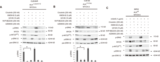
Crizotinib purchased from MedChemExpress. Usage Cited in: Sci Signal. 2014 Oct 28;7(349):ra102. [Abstract]
Activation of ERK5 in neuroblastoma cell lines expressing activated ALK. (A to C) Immunoblotting for the indicated proteins in neuroblastoma cells CLB-BAR (A), CLB-GE (B), and IMR32 (C) cultured on six-well plates in complete growth medium and treated with inhibitors as indicated for 6 hours alone (A and B) or before (C) stimulation with mAb46 for 30 min.
-
Mol Cancer Ther
2025 Jul 2;24(7):1005-1019. PMID: 40299789 -
Talanta
Development and comparative evaluation of two immunoassay platforms for bioanalysis of crizotinib: A potent drug used for the treatment of non-small cell lung cancer. [Abstract]2019 Aug 15:201:217-225. PMID: 31122414 -
Eur J Med Chem
Discovery of N-(5-((5-chloro-4-((2-(isopropylsulfonyl)phenyl)amino)pyrimidin-2-yl)amino)-4-methoxy-2-(4-methyl-1,4-diazepan-1-yl)phenyl)acrylamide (CHMFL-ALK/EGFR-050) as a potent ALK/EGFR dual kinase inhibitor capable of overcoming a variety of ALK/EGFR associated drug resistant mutants in NSCLC. [Abstract]2017 Oct 20:139:674-697. PMID: 28850922 -
-
Cell Rep Methods
RECOVER identifies synergistic drug combinations in vitro through sequential model optimization. [Abstract]2023 Oct 23;3(10):100599. PMID: 37797618 -
Pharmaceuticals (Basel)
Synthesis and Development of 3-((2,4-Difluorophenyl)Amino)Propanoic Acid Derivatives as an Antiproliferative Medicinal Chemistry Scaffold Targeting Growth Factor Receptors. [Abstract]2026 Feb 27;19(3):381. PMID: 41901229 -
Int J Mol Sci
Doxorubicin-Induced TrkAIII Activation: A Selection Mechanism for Resistant Dormant Neuroblastoma Cells. [Abstract]2022 Sep 17;23(18):10895. PMID: 36142807 -
Stem Cell Reports
Inhibition of Farnesyltransferase Potentiates NOTCH-Targeted Therapy against Glioblastoma Stem Cells. [Abstract]2017 Dec 12;9(6):1948-1960. PMID: 29198824 -
ACS Omega
2023 Jun 14;8(25):22603-22612. PMID: 37387790 -
Int J Antimicrob Agents
A new perspective on the antimicrobial mechanism of linezolid against Staphylococcus aureus revealed by proteomics and metabolomics analysis. [Abstract]2025 Jun;65(6):107470. PMID: 40049374 -
Cancer Sci
APE1 Attenuates ALK Tyrosine Kinase Inhibitors Sensitivity in NPM1-ALK Positive Anaplastic Large Cell Lymphoma. [Abstract]2025 Jul 23. PMID: 40702700 -
Transl Oncol
2022 Jan;15(1):101272. PMID: 34823094 -
Transl Oncol
Anti-epidermal growth factor vaccine antibodies increase the antitumor activity of kinase inhibitors in ALK and RET rearranged lung cancer cells. [Abstract]2021 Jan;14(1):100887. PMID: 33129112 -
-
Spectrochim Acta A Mol Biomol Spectrosc
Innovative use of σ and π electron acceptors in the development of three high throughput 96-microwell spectrophotometric assays for crizotinib. [Abstract]2021 Oct 5:259:119884. PMID: 33971436 -
Cancers (Basel)
Novel Mechanisms of ALK Activation Revealed by Analysis of the Y1278S Neuroblastoma Mutation. [Abstract]2017 Oct 30;9(11). pii: E149. PMID: 29084134 -
Spectrochim Acta A Mol Biomol Spectrosc
Charge-transfer reaction of 1,4-benzoquinone with crizotinib: spectrophotometric study, computational molecular modeling and use in development of microwell assay for crizotinib. [Abstract]2014 Oct 15;131:347-54. PMID: 24835938 -
-
Mol Oncol
Immunoassays for the quantification of ALK and phosphorylated ALK support the evaluation of on-target ALK inhibitors in neuroblastoma. [Abstract]2017 Aug;11(8):996-1006. PMID: 28432815
Crizotinib purchased from MedChemExpress. Usage Cited in: Mol Oncol. 2017 Aug;11(8):996-1006. [Abstract]
Dose-response and time course comparison of ALK inhibition by Crizotinib or Ceritinib.
-
Bioengineering (Basel)
Precision Oncology for High-Grade Gliomas: A Tumor Organoid Model for Adjuvant Treatment Selection. [Abstract]2025 Oct 19;12(10):1121. PMID: 41155119 -
Proteomes
Mass Spectrometry and Pharmacological Approaches to Measuring Cooption and Reciprocal Activation of Receptor Tyrosine Kinases. [Abstract]2023 Jun 2;11(2):20. PMID: 37368466 -
Biochim Biophys Acta Mol Cell Res
ZX-29, a novel ALK inhibitor, induces apoptosis via ER stress in ALK rearrangement NSCLC cells and overcomes cell resistance caused by an ALK mutation. [Abstract]2020 Jul;1867(7):118712. PMID: 32224191 -
Saudi Pharm J
Charge-transfer reaction of 2,3-dichloro-1,4-naphthoquinone with crizotinib: Spectrophotometric study, computational molecular modeling and use in development of microwell assay for crizotinib. [Abstract]2015 Jan;23(1):75-84. PMID: 25685046 -
Dis Model Mech
The ALK inhibitor PF-06463922 is effective as a single agent in neuroblastoma driven by expression of ALK and MYCN. [Abstract]2016 Sep 1;9(9):941-52. PMID: 27483357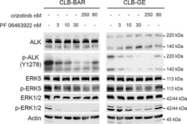
Crizotinib purchased from MedChemExpress. Usage Cited in: Dis Model Mech. 2016 Sep 1;9(9):941-52. [Abstract]
CLB-BAR, CLB-GE neuroblastoma cells are treated for 6 h with either Crizotinib or PF-04643922. Cells are harvested and pre-cleared cell lysates are analyzed on SDS PAGE followed by western blotting for ALK, phospho-ALK-Y1278, phospho-ERK5, pan-ERK5 phospho-ERK1/2 and pan-ERK. Actin is employed as a loading control. Protein band intensities are quantified by Image Studio Lite 3.1 and normalized to untreated samples.
-
Arch Biochem Biophys
Collagen I protects human keratinocytes HaCaT against UVB injury via restoring PINK1/parkin-mediated mitophagy. [Abstract]2024 Mar:753:109905. PMID: 38281543 -
Exp Cell Res
Network-based analysis with primary cells reveals drug response landscape of acute myeloid leukemia. [Abstract]2020 Aug 1;393(1):112054. PMID: 32376287 -
Cancer Med
The underlying mechanisms of lorlatinib penetration across the blood-brain barrier and the distribution characteristics of lorlatinib in the brain. [Abstract]2020 Jun;9(12):4350-4359. PMID: 32347012 -
Invest New Drugs
Iruplinalkib (WX‑0593), a novel ALK/ROS1 inhibitor, overcomes crizotinib resistance in preclinical models for non-small cell lung cancer. [Abstract]2023 Apr;41(2):254-266. PMID: 37036582 -
Front Oncol
Low-Dose Crizotinib, a Tyrosine Kinase Inhibitor, Highly and Specifically Sensitizes P-Glycoprotein-Overexpressing Chemoresistant Cancer Cells Through Induction of Late Apoptosis in vivo and in vitro. [Abstract]2020 May 12;10:696. PMID: 32528877 -
J Cancer Res Clin Oncol
MET canonical transcript expression is a predictive biomarker for chemo-sensitivity to MET-inhibitors in hepatocellular carcinoma cell lines. [Abstract]2021 Jan;147(1):167-175. PMID: 32980960 -
PLoS One
Inhibition of the anti-apoptotic protein BCL2 in EML4-ALK cell models as a second proposed therapeutic target for non-small cell lung cancer. [Abstract]2025 Jan 21;20(1):e0308747. PMID: 39836700 -
PLoS One
A new method for the study of biophysical and morphological parameters in 3D cell cultures: Evaluation in LoVo spheroids treated with crizotinib. [Abstract]2021 Jun 8;16(6):e0252907. PMID: 34101765 -
PLoS One
Synthesis of hapten, generation of specific polyclonal antibody and development of ELISA with high sensitivity for therapeutic monitoring of crizotinib. [Abstract]2019 Feb 11;14(2):e0212048. PMID: 30742664 -
Fundam Clin Pharmacol
2021 Oct;35(5):919-929. PMID: 33523504 -
Biol Methods Protoc
Optimizing drug sensitivity assays in patient-derived tumor organoids: a comparison of IC50 estimation methods and experimental parameters. [Abstract]2025 Feb 13;10(1):bpaf012. PMID: 40060949 -
Eur J Drug Metab Pharmacokinet
Differential Inhibition of Equilibrative Nucleoside Transporter 1 (ENT1) Activity by Tyrosine Kinase Inhibitors. [Abstract]2021 Sep;46(5):625-635. PMID: 34275128 -
Biomed Chromatogr
Development and validation of an ultra-performance liquid chromatography-tandem mass spectrometry method to quantify the small molecule inhibitors adagrasib, alectinib, brigatinib, capmatinib, crizotinib, lorlatinib, selpercatinib, and sotorasib in human plasma. [Abstract]2024 Oct;38(10):e5986. PMID: 39136165 -
Biomed Chromatogr
Pharmacokinetics of cligosiban in dog plasma after oral administration by liquid chromatography electrospray ionization tandem mass spectrometry. [Abstract]2019 Oct;33(10):e4611. PMID: 31145820 -
-
-
-
-
bioRxiv
PAIRWISE: Deep Learning-based Prediction of Effective Personalized Drug Combinations in Cancer. [Abstract]2024 Nov 6:2024.11.04.621884. PMID: 39574568 -
-
-
-
-
Evid Based Complement Alternat Med
In a Rat Model of Acute Liver Failure, Icaritin Improved the Therapeutic Effect of Mesenchymal Stem Cells by Activation of the Hepatocyte Growth Factor/c-Met Pathway. [Abstract]2019 Nov 7;2019:4253846. PMID: 31915446
Crizotinib purchased from MedChemExpress. Usage Cited in: Evid Based Complement Alternat Med. 2019 Nov 7;2019:4253846. [Abstract]
Western blot analysis of cleaved caspase 3, Bax, and Bcl-2 expression after serum-free culture for 72 h with or without Crizotinib.
-
Oncotarget
ALK mutation dynamics and clonal evolution in a neuroblastoma model exhibiting two ALK mutations. [Abstract]2019 Aug 13;10(48):4937-4950. PMID: 31452835 -
-
Oncotarget
Brigatinib, an anaplastic lymphoma kinase inhibitor, abrogates activity and growth in ALK-positive neuroblastoma cells, Drosophila and mice. [Abstract]2016 May 17;7(20):29011-22. PMID: 27049722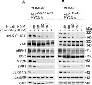
Crizotinib purchased from MedChemExpress. Usage Cited in: Oncotarget. 2016 May 17;7(20):29011-22. [Abstract]
A, B. CLB-BAR (ALK-Δ4-11) and CLB-GE (ALK-F1174V), both ALK addicted cell lines, are treated with increasing doses of Brigatinib for 6 hours. Crizotinib (250 nM) is employed as a positive control. Cells lysates are resolved on SDS/PAGE followed by immunoblotting for pALK (Y1604) and additional downstream targets as indicated.
-
Oncotarget
Activated Alk triggers prolonged neurogenesis and Ret upregulation providing a therapeutic target in ALK-mutated neuroblastoma. [Abstract]2014 May 15;5(9):2688-702. PMID: 24811913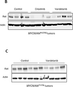
Crizotinib purchased from MedChemExpress. Usage Cited in: Oncotarget. 2014 May 15;5(9):2688-702. [Abstract]
The Ret expression level is investigated by Western blot in MYCN/KI AlkR1279Q and MYCN/KI AlkF1178L treated tumors and controls using the anti-Ret antibody EPR2871. Actin is used as a standard for quantification.


Solvent & Solubility
DMSO : 20 mg/mL (44.41 mM; ultrasonic and warming and heat to 60°C; Hygroscopic DMSO has a significant impact on the solubility of product, please use newly opened DMSO)
H2O : < 0.1 mg/mL (insoluble)
Please refer to the solubility information to select the appropriate solvent. Once prepared, please aliquot and store the solution to prevent product inactivation from repeated freeze-thaw cycles.
Storage method and period of stock solution: -80°C, 1 year; -20°C, 6 months. When stored at -80°C, please use it within 1 year. When stored at -20°C, please use it within 6 months.
Concentration (start) × Volume (start) = Concentration (final) × Volume (final)
Select the appropriate dissolution method based on your experimental animal and administration route.
- For the following dissolution methods, please ensure to first prepare a clear stock solution using an In Vitro approach and then sequentially add co-solvents:
- To ensure reliable experimental results, the clarified stock solution can be appropriately stored based on storage conditions. As for the working solution for In Vivo experiments, it is recommended to prepare freshly and use it on the same day.
- The percentages shown for the solvents indicate their volumetric ratio in the final prepared solution. If precipitation or phase separation occurs during preparation, heat and/or sonication can be used to aid dissolution.
Add each solvent one by one: 10% DMSO 40% PEG300 5% Tween-80 45% Saline
Solubility: ≥ 1 mg/mL (2.22 mM); Clear solution
This protocol yields a clear solution of ≥ 1 mg/mL (saturation unknown).
Taking 1 mL working solution as an example, add 100 μL DMSO stock solution (10.0 mg/mL) to 400 μL PEG300, and mix evenly; then add 50 μL Tween-80 and mix evenly; then add 450 μL Saline to adjust the volume to 1 mL.
Preparation of Saline: Dissolve 0.9 g sodium chloride in ddH₂O and dilute to 100 mL to obtain a clear Saline solution.
Add each solvent one by one: 10% DMSO 90% (20% SBE-β-CD in Saline)
Solubility: ≥ 1 mg/mL (2.22 mM); Clear solution
This protocol yields a clear solution of ≥ 1 mg/mL (saturation unknown).
Taking 1 mL working solution as an example, add 100 μL DMSO stock solution (10.0 mg/mL) to 900 μL 20% SBE-β-CD in Saline, and mix evenly.
Preparation of 20% SBE-β-CD in Saline (4°C, storage for one week): 2 g SBE-β-CD powder is dissolved in 10 mL Saline, completely dissolve until clear.
For the following dissolution methods, please prepare the working solution directly:
It is recommended to prepare fresh solutions and use them promptly within a short period of time.
The percentages shown for the solvents indicate their volumetric ratio in the final prepared solution. If precipitation or phase separation occurs during preparation, heat and/or sonication can be used to aid dissolution.
Add each solvent one by one: 50% PEG300 50% Saline
Solubility: 20 mg/mL (44.41 mM); Suspended solution; Need ultrasonic and warming and heat to 40°C
Please enter the basic information of animal experiments:
-
-
-
-
Recommended: Prepare an additional quantity of animals to account for potential losses during experiments.
Please enter your animal formula composition:
-
%DMSO +
Recommended: Keep the proportion of DMSO in working solution below 2% if your animal is weak.
-
%+
-
+%Tween-80 + +
-
%Saline +
The co-solvents required include: DMSO, . All of co-solvents are available by MedChemExpress (MCE). , Tween 80. All of co-solvents are available by MedChemExpress (MCE).
Working solution concentration: 0.22 mg/mL
Method for preparing stock solution: mg drug dissolved in μL DMSO. Stock solution concentration: mg/mL.
1. Take μL DMSO stock solution;
2. Add μL .
μL , mix evenly;
3. Then add μL Tween 80, mix evenly;
4. Then add μL
Please ensure that the stock solution in the first step is dissolved to a clear state, and add co-solvents in sequence. You can use ultrasonic heating (ultrasonic cleaner, recommended frequency 20-40 kHz), vortexing, etc. to assist dissolution.

Protocol
Cells are seeded in 96-well plates in media supplemented with 10% fetal bovine serum (FBS) and transferred to serum-free media [with 0.04% bovine serum albumin (BSA)] after 24 h. In experiments investigating ligand-dependent RTK phosphorylation, corresponding growth factors are added for up to 20 min. After incubation of cells with PF-2341066 for 1 h and/or appropriate ligands for the designated times, cells are washed once with HBSS supplemented with 1 mM Na3VO4, and protein lysates are generated from cells. Subsequently, phosphorylation of selected protein kinases is assessed by a sandwich ELISA method using specific capture antibodies used to coat 96-well plates and a detection antibody specific for phosphorylated tyrosine residues. Antibody-coated plates are (a) incubated in the presence of protein lysates at 4°C overnight; (b) washed seven times in 1% Tween 20 in PBS; (c) incubated in a horseradish peroxidase-conjugated anti-total-phosphotyrosine (PY-20) antibody (1:500) for 30 min; (d) washed seven times again; (e) incubated in 3,3′,5,5′-tetramethyl benzidine peroxidase substrate to initiate a colorimetric reaction that is stopped by adding 0.09 N H2SO4; and (f) measured for absorbance in 450 nm using a spectrophotometer.
MedChemExpress (MCE) has not independently confirmed the accuracy of these methods. They are for reference only.
Tumor cells are seeded in 96-well plates at low density in media supplemented with 10% FBS (growth media) and transferred to serum-free media (0% FBS and 0.04% BSA) after 24 h. Appropriate controls or designated concentrations of PF-2341066 are added to each well, and cells are incubated for 24 to 72 h. Human umbilical vascular endothelial cells (HUVEC) are seeded in 96-well plates in EGM2 media for 5 to 6 h at > 20,000 cells per well and transferred to serum-free media overnight. The following day, appropriate controls or designated concentrations of PF-2341066 are added to each well, and after 1 h incubation, HGF is added to designated wells at 100 ng/mL. A 3-(4,5-dimethylthiazol-2-yl)-2,5-diphenyltetrazolium bromide assay is done to determine the relative tumor cell or HUVEC numbers.
MedChemExpress (MCE) has not independently confirmed the accuracy of these methods. They are for reference only.
Athymic mice bearing xenografts (300-800 mm3) are given PF-2341066 in water by oral gavage at designated dose levels. At designated times following PF-2341066 administration, mice are humanely euthanized, and tumors are resected. Tumors are snap frozen and pulverized using a liquid nitrogen-cooled cryomortar and pestle, protein lysates are generated, and protein concentrations are determined using a BSA assay. The level of total and phosphorylated protein is determined using a capture ELISA or immunoprecipitation-immunoblotting method.
MedChemExpress (MCE) has not independently confirmed the accuracy of these methods. They are for reference only.


Purity & Documentation
-
Data Sheet (282 KB)
-
SDS (623 KB)
- English - EN (623 KB)
- Français - FR (623 KB)
- Deutsch - DE (623 KB)
- Norwegian - NO (623 KB)
- Español - ES (623 KB)
- Swedish - SV (623 KB)
- Italian - IT (623 KB)
- Korean - KR (623 KB)
- Portuguese - PT (623 KB)
-
Handling Instructions (2659 KB)
References
[1]. Zou HY, et al. An orally available small-molecule inhibitor of c-Met, PF-2341066, exhibits cytoreductive antitumor efficacy through antiproliferative and antiangiogenic mechanisms. Cancer Res. 2007, 67(9), 4408-4417. [Content Brief]
[2]. Christensen JG, et al. Cytoreductive antitumor activity of PF-2341066, a novel inhibitor of anaplastic lymphoma kinase and c-Met, in experimental models of anaplastic large-cell lymphoma. Mol Cancer Ther. 2007, 6(12 Pt 1), 3314-3322. [Content Brief]
[3]. Cui JJ, et al. Structure based drug design of crizotinib (PF-02341066), a potent and selective dual inhibitor of mesenchymal-epithelial transition factor (c-MET) kinase and anaplastic lymphoma kinase (ALK). J Med Chem. 2011 Sep 22;54(18):6342-63. [Content Brief]
[4]. Cullinane C, et al. Differential (18)F-FDG and 3'-deoxy-3'-(18)F-fluorothymidine PET responses to pharmacologic inhibition of the c-MET receptor in preclinical tumor models. J Nucl Med. 2011 Aug;52(8):1261-7 [Content Brief]
[5]. Shen A, et al. c-Myc alterations confer therapeutic response and acquired resistance to c-Met inhibitors in MET-addicted cancers. Cancer Res. 2015 Nov 1;75(21):4548-59. [Content Brief]
[6]. Umapathy G, et al. The kinase ALK stimulates the kinase ERK5 to promote the expression of the oncogene MYCN in neuroblastoma. Sci Signal. 2014 Oct 28;7(349):ra102. [Content Brief]
[7]. Tucker ER, et al. Immunoassays for the quantification of ALK and phosphorylated ALK support the evaluation of on-target ALK inhibitors in neuroblastoma. Mol Oncol. 2017 Aug;11(8):996-1006. [Content Brief]
[8]. Liu H, et al. Identifying and Targeting Sporadic Oncogenic GeneticLiu H, et al. Identifying and Targeting Sporadic Oncogenic Genetic Aberrations in Mouse Models of Triple Negative Breast Cancer. Cancer Discov. 2018 Mar;8(3):354-369. [Content Brief]


Complete Stock Solution Preparation Table
Please refer to the solubility information to select the appropriate solvent. Once prepared, please aliquot and store the solution to prevent product inactivation from repeated freeze-thaw cycles.
Storage method and period of stock solution: -80°C, 1 year; -20°C, 6 months. When stored at -80°C, please use it within 1 year. When stored at -20°C, please use it within 6 months.
| Optional Solvent | Concentration Solvent Mass | 1 mg | 5 mg | 10 mg | 25 mg |
|---|---|---|---|---|---|
| DMSO | 1 mM | 2.2205 mL | 11.1027 mL | 22.2054 mL | 55.5136 mL |
| 5 mM | 0.4441 mL | 2.2205 mL | 4.4411 mL | 11.1027 mL | |
| 10 mM | 0.2221 mL | 1.1103 mL | 2.2205 mL | 5.5514 mL | |
| 15 mM | 0.1480 mL | 0.7402 mL | 1.4804 mL | 3.7009 mL | |
| 20 mM | 0.1110 mL | 0.5551 mL | 1.1103 mL | 2.7757 mL | |
| 25 mM | 0.0888 mL | 0.4441 mL | 0.8882 mL | 2.2205 mL | |
| 30 mM | 0.0740 mL | 0.3701 mL | 0.7402 mL | 1.8505 mL | |
| 40 mM | 0.0555 mL | 0.2776 mL | 0.5551 mL | 1.3878 mL |


 Powered by Bioz