



SC66
Based on 5 publication(s) in Google Scholar
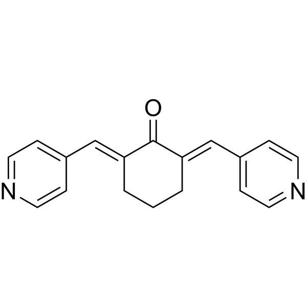
SC66 is an Akt inhibitor, reduces cell viability in a dose- and time-dependent manner, inhibits colony formation and induces apoptosis in hepatocellular carcinoma (HCC) cells.
For research use only. We do not sell to patients.
- Purity : 99.65%
- CAS No.: 871361-88-5
- Formula: C18H16N2O
- Molecular Weight:276.33
-
Storage:Powder -20°C, 3 years , 4°C, 2 years ; In solvent -80°C, 2 years , -20°C, 1 year

To place orders, for customer services and technical support, please contact: MedChemExpress USA
Tel: 609-228-6898 E-mail: [email protected] [email protected]


-
 Biological Activity
-
Chemical Information
-
Solvent & Solubility
- Protocols
- Purity & Documentation
- References
-
 Help & FAQs


-
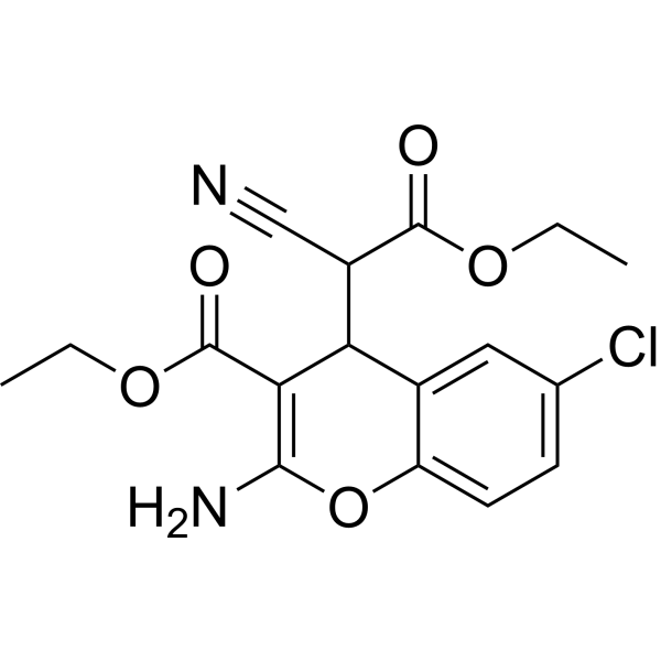
HY-18749
SC79Akt Activator -
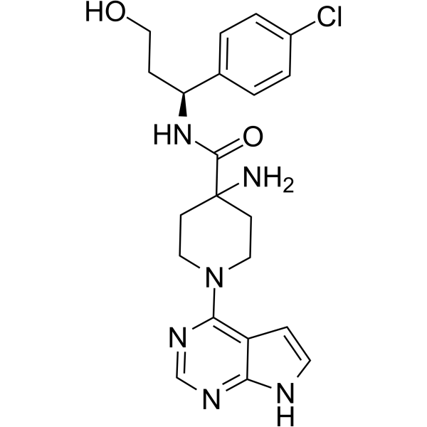
HY-15431
CapivasertibAkt Inhibitor -
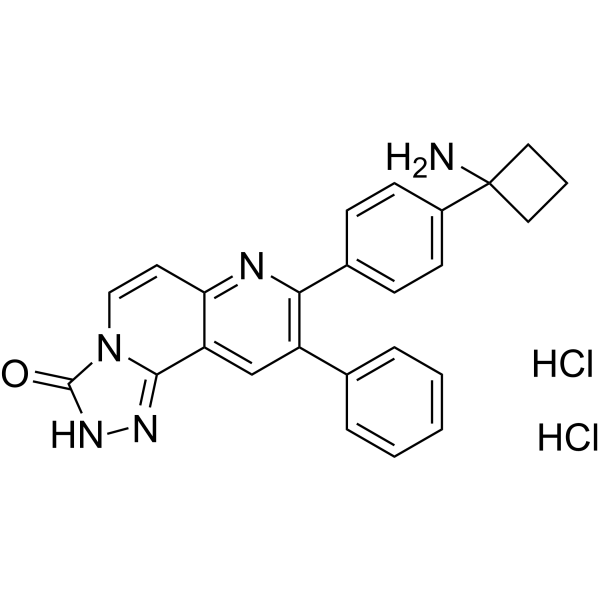
HY-10358
MK-2206 dihydrochlorideAllosteric Akt Inhibitor -
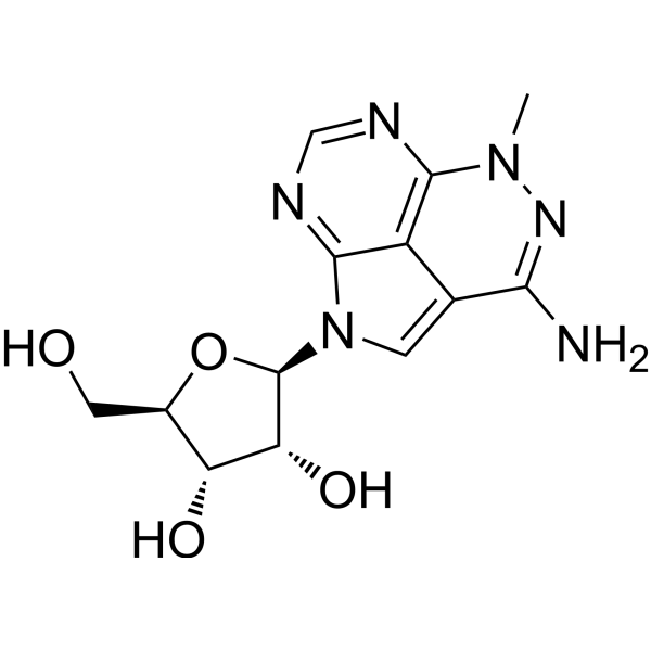
HY-15457
TriciribineHIV Inhibitor -
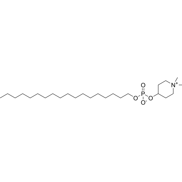
HY-50909
PerifosineAkt Inhibitor -
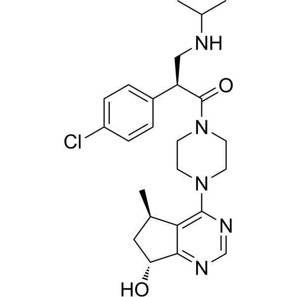
HY-15186
IpatasertibPan-Akt Inhibitor -
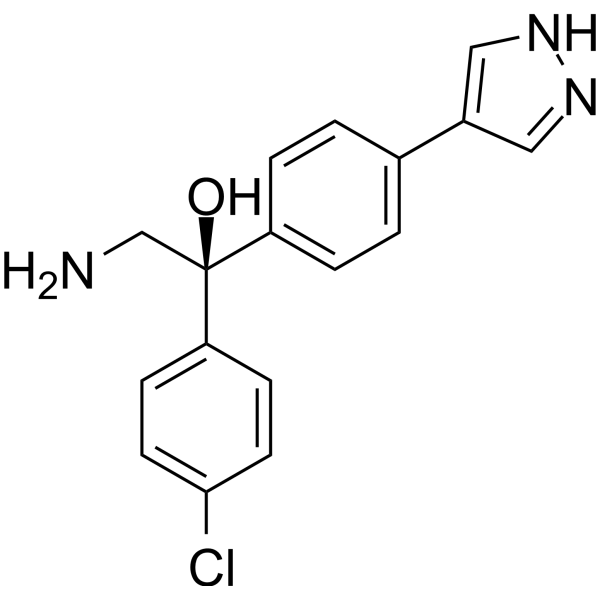
HY-16071
AT13148AGC Kinase Inhibitor -
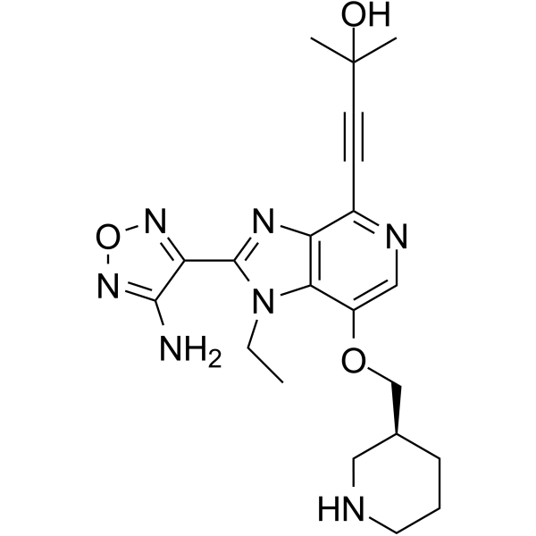
HY-10249
GSK-690693Akt Inhibitor -
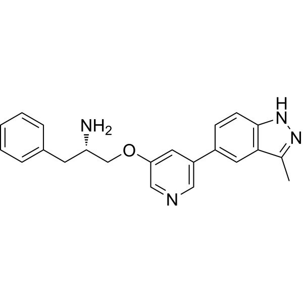
HY-13254
A-674563Akt1 Inhibitor -
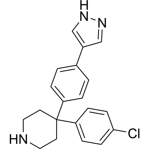
HY-12059
AT7867 -
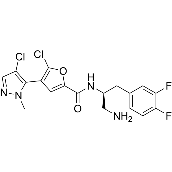
HY-15965
UprosertibAkt Inhibitor
Publications Citing Use of MedChemExpress (MCE) SC66
More Customer Validation & Images

-
WB


Biological Activity
Description
IC50 & Target
Akt[1]
Cellular Effect
|
Cell Line

|
Type | Value | Description | References |
|---|---|---|---|---|
| HT-29 | IC50 |
2.35 μM
Compound: AN3
|
Cytotoxicity against human HT-29 cells after 72 hrs by MTT assay
Cytotoxicity against human HT-29 cells after 72 hrs by MTT assay
|
[PMID: 22551677] |
| K562 | IC50 |
2.5 μM
Compound: A1
|
Inhibition of NF-kappaB activation in human K562 cells
Inhibition of NF-kappaB activation in human K562 cells
|
[PMID: 20728364] |
| L929 | IC50 |
>250 μM
Compound: 10
|
Trypanocidal activity against Trypanosoma cruzi Tulahuen amastigotes infected in mouse L929 cells assessed as parasite growth inhibition after 7 days by beta-galactosidase assay
Trypanocidal activity against Trypanosoma cruzi Tulahuen amastigotes infected in mouse L929 cells assessed as parasite growth inhibition after 7 days by beta-galactosidase assay
|
[PMID: 24321832] |
| MDA-MB-231 | EC50 |
1.1 μM
Compound: A1
|
Cytotoxicity against human MDA-MB-231 cells after 72 hrs by SRB assay
Cytotoxicity against human MDA-MB-231 cells after 72 hrs by SRB assay
|
[PMID: 20728364] |
| MDA-MB-468 | EC50 |
1.9 μM
Compound: A1
|
Cytotoxicity against human MDA-MB-468 cells after 72 hrs by SRB assay
Cytotoxicity against human MDA-MB-468 cells after 72 hrs by SRB assay
|
[PMID: 20728364] |
| NB-4 | IC50 |
0.27 μM
Compound: 26
|
Cytotoxicity against human NB4 cells assessed as reduction in cell viability after 72 hrs by MTS assay
Cytotoxicity against human NB4 cells assessed as reduction in cell viability after 72 hrs by MTS assay
|
[PMID: 24960549] |
| PANC-1 | IC50 |
2.18 μM
Compound: AN3
|
Cytotoxicity against human PANC1 cells after 72 hrs by MTT assay
Cytotoxicity against human PANC1 cells after 72 hrs by MTT assay
|
[PMID: 22551677] |
| PC-3 | IC50 |
2.12 μM
Compound: AN3
|
Cytotoxicity against human PC3 cells after 72 hrs by MTT assay
Cytotoxicity against human PC3 cells after 72 hrs by MTT assay
|
[PMID: 22551677] |
| Peritoneal macrophage cell | CC50 |
23.9 μM
Compound: 10
|
Cytotoxicity against mouse peritoneal macrophages after 24 hrs by MTT assay
Cytotoxicity against mouse peritoneal macrophages after 24 hrs by MTT assay
|
[PMID: 24321832] |
| SK-BR-3 | IC50 |
0.2 μM
Compound: A1
|
Cytotoxicity against human SKBR3 cells after 72 hrs by SRB assay
Cytotoxicity against human SKBR3 cells after 72 hrs by SRB assay
|
[PMID: 20728364] |
| SUP-T1 | CC50 |
0.82 μM
Compound: UHLMT-70
|
Cytotoxicity against human SUP-T1 cells assessed as reduction in cell viability incubated for 16 hrs by MTT assay
Cytotoxicity against human SUP-T1 cells assessed as reduction in cell viability incubated for 16 hrs by MTT assay
|
[PMID: 37473689] |
In Vitro
SC66 inhibits cell viability and colony forming capacity of HCC cells with IC50s of 0.77,0.47,0.92,0.75 and 2.85 μg/mL at 72 hours for HepG2, Hep3B, PLC/PRF/5,HA22T/VGH and Huh7 cells. HepG2, HA22T/VGH and PLC/PRF/5 cells have similar IC50s of approximately 0.85 and 0.75 μg/mL at 48 and 72 hours, respectively. To determine whether the decrease in cell viability is related to apoptosis induction, TUNEL assays are performed in Hep3B and Huh7 cells treated with 1, 2 and 4 μg/mL of SC66 for 24 hours. In Hep3B cells the number of TUNEL-positive cells increased with increasing concentrations of SC66, whereas in Huh7 cells very few light brown-colored cells are observed only after treatment with 4 μg/mL SC66[1].
MedChemExpress (MCE) has not independently confirmed the accuracy of these methods. They are for reference only.
In Vivo
MedChemExpress (MCE) has not independently confirmed the accuracy of these methods. They are for reference only.
- Detection of Apoptosis
- TUNEL staining for apoptotic DNA fragmentation
- Cell Cytotoxicity Assay
- Mammalian live/dead viability and cytotoxicity staining
- Annexin V plus membrane-impermeant dye apoptosis staining
- Apoptosis Solutions
- CCK-8/WST-8 Cell Proliferation Assay
- Soft Agar Colony Formation Assay
- Liver Cancer Modeling
- MTT Cell Proliferation Assay
- Colony Formation (Clonogenic) Assay
- Cell Viability Determination by MTT Colorimetric Assay


Chemical Information
-
CAS No. 871361-88-5
-
Appearance Solid
-
Molecular Weight 276.33
-
Formula C18H16N2O
-
Color Light yellow to yellow
-
SMILES
O=C1/C(CCC/C1=C\C2=CC=NC=C2)=C/C3=CC=NC=C3
-
Shipping
Room temperature in continental US; may vary elsewhere.
-
Storage
Powder -20°C 3 years 4°C 2 years In solvent -80°C 2 years -20°C 1 year


Publications (5)
-
Journal Impact Factor
-
Most Recent
-
Cell Death Dis
2020 May 11;11(5):353. PMID: 32393791
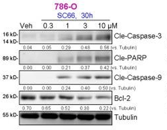
SC66 purchased from MedChemExpress. Usage Cited in: Cell Death Dis. 2020 May 11;11(5):353. [Abstract]
786-O RCC cellsare treated with indicated concentration of SC66, cells are further cultured for applied time periods expression of apoptosis-associated proteins.
-
Cancer Biol Ther
RND1 inhibits epithelial-mesenchymal transition and temozolomide resistance of glioblastoma via AKT/GSK3-β pathway. [Abstract]2024 Dec 31;25(1):2321770. PMID: 38444223 -
Front Pharmacol
AKT Inhibitor SC66 Inhibits Proliferation and Induces Apoptosis in Human Glioblastoma Through Down-Regulating AKT/β-Catenin Pathway. [Abstract]2020 Jul 31;11:1102. PMID: 32848734 -
Toxicology
SIRT4 Suppresses Doxorubicin-Induced Cardiotoxicity by Regulating the AKT/mTOR/Autophagy Pathway. [Abstract]2022 Feb 5;469:153119. PMID: 35134463 -
Molecules
Geniposidic Acid from Eucommia ulmoides Oliver Staminate Flower Tea Mitigates Cellular Oxidative Stress via Activating AKT/NRF2 Signaling. [Abstract]2022 Dec 5;27(23):8568. PMID: 36500666


Solvent & Solubility
In Vitro:
DMSO : 25 mg/mL (90.47 mM; Need ultrasonic; Hygroscopic DMSO has a significant impact on the solubility of product, please use newly opened DMSO)
Please refer to the solubility information to select the appropriate solvent. Once prepared, please aliquot and store the solution to prevent product inactivation from repeated freeze-thaw cycles.
Storage method and period of stock solution: -80°C, 2 years; -20°C, 1 year. When stored at -80°C, please use it within 2 years. When stored at -20°C, please use it within 1 year.
Please refer to the solubility information to select the appropriate solvent. Once prepared, please aliquot and store the solution to prevent product inactivation from repeated freeze-thaw cycles.
Storage method and period of stock solution: -80°C, 2 years; -20°C, 1 year. When stored at -80°C, please use it within 2 years. When stored at -20°C, please use it within 1 year.
Concentration (start) × Volume (start) = Concentration (final) × Volume (final)
In Vivo:
Select the appropriate dissolution method based on your experimental animal and administration route.
- For the following dissolution methods, please ensure to first prepare a clear stock solution using an In Vitro approach and then sequentially add co-solvents:
- To ensure reliable experimental results, the clarified stock solution can be appropriately stored based on storage conditions. As for the working solution for In Vivo experiments, it is recommended to prepare freshly and use it on the same day.
- The percentages shown for the solvents indicate their volumetric ratio in the final prepared solution. If precipitation or phase separation occurs during preparation, heat and/or sonication can be used to aid dissolution.
Add each solvent one by one: 10% DMSO 40% PEG300 5% Tween-80 45% Saline
Solubility: ≥ 2.5 mg/mL (9.05 mM); Clear solution
This protocol yields a clear solution of ≥ 2.5 mg/mL (saturation unknown).
Taking 1 mL working solution as an example, add 100 μL DMSO stock solution (25.0 mg/mL) to 400 μL PEG300, and mix evenly; then add 50 μL Tween-80 and mix evenly; then add 450 μL Saline to adjust the volume to 1 mL.
Preparation of Saline: Dissolve 0.9 g sodium chloride in ddH₂O and dilute to 100 mL to obtain a clear Saline solution.
Add each solvent one by one: 10% DMSO 90% (20% SBE-β-CD in Saline)
Solubility: ≥ 2.5 mg/mL (9.05 mM); Clear solution
This protocol yields a clear solution of ≥ 2.5 mg/mL (saturation unknown).
Taking 1 mL working solution as an example, add 100 μL DMSO stock solution (25.0 mg/mL) to 900 μL 20% SBE-β-CD in Saline, and mix evenly.
Preparation of 20% SBE-β-CD in Saline (4°C, storage for one week): 2 g SBE-β-CD powder is dissolved in 10 mL Saline, completely dissolve until clear.
In Vivo Dissolution Calculator
Please enter the basic information of animal experiments:
-
-
-
-
Recommended: Prepare an additional quantity of animals to account for potential losses during experiments.
Please enter your animal formula composition:
-
%DMSO +
Recommended: Keep the proportion of DMSO in working solution below 2% if your animal is weak.
-
%+
-
+%Tween-80 + +
-
%Saline +
The co-solvents required include: DMSO, . All of co-solvents are available by MedChemExpress (MCE). , Tween 80. All of co-solvents are available by MedChemExpress (MCE).
Working solution concentration: 0.22 mg/mL
Method for preparing stock solution: mg drug dissolved in μL DMSO. Stock solution concentration: mg/mL.
1. Take μL DMSO stock solution;
2. Add μL .
μL , mix evenly;
3. Then add μL Tween 80, mix evenly;
4. Then add μL
Please ensure that the stock solution in the first step is dissolved to a clear state, and add co-solvents in sequence. You can use ultrasonic heating (ultrasonic cleaner, recommended frequency 20-40 kHz), vortexing, etc. to assist dissolution.


Purity & Documentation
-
Data Sheet (276 KB)
-
SDS (396 KB)
- English - EN (396 KB)
- Français - FR (396 KB)
- Deutsch - DE (396 KB)
- Norwegian - NO (396 KB)
- Español - ES (396 KB)
- Swedish - SV (396 KB)
- Italian - IT (396 KB)
- Korean - KR (396 KB)
- Portuguese - PT (396 KB)
-
Handling Instructions (2659 KB)
References


Complete Stock Solution Preparation Table
Please refer to the solubility information to select the appropriate solvent. Once prepared, please aliquot and store the solution to prevent product inactivation from repeated freeze-thaw cycles.
Storage method and period of stock solution: -80°C, 2 years; -20°C, 1 year. When stored at -80°C, please use it within 2 years. When stored at -20°C, please use it within 1 year.
| Optional Solvent | Concentration Solvent Mass | 1 mg | 5 mg | 10 mg | 25 mg |
|---|---|---|---|---|---|
| DMSO | 1 mM | 3.6189 mL | 18.0943 mL | 36.1886 mL | 90.4715 mL |
| 5 mM | 0.7238 mL | 3.6189 mL | 7.2377 mL | 18.0943 mL | |
| 10 mM | 0.3619 mL | 1.8094 mL | 3.6189 mL | 9.0472 mL | |
| 15 mM | 0.2413 mL | 1.2063 mL | 2.4126 mL | 6.0314 mL | |
| 20 mM | 0.1809 mL | 0.9047 mL | 1.8094 mL | 4.5236 mL | |
| 25 mM | 0.1448 mL | 0.7238 mL | 1.4475 mL | 3.6189 mL | |
| 30 mM | 0.1206 mL | 0.6031 mL | 1.2063 mL | 3.0157 mL | |
| 40 mM | 0.0905 mL | 0.4524 mL | 0.9047 mL | 2.2618 mL | |
| 50 mM | 0.0724 mL | 0.3619 mL | 0.7238 mL | 1.8094 mL | |
| 60 mM | 0.0603 mL | 0.3016 mL | 0.6031 mL | 1.5079 mL | |
| 80 mM | 0.0452 mL | 0.2262 mL | 0.4524 mL | 1.1309 mL |


SC66 Related Classifications
HY-19832 Related Classifications
 Powered by Bioz