



EGFR Protein, Human (Active, sf9, His-GST)
Based on 2 publication(s) in Google Scholar
EGFR is a transmembrane glycoprotein. EGFR binds to ligands, triggering receptor dimerization, inducing intracellular TK domain autophosphorylation, activating downstream Ras/MAPK, PI3K/Akt and other signaling pathways, and regulating cell proliferation, survival, migration and angiogenesis. EGFR Protein, Human (sf9, His-GST) is a recombinant EGFR protein expressed by Sf9 insect cells with N-His, N-GST tags.
- Species: Human
- Source: Sf9 insect cells
-
Storage:Stored at -80°C for 1 year from date of receipt. It is stable at -20°C for 3 months after opening. It is recommended to freeze aliquots at -80°C for extended storage. Avoid repeated freeze-thaw cycles.

To place orders, for customer services and technical support, please contact: MedChemExpress USA
Tel: 609-228-6898 E-mail: [email protected] [email protected]


-
 Biological Activity
-
Technical Parameters
-
Product Properties
- Documentation
- References
- Help & FAQs


-

HY-P702832
EGFR Protein, Human (Active, T790M, L858R, sf9, Strep II, GST, His) -
HY-B2176
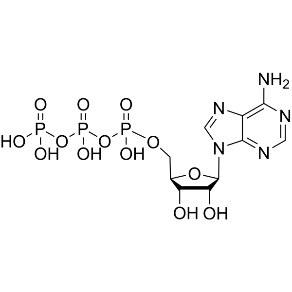
ATP -
HY-P705562
EGFR Protein, Human (Active, L858R, sf9, His) -
HY-P70533
FGF-8b Protein, Human/Mouse -
HY-P74177
EGFR Protein, Human (621a.a, HEK293, Fc) -
HY-P77507
WWP2 Protein, Human (sf9, His-GST) -
HY-P70613
EGFR Protein, Human (621a.a, HEK293, His)


Biological Activity
Description
EGFR is a transmembrane glycoprotein. EGFR binds to ligands, triggering receptor dimerization, inducing intracellular TK domain autophosphorylation, activating downstream Ras/MAPK, PI3K/Akt and other signaling pathways, and regulating cell proliferation, survival, migration and angiogenesis. EGFR Protein, Human (sf9, His-GST) is a recombinant EGFR protein expressed by Sf9 insect cells with N-His, N-GST tags.
Background
EGFR belongs to the epidermal growth factor receptor family[1][2][3][4][5]. EGFR is a transmembrane glycoprotein. EGFR has an extracellular domain of 621 amino acids, a single transmembrane domain of 23 amino acids, and an enzymatically active intracellular domain of 542 amino acids[6]. EGFR binds to its ligand, triggers receptor dimerization, induces autophosphorylation of the intracellular TK domain, activates downstream Ras/MAPK, PI3K/Akt and other signaling pathways, and regulates cell proliferation, survival, migration and angiogenesis[1][3][4][5]. EGFR is expressed in a variety of normal epithelial tissues and tumor tissues, such as breast, lung, stomach, colon, head and neck, and its abnormal expression is associated with the malignant progression of tumors[1][2][3][4][5].
Verified Bioactivity
The specific activity was determined to be ≥4 nmol/min/mg using Poly(Glu:Tyr) 4:1 as substrate.

Assay Procedure
Materials
Assay buffer A(5X): 200 mM Tris-HCl, pH 7.5, 100 mM MgCl2 and 0.5 mg/mL BSA
Buffer 1(2X): 1 mL assay buffer A, 2.5 μL DTT (0.1 M), 10 μL MnCl2 (1 M) and 1.5 mL of dH20
Buffer 2 (1X): 1 mL assay buffer 1, and 1 mL dH2O
Test protein: EGFR Protein, Human (Active, sf9, His-GST) (HY-P72987)
Substrate: Poly (Glu : Tyr) 4:1
ADP-Glo kit
Procedure
1. Thaw the ADP-GloTM Reagents at ambient temperature. Then prepare Kinase Detection Reagent by mixing Kinase assay buffer with the Lyophilized Kinase Detection Substrate.
2. Thaw the components of enzyme system, ADP and ATP on ice.
3. Prepare 1 mL of 250 μM ATP Sssay Solution by adding 25 μL ATP solution (10 mM) to 500 μL of 2X buffer and 475 μL of dH2O.
4. In a white 96 well plate, add the following reaction components bringing the initial reaction volume up to 20 μL:
a. 10 μL of diluted active enzyme.
b. 5 μL of 1mg/mL stock solution of substrate (final Conc.: 0.2 mg/mL).
5. Set up the blank control as outlined in step (4), excluding the addition of the substrate. Replace the substrate with an equal volume of dH2O.
6. Initiate the reactions by the addition of 5 μL of 250 μM ATP assay solution thereby bringing the final volume up to 25 μL. Shake the plate and incubate the reaction mixture at R.T. for 60 minutes.
| Components | Volume (μL) | Blank Control (μL) |
|---|---|---|
| Active enzyme | 10 | 10 |
| Substrate stock solution (1 mg/mL) | 5 | 5 μL dH2O |
| 2X buffer | 5 | 5 |
| ATP assay solution (250 μM) | 5 | 5 |
8. Terminate the reaction and deplete the remaining ATP by adding 25 μL of ADP- GloTM Reagent. Shake the 96 well plate and then incubate the reaction mixture for another 40 minutes at R.T.
9. Add 50 μL of the Kinase Detection Reagent, shake the plate and then incubate the reaction mixture for another 30 minutes at R.T.
10. Read the 96-well reaction plate using Microplate Luminomet.
11. Calculate specific activity:
Specific Activity (nmol/min/μg) = | Phosphate released* (nmol) |
| Reaction time (min) × amount of enzyme (μg) |
*Derived from the phosphate standard curve and adjusted for Control.


Publications (2)
-
Journal Impact Factor
-
Most Recent
-
Phytomedicine
Quercetin ameliorates doxorubicin-induced atrial fibrillation via EGFR-mediated restoration of autophagic flux. [Abstract]2025 Nov 25:148:157357. PMID: 41056856 -
Phytomedicine
Artificial intelligence combined with affinity chromatography discover that 7-Epitaxol promotes autophagy in NSCLC cells by interacting with EGFR: Discovery of novel EGFR antagonist based on DL and CMC. [Abstract]2025 Oct:146:157127. PMID: 40768807


Technical Parameters
-
Species Human
-
Source Sf9 insect cells
-
Tag N-GST;N-10*His
-
Accession
P00533-1 (M668-A1210)
-
Molecular Construction
-
N-term
-
10*His-GST
-
EGFR (M668-A1210)
Accession # P00533-1 -
C-term
-
-
Protein Length
Cytoplasmic Domain
-
Synonyms
EGFR; Epidermal Growth Factor Receptor Variant EX12_14del; Prev. ERBB; Cell Proliferation-Inducing Protein 61; ERBB1; Cell Growth Inhibiting Protein 40; ERRP; Receptor Protein-Tyrosine Kinase; Receptor Tyrosine-Protein Kinase ErbB-1; Alternative Protein E
-
AA Sequence
MRRRHIVRKRTLRRLLQERELVEPLTPSGEAPNQALLRILKETEFKKIKVLGSGAFGTVYKGLWIPEGEKVKIPVAIKELREATSPKANKEILDEAYVMASVDNPHVCRLLGICLTSTVQLITQLMPFGCLLDYVREHKDNIGSQYLLNWCVQIAKGMNYLEDRRLVHRDLAARNVLVKTPQHVKITDFGLAKLLGAEEKEYHAEGGKVPIKWMALESILHRIYTHQSDVWSYGVTVWELMTFGSKPYDGIPASEISSILEKGERLPQPPICTIDVYMIMVKCWMIDADSRPKFRELIIEFSKMARDPQRYLVIQGDERMHLPSPTDSNFYRALMDEEDMDDVVDADEYLIPQQGFFSSPSTSRTPLLSSLSATSNNSTVACIDRNGLQSCPIKEDSFLQRYSSDPTGALTEDSIDDTFLPVPEYINQSVPKRPAGSVQNPVYHNQPLNPAPSRDPHYQDPHSTAVGNPEYLNTVQPTCVNSTFDSPAHWAQKGSHQISLDNPDYQQDFFPKEAKPNGIFKGSTAENAEYLRVAPQSSEFIGA
-
Predicted Molecular Mass
89.1 kDa
-
Molecular Weight
89.1 kDa
-
Glycosylation
Yes
-
Purity
≥ 75%, as determined by reducing SDS-PAGE.


Product Properties
Solution.
1.Supplied as a 0.22 μm filtered solution of 20 mM Tris, 300 mM NaCl, 10% glycerol, 0.02% Triton X-100, 0.4 mM β-Me, 0.5 mM GSH, 0.5 mM PMSF, pH 7.5.
2.Supplied as a 0.22 μm filtered solution of 20 mM Tris, 300 mM NaCl, 10% glycerol, pH 7.5, 0.02% Triton X-100, 0.4 mM β-mercaptoethanol, 0.5 mM PMSF, 2 mM GSH.
Please refer to the lot-specific COA for specific buffer information.
Note: For SPR assay, please replace the buffer. Primary amine components (e.g., Tris, imidazole) can affect protein-coupled chips.
<1 EU/μg, determined by LAL method.
Please use rapid thawing with running water to thaw the protein.
Stored at -80°C for 1 year from date of receipt. It is stable at -20°C for 3 months after opening. It is recommended to freeze aliquots at -80°C for extended storage. Avoid repeated freeze-thaw cycles.
Shipping with dry ice.


Documentation
-
Data Sheet (266 KB)
-
SDS (251 KB)
- English - EN (251 KB)
- Français - FR (251 KB)
- Deutsch - DE (251 KB)
- Norwegian - NO (251 KB)
- Español - ES (251 KB)
- Swedish - SV (251 KB)
- Italian - IT (251 KB)
- Korean - KR (251 KB)
- Portuguese - PT (251 KB)
-
Handling Instructions (2659 KB)


References
[1]. Lemmon M A, et al. Cell signaling by receptor tyrosine kinases. Cell, 2010, 141(7): 1117-1134.
[2]. Giaccone G. HER1/EGFR-targeted agents: predicting the future for patients with unpredictable outcomes to therapy. Ann Oncol. 2005 Apr;16(4):538-48. [Content Brief]
[3]. Yarden Y, et al. Untangling the ErbB signalling network. Nat Rev Mol Cell Biol. 2001 Feb;2(2):127-37. [Content Brief]
[4]. Shepard HM, et al. Signal integration: a framework for understanding the efficacy of therapeutics targeting the human EGFR family. J Clin Invest. 2008 Nov;118(11):3574-81. [Content Brief]
[5]. Okines A, et al. Targeting the human EGFR family in esophagogastric cancer. Nat Rev Clin Oncol. 2011 Apr 5;8(8):492-503. [Content Brief]
[6]. Ho ECH, et al. Characterization of monoclonal antibodies generated to the 287-302 amino acid loop of the human epidermal growth factor receptor'. Antib Ther. 2019 Oct;2(4):88-98. [Content Brief]

Calculators
Concentration (start) × Volume (start) = Concentration (final) × Volume (final)


 Powered by Bioz

