



EGFR Protein, Rat (HEK293, His)
Based on 1 Customer Validation
EGFR Protein is a receptor tyrosine kinase that binds to ligands of the EGF family.EGFR Protein is involved in the regulation of NF-kappa-B , RAS-RAF-MEK-ERK, PI3 kinase-AKT, PLCgamma-PKC and STATs signaling pathways.EGFR Protein has calmodulin binding activity, epidermal growth factor binding activity and epidermal growth factor activating receptor activity.EGFR Protein, Rat (HEK293, His) is the recombinant rat-derived EGFR protein, expressed by HEK293 , with C-His labeled tag.
- Species: Rat
- Source: HEK293
-
Storage:Stored at -20°C for 2 years from date of receipt. After reconstitution, it is stable at 4°C for 1 week or -20°C for longer (with carrier protein). It is recommended to freeze aliquots at -20°C or -80°C for extended storage.


To place orders, for customer services and technical support, please contact: MedChemExpress USA
Tel: 609-228-6898 E-mail: [email protected] [email protected]


-
 Biological Activity
-
Technical Parameters
-
Product Properties
- Documentation
- Help & FAQs




Biological Activity
Description
EGFR Protein is a receptor tyrosine kinase that binds to ligands of the EGF family.EGFR Protein is involved in the regulation of NF-kappa-B , RAS-RAF-MEK-ERK, PI3 kinase-AKT, PLCgamma-PKC and STATs signaling pathways.EGFR Protein has calmodulin binding activity, epidermal growth factor binding activity and epidermal growth factor activating receptor activity.EGFR Protein, Rat (HEK293, His) is the recombinant rat-derived EGFR protein, expressed by HEK293 , with C-His labeled tag.
Background
The EGFR protein, a receptor tyrosine kinase, binds ligands of the EGF family, including EGF, TGFA/TGF-alpha, AREG, epigen/EPGN, BTC/betacellulin, epiregulin/EREG, and HBEGF/heparin-binding EGF. This interaction initiates cascades that convert extracellular signals into cellular responses, involving receptor homo- and/or heterodimerization and autophosphorylation on key cytoplasmic residues. The phosphorylated receptor recruits adapter proteins like GRB2, activating downstream signaling cascades, including RAS-RAF-MEK-ERK, PI3 kinase-AKT, PLCgamma-PKC, and STATs modules. Additionally, EGFR may trigger the NF-kappa-B signaling cascade and directly phosphorylate proteins like RGS16, activating its GTPase activity, and potentially linking EGF receptor signaling to G protein-coupled receptor signaling. Furthermore, EGFR phosphorylates MUC1, enhancing its interaction with SRC and CTNNB1/beta-catenin. It positively regulates cell migration through interaction with CCDC88A/GIV, retaining EGFR at the cell membrane post-ligand stimulation, thereby promoting EGFR signaling and triggering cell migration. Beyond its canonical functions, EGFR contributes to enhancing learning and memory performance and plays a role in mammalian pain signaling, with isoform 2 potentially acting as an antagonist to EGF action[1][2][3][4][5].
Verified Bioactivity
1.This product does not contain protein kinase domain.
2.Measured by its ability to bind human EGF-Fc in a functional ELISA.
3.Measured by its binding ability in a functional ELISA. Immobilized EGFR at 1 μg/ml can bind Anti-EGFR antibody, the ED50 of human EGFR protein is 1.873 ng/mL, corresponding to a specific activity is 5.34×10⁵ units/mg.
MCE Validation Data
-
Purity - SDS-PAGE
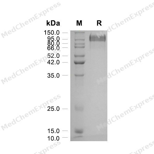
Purity - SDS-PAGE
-
Bioactivity - ELISA
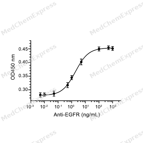
Bioactivity - ELISA


Technical Parameters
-
Species Rat
-
Source HEK293
-
Tag C-His
-
Accession
E7CXR8 (L25-S646)
-
Molecular Construction
-
N-term
-
EGFR (L25-S646)
Accession # E7CXR8 -
His
-
C-term
-
-
Protein Length
Partial
-
Synonyms
EGFR; Epidermal Growth Factor Receptor Variant EX12_14del; Prev. ERBB; Cell Proliferation-Inducing Protein 61; ERBB1; Cell Growth Inhibiting Protein 40; ERRP; Receptor Protein-Tyrosine Kinase; Receptor Tyrosine-Protein Kinase ErbB-1; Alternative Protein E
-
AA Sequence
LEEKKVCQGTSNRLTQLGTFEDHFLSLQRMFNNCEVVLGNLEITYVQRNYDLSFLKTIQEVAGYVLIALNTVERIPLENLQIIRGNALYENTYALAVLSNYGTNKTGLRELPMRNLQEILIGAVRFSNNPILCNMETIQWRDIVQDVFLSNMSMDVQRHLTGCPKCDPSCPNGSCWGRGEENCQKLTKIICAQQCSRRCRGRSPSDCCHNQCAAGCTGPRESDCLVCHRFRDEATCKDTCPPLMLYNPTTYQMDVNPEGKYSFGATCVKKCPRNYVVTDHGSCVRACGPDYYEVEEDGVSKCKKCDGPCRKVCNGIGIGEFKDTLSINATNIKHFKYCTAISGDLHILPVAFKGDSFTRTPPLDPRELEILKTVKEITGFLLIQAWPENWTDLHAFENLEIIRGRTKQHGQFSLAVVGLNITSLGLRSLKEISDGDVIISGNRNLCYANTINWKKLFGTPNQKTKIMNNRAEKDCKATNHVCNPLCSSEGCWGPEPTDCVSCQNVSRGRECVDKCNILEGEPREFVENSECIQCHPECLPQTMNITCTGRGPDNCIKCAHYVDGPHCVKTCPSGIMGENNTLVWKFADANNVCHLCHANCTYGCAGPGLKGCQQPEGPKIPS
-
Molecular Weight
Approximately 93-110 kDa, based on SDS-PAGE under reducing conditions, due to the glycosylation.
-
Purity
≥ 95%, as determined by reducing SDS-PAGE.


Product Properties
Lyophilized powder.
1.Lyophilized from a 0.22 μm filtered solution of PBS, pH 7.4, 5% trehalose, 5% mannitol, 0.01% tween 80.
2.Lyophilized from a 0.22 μm filtered solution of 20 mM PB, 150 mM NaCl, pH 7.4.
Please refer to the lot-specific COA for specific buffer information.
<1 EU/μg, determined by LAL method.
It is not recommended to reconstitute to a concentration less than 100 μg/mL in ddH2O. For long term storage it is recommended to add a carrier protein (0.1% BSA, 5% HSA, 10% FBS or 5% Trehalose).
Stored at -20°C for 2 years from date of receipt. After reconstitution, it is stable at 4°C for 1 week or -20°C for longer (with carrier protein). It is recommended to freeze aliquots at -20°C or -80°C for extended storage.
Room temperature in continental US; may vary elsewhere.


Documentation
-
Data Sheet (240 KB)
-
SDS (252 KB)
- English - EN (252 KB)
- Français - FR (252 KB)
- Deutsch - DE (252 KB)
- Norwegian - NO (252 KB)
- Español - ES (252 KB)
- Swedish - SV (252 KB)
- Italian - IT (252 KB)
- Korean - KR (252 KB)
- Portuguese - PT (252 KB)
-
Handling Instructions (2659 KB)

Calculators
Concentration (start) × Volume (start) = Concentration (final) × Volume (final)


 Powered by Bioz

