



Echistatin TFA

Based on 1 Customer Validation
Echistatin TFA is a naturally derived RGD-containing snake venom peptide. Echistatin TFA exhibits an IC50 of 0.6 nM against mouse αvβ3 integrin, and acts as an antagonist against αvβ3, αIIbβ3, α5β1 integrins, pp125FAK and paxillin. Echistatin TFA reduces the phosphorylation of pp125FAK and paxillin, inhibits the autophosphorylation and kinase activity of pp125FAK, and weakens its binding to pp60src and paxillin. Echistatin TFA disrupts the actin cytoskeleton and focal adhesions, induces melanoma cell detachment from fibronectin, and regulates cell adhesion and motility. Echistatin TFA inhibits osteoclast maturation and migration, bone resorption, platelet aggregation, bone loss, as well as adhesion and metastasis of Lewis lung cancer cells; it also increases the number of osteoclasts and bone coverage area in mice with secondary hyperparathyroidism. Echistatin TFA can be used in research related to melanoma, lung cancer and secondary hyperparathyroidism.
For research use only. We do not sell to patients.
- Purity : 99.32%
- Formula: C217H341N71O74S9.xC2HF3O2
- Molecular Weight:5417.00 (free acid)
-
Storage:
Sealed storage, away from moisture.
Powder -80°C, 2 years , -20°C, 1 year* In solvent : -80°C, 6 months; -20°C, 1 month (sealed storage, away from moisture)

To place orders, for customer services and technical support, please contact: MedChemExpress USA
Tel: 609-228-6898 E-mail: [email protected] [email protected]


-
 Biological Activity
-
Chemical Information
-
Solvent & Solubility
- Protocols
- Purity & Documentation
- References
-
 Help & FAQs




Biological Activity
Description
IC50 & Target
[4]|
αvβ3 |
α5β1 |
αIIbβ3 |
In Vitro
Echistatin (100 μg/mL; 30 min) induces detachment of fibronectin-adherent B16-BL6 mouse melanoma cells and causes loss of pp125FAK and paxillin from focal adhesions[1].
Echistatin (10 nCi/tube; 1 h) binds specifically to αvβ3 integrin in membrane fractions enriched from mouse osteoclast-like cells, with an IC50 of 0.6 nM, and this binding is selectively blocked by anti-αvβ3 integrin antibody[2].
Echistatin (100 nM; 7 days, days 0-4, days 4-7) potently inhibits the formation of TRAP-positive multinucleated osteoclast-like cells in a mouse MB1.8 cell-bone marrow co-culture system, with an IC50 of 0.7 nM[2].
Echistatin (10-11 to 10-8 M; 15 h) inhibits M-CSF-induced fusion of mouse mononuclear pre-fusion osteoclasts with an IC50 of 0.6 nM, and inhibits M-CSF-induced migration of mouse mononuclear pre-fusion osteoclasts with an IC50 of 1.0 nM[2].
Echistatin enables the quantitative detection of Echistatin levels in biological samples via competitive receptor binding assays, in which Echistatin extracts from HEK 293 cells overexpressing αvβ3 integrin displace radiolabeled Echistatin[3].
MedChemExpress (MCE) has not independently confirmed the accuracy of these methods. They are for reference only.
In Vivo
Echistatin (6-30 µg/kg/min; subcutaneous injection; continuous infusion; 6 h) inhibits the acute calcemic response to exogenous PTH in parathyroidectomized mice[3].
Echistatin (1 μg/g; intraperitoneal injection; once every 72 hours; 4 doses total) inhibits Lewis lung cancer tumor growth and metastasis formation in male C57BL/6NCrlBR mice[4].
MedChemExpress (MCE) has not independently confirmed the accuracy of these methods. They are for reference only.


Chemical Information
-
Appearance Solid
-
Molecular Weight 5417.00 (free acid)
-
Formula C217H341N71O74S9.xC2HF3O2
-
Color White to off-white
-
Sequence
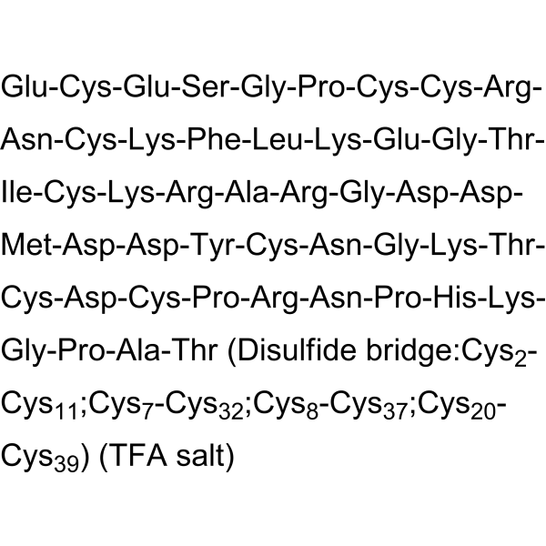
Glu-Cys-Glu-Ser-Gly-Pro-Cys-Cys-Arg-Asn-Cys-Lys-Phe-Leu-Lys-Glu-Gly-Thr-Ile-Cys-Lys-Arg-Ala-Arg-Gly-Asp-Asp-Met-Asp-Asp-Tyr-Cys-Asn-Gly-Lys-Thr-Cys-Asp-Cys-Pro-Arg-Asn-Pro-His-Lys-Gly-Pro-Ala-Thr (Disulfide bridge:Cys2-Cys11;Cys7-Cys32;Cys8-Cys37;Cys20-Cys39)
-
Sequence Shortening
ECESGPCCRNCKFLKEGTICKRARGDDMDDYCNGKTCDCPRNPHKGPAT (Disulfide bridge:Cys2-Cys11;Cys7-Cys32;Cys8-Cys37;Cys20-Cys39)
-
Structure Classification
-
Initial Source
snake venoms
-
Shipping
Room temperature in continental US; may vary elsewhere.
-
Storage
Sealed storage, away from moisture
Powder -80°C 2 years -20°C 1 year * In solvent : -80°C, 6 months; -20°C, 1 month (sealed storage, away from moisture)


Solvent & Solubility
In Vitro:
H2O : 50 mg/mL (Need ultrasonic)
In Vivo:
For the following dissolution methods, please prepare the working solution directly:
It is recommended to prepare fresh solutions and use them promptly within a short period of time.
The percentages shown for the solvents indicate their volumetric ratio in the final prepared solution. If precipitation or phase separation occurs during preparation, heat and/or sonication can be used to aid dissolution.
Add each solvent one by one: PBS
Solubility: 50 mg/mL; Clear solution; Need ultrasonic


Purity & Documentation
-
Data Sheet (336 KB)
-
SDS (252 KB)
- English - EN (252 KB)
- Français - FR (252 KB)
- Deutsch - DE (252 KB)
- Norwegian - NO (252 KB)
- Español - ES (252 KB)
- Swedish - SV (252 KB)
- Italian - IT (252 KB)
- Korean - KR (252 KB)
- Portuguese - PT (252 KB)
-
Handling Instructions (2659 KB)
References
[1]. Della Morte R, et al. Echistatin inhibits pp125FAK autophosphorylation, paxillin phosphorylation and pp125FAK-paxillin interaction in fibronectin-adherent melanoma cells. European journal of biochemistry. 2000 Aug;267(16):5047-54. [Content Brief]
[2]. Nakamura I, et al. Echistatin inhibits the migration of murine prefusion osteoclasts and the formation of multinucleated osteoclast-like cells. Endocrinology. 1998 Dec;139(12):5182-93. [Content Brief]
[3]. Masarachia P, et al. Histomorphometric evidence for echistatin inhibition of bone resorption in mice with secondary hyperparathyroidism. Endocrinology. 1998 Mar;139(3):1401-10. [Content Brief]
[4]. Spalleticernia D, et al. Echistatin inhibits Lewis lung carcinoma cell-matrix adhesion in vitro and experimental metastasis in vivo. International journal of oncology. 1997 Oct;11(4):757-63. [Content Brief]

Calculators
Concentration (start) × Volume (start) = Concentration (final) × Volume (final)


Echistatin TFA Related Classifications
HY-P1189A Related Classifications
 Powered by Bioz