From 11:00 pm to 12:00 pm EST ( 8:00 pm to 9:00 pm PST ) on January 6th, the website will be under maintenance. We are sorry for the inconvenience. Please arrange your schedule properly.
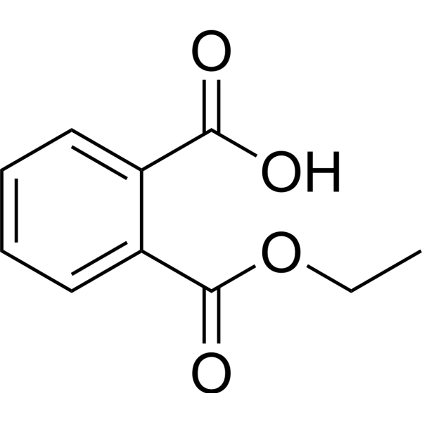
Monoethyl phthalate is an orally active PDX-1 activator and the major hydrolytic metabolite of Diethyl phthalate (HY-Y0284) in vivo, with reproductive toxicity. Monoethyl phthalate targets aromatase (aromatase/CYP19A1) and PPAR to induce cell proliferation. The plasmaproteinbinding rate of Monoethyl phthalate in rats and humans is lower than that of Diethyl phthalate. It exhibits significant enterohepatic circulation in rats and mainly accumulates in liver tissues. Monoethyl phthalate shows no estrogenic activity in estrogen-dependent human breast cancer cells. Monoethyl phthalate can be used in studies of reproductive toxicity and related environmental endocrine disruption mechanisms .
Canine Serum Albumin is a plasmaprotein derived from dogs. Canine Serum Albumin exhibits greater flexibility than human serum albumin and bovine serum albumin. Serum albumin is a multifunctional protein with exceptional ligand-binding capacity, which enables it to act as a transport molecule for various metabolites, drugs, nutrients, metals and other molecules .
Zotiraciclib (TG02; SB1317) is an orally active JAK2/FLT3/CDK2 inhibitor with IC50 values of 13 nM, 73 nM and 56 nM , respectively. Zotiraciclib inhibits cancer cell proliferation, tumor growth and the activity of CYP2D6. Zotiraciclib exhibits high plasmaproteinbinding rate, Caco-2 permeability and tissue distribution capacity, as well as metabolic stability in human and canine liver microsomes. Zotiraciclib achieves tumor growth inhibition in nude mouse models of colon cancer and lymphoma xenografts. Zotiraciclib can be used for research related to colon cancer, B-cell lymphoma, advanced leukemia, acute leukemia and multiple myeloma .
Progesterone/BSA is a conjugate of Progesterone (HY-N0437) and bovine serum albumin (BSA). Progesterone/BSA cannot penetrate the plasma membrane of human sperm, but still rapidly elevates intracellular free calcium and induces the acrosome reaction. Progesterone/BSA can also act as a probe to specifically bind to progesterone-bindingproteins on the membrane of rat brain synaptosomes .
C1 Esterase Inhibitor (Human) is a C1 Esterase inhibitor derived from humanplasma. C1 Esterase Inhibitor (Human), a glycoprotein, is a serum protease inhibitor (serpin) that binds covalently and inactivates C1r, C1s, and mannan-bindingprotein-associated proteases (MASPs). C1 Esterase Inhibitor (Human) has anti-inflammatory effects. C1 Esterase Inhibitor (Human) can be used to prevent angioedema attacks associated with hereditary angioedema .
SPSB2-iNOS inhibitory cyclic peptide-1 is an inhibitor for the interaction of SPRY domain and SOCS-box protein 2 (SPSB2) and iNOS, through binding SPSB2 on iNOS site with KD of 4.4 nM. SPSB2-iNOS inhibitory cyclic peptide-1 is resistant to the proteases pepsin, trypsin and α-chymotrypsin. SPSB2-iNOS inhibitory cyclic peptide-1 is stable in humanplasma and in oxidative environment .
AGU661 is a Microsomal prostaglandin E2 synthase 1 (mPGES-1) inhibitor with an IC50 of 0.22 nM. AGU661 lowers PGE2 formation in human pro-inflammatory M1 macrophages and activated monocytes without affecting other lipid mediator pathways. AGU661 has unfavorable physicochemical properties with poor metabolic stability and strong plasmaproteinbinding tendencies. AGU661 into PLGA-based NPs significantly enhances its bioactivity. AGU661 can be used for inflammatory disorders research .
EGFR is a driver of tumorigenesis. EGFR is mainly found in an auto-inhibited, dimerization-incompetent, state at the plasma membrane (PM). Ligand binding promotes receptor dimerization, which determines a series of structural rearrangements that are conveyed to the cytoplasmic domain allowing the formation of asymmetric dimers between the two juxtaposed catalytic domains. EGFR has multiple mutants. EGFR C797S/L858R Recombinant Human Active Protein Kinase is a recombinant EGFR C797S/L858R protein that can be used to study EGFR C797S/L858R-related functions .
Human IGFBP4 mRNA encodes the human insulin like growth factor bindingprotein 4 (IGFBP4) protein, a member of the insulin-like growth factor bindingprotein (IGFBP) family. IGFBP4 can bind both insulin-like growth factors (IGFs) I and II and circulates in the plasma in both glycosylated and non-glycosylated forms. Binding of this protein prolongs the half-life of the IGFs and alters their interaction with cell surface receptors.
Human IGFBP3 mRNA encodes the human insulin like growth factor bindingprotein 3 (IGFBP3) protein, a member of the insulin-like growth factor bindingprotein (IGFBP) family. IGFBP3 can form a ternary complex with insulin-like growth factor acid-labile subunit (IGFALS) and either insulin-like growth factor (IGF) I or II. In this form, it circulates in the plasma, prolonging the half-life of IGFs and altering their interaction with cell surface receptors.
AMG-222 is a dipeptidyl peptidase IV (DPP-IV) inhibitor that exerts its inhibitory effect by tightly and reversibly binding to DPPIV. AMG 222 binds to humanplasmaproteins in a saturable and concentration-dependent manner, with a binding rate of 80.8% at 1 nM, while the binding rate decreases to 29.4% at concentrations above 100 nM. AMG-222 can be used in research related to diabetes .
EGFR is a driver of tumorigenesis. EGFR is mainly found in an auto-inhibited, dimerization-incompetent, state at the plasma membrane (PM). Ligand binding promotes receptor dimerization, which determines a series of structural rearrangements that are conveyed to the cytoplasmic domain allowing the formation of asymmetric dimers between the two juxtaposed catalytic domains. EGFR has multiple mutants. EGFR G719C Recombinant Human Active Protein Kinase is a recombinant EGFR G719C protein that can be used to study EGFR G719C-related functions .
EGFR is a driver of tumorigenesis. EGFR is mainly found in an auto-inhibited, dimerization-incompetent, state at the plasma membrane (PM). Ligand binding promotes receptor dimerization, which determines a series of structural rearrangements that are conveyed to the cytoplasmic domain allowing the formation of asymmetric dimers between the two juxtaposed catalytic domains. EGFR has multiple mutants. EGFR L718Q Recombinant Human Active Protein Kinase is a recombinant EGFR L718Q protein that can be used to study EGFR L718Q-related functions .
EGFR is a driver of tumorigenesis. EGFR is mainly found in an auto-inhibited, dimerization-incompetent, state at the plasma membrane (PM). Ligand binding promotes receptor dimerization, which determines a series of structural rearrangements that are conveyed to the cytoplasmic domain allowing the formation of asymmetric dimers between the two juxtaposed catalytic domains. EGFR has multiple mutants. EGFR C797S Recombinant Human Active Protein Kinase is a recombinant EGFR C797S protein that can be used to study EGFR C797S-related functions .
EGFR is a driver of tumorigenesis. EGFR is mainly found in an auto-inhibited, dimerization-incompetent, state at the plasma membrane (PM). Ligand binding promotes receptor dimerization, which determines a series of structural rearrangements that are conveyed to the cytoplasmic domain allowing the formation of asymmetric dimers between the two juxtaposed catalytic domains. EGFR has multiple mutants. EGFR L858R Recombinant Human Active Protein Kinase is a recombinant EGFR L858R protein that can be used to study EGFR L858R-related functions .
EGFR is a driver of tumorigenesis. EGFR is mainly found in an auto-inhibited, dimerization-incompetent, state at the plasma membrane (PM). Ligand binding promotes receptor dimerization, which determines a series of structural rearrangements that are conveyed to the cytoplasmic domain allowing the formation of asymmetric dimers between the two juxtaposed catalytic domains. EGFR has multiple mutants. EGFR L861Q Recombinant Human Active Protein Kinase is a recombinant EGFR L861Q protein that can be used to study EGFR L861Q-related functions .
EGFR is a driver of tumorigenesis. EGFR is mainly found in an auto-inhibited, dimerization-incompetent, state at the plasma membrane (PM). Ligand binding promotes receptor dimerization, which determines a series of structural rearrangements that are conveyed to the cytoplasmic domain allowing the formation of asymmetric dimers between the two juxtaposed catalytic domains. EGFR has multiple mutants. EGFR G719S Recombinant Human Active Protein Kinase is a recombinant EGFR G719S protein that can be used to study EGFR G719S-related functions .
EGFR is a driver of tumorigenesis. EGFR is mainly found in an auto-inhibited, dimerization-incompetent, state at the plasma membrane (PM). Ligand binding promotes receptor dimerization, which determines a series of structural rearrangements that are conveyed to the cytoplasmic domain allowing the formation of asymmetric dimers between the two juxtaposed catalytic domains. EGFR has multiple mutants. EGFR T790M Recombinant Human Active Protein Kinase is a recombinant EEGFR T790M protein that can be used to study EGFR T790M-related functions .
EGFR is a driver of tumorigenesis. EGFR is mainly found in an auto-inhibited, dimerization-incompetent, state at the plasma membrane (PM). Ligand binding promotes receptor dimerization, which determines a series of structural rearrangements that are conveyed to the cytoplasmic domain allowing the formation of asymmetric dimers between the two juxtaposed catalytic domains. EGFR has multiple mutants. EGFR d752-759 Recombinant Human Active Protein Kinase is a recombinant EGFR d752-759 protein that can be used to study EGFR d752-759-related functions .
EGFR is a driver of tumorigenesis. EGFR is mainly found in an auto-inhibited, dimerization-incompetent, state at the plasma membrane (PM). Ligand binding promotes receptor dimerization, which determines a series of structural rearrangements that are conveyed to the cytoplasmic domain allowing the formation of asymmetric dimers between the two juxtaposed catalytic domains. EGFR has multiple mutants. EGFR d746-750 Recombinant Human Active Protein Kinase is a recombinant EGFR d746-750 protein that can be used to study EGFR d746-750-related functions .
EGFR is a driver of tumorigenesis. EGFR is mainly found in an auto-inhibited, dimerization-incompetent, state at the plasma membrane (PM). Ligand binding promotes receptor dimerization, which determines a series of structural rearrangements that are conveyed to the cytoplasmic domain allowing the formation of asymmetric dimers between the two juxtaposed catalytic domains. EGFR has multiple mutants. EGFR T790M/L858R Recombinant Human Active Protein Kinase is a recombinant EGFR T790M/L858R protein that can be used to study EGFR T790M/L858R-related functions .
EGFR is a driver of tumorigenesis. EGFR is mainly found in an auto-inhibited, dimerization-incompetent, state at the plasma membrane (PM). Ligand binding promotes receptor dimerization, which determines a series of structural rearrangements that are conveyed to the cytoplasmic domain allowing the formation of asymmetric dimers between the two juxtaposed catalytic domains. EGFR has multiple mutants. EGFR d747-749/A750P Recombinant Human Active Protein Kinase is a recombinant EGFR d747-749/A750P protein that can be used to study EGFR d747-749/A750P-related functions .
EGFR is a driver of tumorigenesis. EGFR is mainly found in an auto-inhibited, dimerization-incompetent, state at the plasma membrane (PM). Ligand binding promotes receptor dimerization, which determines a series of structural rearrangements that are conveyed to the cytoplasmic domain allowing the formation of asymmetric dimers between the two juxtaposed catalytic domains. EGFR has multiple mutants. EGFR d746-750/C797S Recombinant Human Active Protein Kinase is a recombinant EGFR d746-750/C797S protein that can be used to study EGFR d746-750/C797S-related functions .
EGFR is a driver of tumorigenesis. EGFR is mainly found in an auto-inhibited, dimerization-incompetent, state at the plasma membrane (PM). Ligand binding promotes receptor dimerization, which determines a series of structural rearrangements that are conveyed to the cytoplasmic domain allowing the formation of asymmetric dimers between the two juxtaposed catalytic domains. EGFR has multiple mutants. EGFR d747-752/P753S Recombinant Human Active Protein Kinase is a recombinant EGFR d747-752/P753S protein that can be used to study EGFR d747-752/P753S-related functions .
ML350 (CYM50202) is a highly potent OPRK1 antagonist with selectivity and broad biological applications. With IC50 values of 9-16 nM, ML350 shows high selectivity for OPRK1, with selectivity of 219-382-fold and 20-35-fold relative to OPRD1 and OPRM1, respectively. ML350 exhibited favorable characteristics in in vivo pharmacokinetic analysis, including high passive membrane permeability and moderate humanplasmaproteinbinding. Extensive screening of ML350 against multiple ion channels, receptors, and transporters showed that it does not have adverse off-target effects .
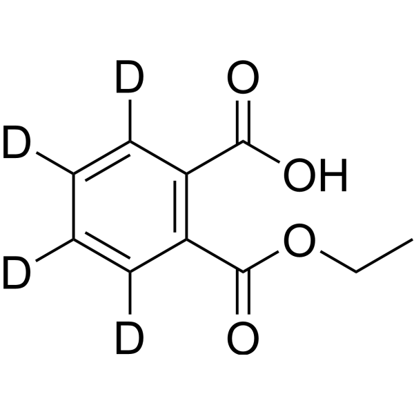
Monoethyl phthalate-d4 is the deuterium labeled Monoethyl phthalate. Monoethyl phthalate is an orally active PDX-1 activator and the major hydrolytic metabolite of Diethyl phthalate (HY-Y0284) in vivo, with reproductive toxicity. Monoethyl phthalate targets aromatase (aromatase/CYP19A1) and PPAR to induce cell proliferation. The plasmaproteinbinding rate of Monoethyl phthalate in rats and humans is lower than that of Diethyl phthalate. It exhibits significant enterohepatic circulation in rats and mainly accumulates in liver tissues. Monoethyl phthalate shows no estrogenic activity in estrogen-dependent human breast cancer cells. Monoethyl phthalate can be used in studies of reproductive toxicity and related environmental endocrine disruption mechanisms .
SPSB2-iNOS inhibitory cyclic peptide-1 TFA is the TFA salt form of SPSB2-iNOS inhibitory cyclic peptide-1(HY-P10383). SPSB2-iNOS inhibitory cyclic peptide-1 is an inhibitor for the interaction of SPRY domain and SOCS-box protein 2 (SPSB2) and iNOS, through binding SPSB2 on iNOS site with KD of 4.4 nM. SPSB2-iNOS inhibitory cyclic peptide-1 is resistant to the proteases pepsin, trypsin and α-chymotrypsin. SPSB2-iNOS inhibitory cyclic peptide-1 is stable in humanplasma and in oxidative environment .
GSK461364 analogue 1 is a thiophene-based PLK1 inhibitor with a PLK1 IC50 of 2 nM and a PLK3 IC50 of 630 nM. GSK461364 analogue 1 also acts as an inhibitor of Nek2 kinase (IC50: 21 nM). GSK461364 analogue 1 has a solubility of ≥190 μM in pH 7.4 PBS and a humanplasmaproteinbinding rate of 91.5%. GSK461364 analogue 1 can be used in studies related to colon cancer, lung cancer, breast cancer and ovarian cancer .
IACS-4619 (compound 4) is a highly selective 2-aminopyrimidine-based MTH1 (MutT homolog 1) inhibitor (IC50=0.2 nM). IACS-4619 inhibits MTH1 by blocking its hydrolysis of oxidized purine nucleotides such as 8-oxo-dGTP, thereby preventing MTH1 from inhibiting the incorporation of oxidized nucleotides into DNA. IACS-4619 significantly inhibits endogenous MTH1 activity in MTH1-overexpressing U2OS cells, but without antiproliferative or cytotoxic effects on various human cancer and normal cell lines. IACS-4619 can be used in oncology research related to the MTH1 target .
Z971169476 is a sulfonamide-based protein-RNA interaction inhibitor targeting the KH34 region of insulin-like growth factor 2 mRNA binding protein 2 (IGF2BP2/IMP2) .
EGFR is a driver of tumorigenesis. EGFR is mainly found in an auto-inhibited, dimerization-incompetent, state at the plasma membrane (PM). Ligand binding promotes receptor dimerization, which determines a series of structural rearrangements that are conveyed to the cytoplasmic domain allowing the formation of asymmetric dimers between the two juxtaposed catalytic domains. EGFR has multiple mutants. EGFR d746-750/T790M/C797S Recombinant Human Active Protein Kinase is a recombinant EGFR d746-750/T790M/C797S protein that can be used to study EGFR d746-750/T790M/C797S-related functions .
EGFR is a driver of tumorigenesis. EGFR is mainly found in an auto-inhibited, dimerization-incompetent, state at the plasma membrane (PM). Ligand binding promotes receptor dimerization, which determines a series of structural rearrangements that are conveyed to the cytoplasmic domain allowing the formation of asymmetric dimers between the two juxtaposed catalytic domains. EGFR has multiple mutants. EGFR d746-750/T790M/C797S/L858R Recombinant Human Active Protein Kinase is a recombinant EGFR d746-750/T790M/C797S/L858R protein that can be used to study EGFR d746-750/T790M/C797S/L858R-related functions .
Membrane receptors, also known cell surface receptors or transmembrane receptors, are transmembrane proteins embedded into the plasma membrane which play an essential role in maintaining communication between the internal processes within the cell and various types of extracellular signals. They act in cell signaling by receiving (binding to) extracellular molecules, which are also called ligands. These extracellular molecules include hormones, cytokines, growth factors, neurotransmitters, lipophilic signaling molecules such as prostaglandins, and cell recognition molecules.
There are three kinds of membrane receptors: ion channel-linked receptors, enzyme-linked receptors and G-protein-linked receptors. They play important roles in keeping human normal physiologic processes. GPCRs and ion channels are important drug targets in drug discovery.
MCE provides a unique collection of 6,857 compounds targeting a variety of membrane receptors. MCE Membrane reeptor-targeted Compound Library can be used for membrane receptor-focused screening and drug discovery.
Canine Serum Albumin is a plasmaprotein derived from dogs. Canine Serum Albumin exhibits greater flexibility than human serum albumin and bovine serum albumin. Serum albumin is a multifunctional protein with exceptional ligand-binding capacity, which enables it to act as a transport molecule for various metabolites, drugs, nutrients, metals and other molecules .
Progesterone/BSA is a conjugate of Progesterone (HY-N0437) and bovine serum albumin (BSA). Progesterone/BSA cannot penetrate the plasma membrane of human sperm, but still rapidly elevates intracellular free calcium and induces the acrosome reaction. Progesterone/BSA can also act as a probe to specifically bind to progesterone-bindingproteins on the membrane of rat brain synaptosomes .
SPSB2-iNOS inhibitory cyclic peptide-1 is an inhibitor for the interaction of SPRY domain and SOCS-box protein 2 (SPSB2) and iNOS, through binding SPSB2 on iNOS site with KD of 4.4 nM. SPSB2-iNOS inhibitory cyclic peptide-1 is resistant to the proteases pepsin, trypsin and α-chymotrypsin. SPSB2-iNOS inhibitory cyclic peptide-1 is stable in humanplasma and in oxidative environment .
SPSB2-iNOS inhibitory cyclic peptide-1 TFA is the TFA salt form of SPSB2-iNOS inhibitory cyclic peptide-1(HY-P10383). SPSB2-iNOS inhibitory cyclic peptide-1 is an inhibitor for the interaction of SPRY domain and SOCS-box protein 2 (SPSB2) and iNOS, through binding SPSB2 on iNOS site with KD of 4.4 nM. SPSB2-iNOS inhibitory cyclic peptide-1 is resistant to the proteases pepsin, trypsin and α-chymotrypsin. SPSB2-iNOS inhibitory cyclic peptide-1 is stable in humanplasma and in oxidative environment .
C1 Esterase Inhibitor (Human) is a C1 Esterase inhibitor derived from humanplasma. C1 Esterase Inhibitor (Human), a glycoprotein, is a serum protease inhibitor (serpin) that binds covalently and inactivates C1r, C1s, and mannan-bindingprotein-associated proteases (MASPs). C1 Esterase Inhibitor (Human) has anti-inflammatory effects. C1 Esterase Inhibitor (Human) can be used to prevent angioedema attacks associated with hereditary angioedema .
Monoethyl phthalate is an orally active PDX-1 activator and the major hydrolytic metabolite of Diethyl phthalate (HY-Y0284) in vivo, with reproductive toxicity. Monoethyl phthalate targets aromatase (aromatase/CYP19A1) and PPAR to induce cell proliferation. The plasmaproteinbinding rate of Monoethyl phthalate in rats and humans is lower than that of Diethyl phthalate. It exhibits significant enterohepatic circulation in rats and mainly accumulates in liver tissues. Monoethyl phthalate shows no estrogenic activity in estrogen-dependent human breast cancer cells. Monoethyl phthalate can be used in studies of reproductive toxicity and related environmental endocrine disruption mechanisms .
RBP4 protein acts as a retinol-binding protein, essential for transporting retinol in blood plasma. It facilitates the delivery of retinol from the liver to peripheral tissues and likely transfers bound all-trans retinol to STRA6 for cell membrane transport. Interactions with TTR prevent kidney glomeruli filtration loss. Direct interaction with STRA6 underscores RBP4's role in intricate retinol transport and distribution processes in the body. RBP4 Protein, Human is the recombinant human-derived RBP4 protein, expressed by E. coli , with tag free.
RBP4 protein acts as a retinol-binding protein, essential for transporting retinol in blood plasma. It facilitates the delivery of retinol from the liver to peripheral tissues and likely transfers bound all-trans retinol to STRA6 for cell membrane transport. Interactions with TTR prevent kidney glomeruli filtration loss. Direct interaction with STRA6 underscores RBP4's role in intricate retinol transport and distribution processes in the body. RBP4 Protein, Human (HEK293, hFc) is the recombinant human-derived RBP4 protein, expressed by HEK293 , with C-hFc labeled tag.
RBP4 protein acts as a retinol-binding protein, essential for transporting retinol in blood plasma. It facilitates the delivery of retinol from the liver to peripheral tissues and likely transfers bound all-trans retinol to STRA6 for cell membrane transport. Interactions with TTR prevent kidney glomeruli filtration loss. Direct interaction with STRA6 underscores RBP4's role in intricate retinol transport and distribution processes in the body. RBP4 Protein, Human (HEK293, C-His) is the recombinant human-derived RBP4 protein, expressed by HEK293 , with C-6*His labeled tag.
HABP2 is a protein that cleaves the alpha chain of fibrinogen as well as the beta chain between "Lys-53" and "Lys-54" at multiple sites, preventing direct formation of a fibrin clot. It activates coagulation factor VII and converts prourokinase into its active form. HABP2 Protein, Human (HEK293, His) is the recombinant human-derived HABP2 protein, expressed by HEK293 , with C-6*His labeled tag.
PSP94/MSMB protein forms homodimers and interacts with PI16. PSP94/MSMB Protein, Human (His) is the recombinant human-derived PSP94/MSMB protein, expressed by E. coli , with C-His labeled tag.
Monoethyl phthalate-d4 is the deuterium labeled Monoethyl phthalate. Monoethyl phthalate is an orally active PDX-1 activator and the major hydrolytic metabolite of Diethyl phthalate (HY-Y0284) in vivo, with reproductive toxicity. Monoethyl phthalate targets aromatase (aromatase/CYP19A1) and PPAR to induce cell proliferation. The plasmaproteinbinding rate of Monoethyl phthalate in rats and humans is lower than that of Diethyl phthalate. It exhibits significant enterohepatic circulation in rats and mainly accumulates in liver tissues. Monoethyl phthalate shows no estrogenic activity in estrogen-dependent human breast cancer cells. Monoethyl phthalate can be used in studies of reproductive toxicity and related environmental endocrine disruption mechanisms .
Human IGFBP4 mRNA encodes the human insulin like growth factor bindingprotein 4 (IGFBP4) protein, a member of the insulin-like growth factor bindingprotein (IGFBP) family. IGFBP4 can bind both insulin-like growth factors (IGFs) I and II and circulates in the plasma in both glycosylated and non-glycosylated forms. Binding of this protein prolongs the half-life of the IGFs and alters their interaction with cell surface receptors.
Human IGFBP3 mRNA encodes the human insulin like growth factor bindingprotein 3 (IGFBP3) protein, a member of the insulin-like growth factor bindingprotein (IGFBP) family. IGFBP3 can form a ternary complex with insulin-like growth factor acid-labile subunit (IGFALS) and either insulin-like growth factor (IGF) I or II. In this form, it circulates in the plasma, prolonging the half-life of IGFs and altering their interaction with cell surface receptors.
Inquiry Online
Your information is safe with us. * Required Fields.
Western blot analysis of extracts from THP-1(lane 2(20μg), Jurkat (lane 3(20μg) and NIH3T3(lane 4(20μg) using FOXO1A (HY-P80132) Rabbit mAb. Proteins were transferred
to a PVDF membrane and blocked with 5% non-fat milk in TBST for 2 hour at room temperature. The primary antibody (1/1000) and Loading control antibody (Beta Actin, HY-P80438, 1/10000) was
used in 5% non-fat milk in TBST at 4°C overnight. Goat Anti-Mouse/Rabbit IgG-HRP Secondary Antibody (1/10000) was used for 1 hour at room temperature.
Western blot analysis of extracts from THP-1(lane 2(20μg), Jurkat (lane 3(20μg) and NIH3T3(lane 4(20μg) using FOXO1A (HY-P80132) Rabbit mAb. Proteins were transferred
to a PVDF membrane and blocked with 5% non-fat milk in TBST for 2 hour at room temperature. The primary antibody (1/1000) and Loading control antibody (Beta Actin, HY-P80438, 1/10000) was
MedchemExpress Validation 03
Western blot analysis of extracts from THP-1(lane 2(20μg), Jurkat (lane 3(20μg) and NIH3T3(lane 4(20μg) using FOXO1A (HY-P80132) Rabbit mAb. Proteins were transferred
MedchemExpress Validation 04
Western blot analysis of extracts from THP-1(lane 2(20μg), Jurkat (lane 3(20μg) and NIH3T3(lane 4(20μg) using FOXO1A (HY-P80132) Rabbit mAb. Proteins were transferred
to a PVDF membrane and blocked with 5% non-fat milk in TBST for 2 hour at room temperature. The primary antibody (1/1000) and Loading control antibody (Beta Actin, HY-P80438, 1/10000) was
used in 5% non-fat milk in TBST at 4°C overnight. Goat Anti-Mouse/Rabbit IgG-HRP Secondary Antibody (1/10000) was used for 1 hour at room temperature.
MedchemExpress Validation
Western blot analysis of extracts from THP-1(lane 2(20μg), Jurkat (lane 3(20μg) and NIH3T3(lane 4(20μg) using FOXO1A (HY-P80132) Rabbit mAb. Proteins were transferred
to a PVDF membrane and blocked with 5% non-fat milk in TBST for 2 hour at room temperature. The primary antibody (1/1000) and Loading control antibody (Beta Actin, HY-P80438, 1/10000) was
used in 5% non-fat milk in TBST at 4°C overnight. Goat Anti-Mouse/Rabbit IgG-HRP Secondary Antibody (1/10000) was used for 1 hour at room temperature.
MedchemExpress Validation
Western blot analysis of extracts from THP-1(lane 2(20μg), Jurkat (lane 3(20μg) and NIH3T3(lane 4(20μg) using FOXO1A (HY-P80132) Rabbit mAb. Proteins were transferred
to a PVDF membrane and blocked with 5% non-fat milk in TBST for 2 hour at room temperature. The primary antibody (1/1000) and Loading control antibody (Beta Actin, HY-P80438, 1/10000) was
used in 5% non-fat milk in TBST at 4°C overnight. Goat Anti-Mouse/Rabbit IgG-HRP Secondary Antibody (1/10000) was used for 1 hour at room temperature.
MedChemExpress values your privacy and your trust is important to us. We use cookies to enhance your website experience. Some cookies are necessary to run the website.
Privacy and Cookie Policy