



Zotiraciclib
Based on 3 publication(s) in Google Scholar
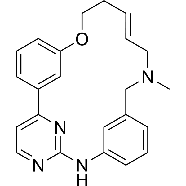
Zotiraciclib (TG02; SB1317) is an orally active JAK2/FLT3/CDK2 inhibitor with IC50 values of 13 nM, 73 nM and 56 nM, respectively. Zotiraciclib inhibits cancer cell proliferation, tumor growth and the activity of CYP2D6. Zotiraciclib exhibits high plasma protein binding rate, Caco-2 permeability and tissue distribution capacity, as well as metabolic stability in human and canine liver microsomes. Zotiraciclib achieves tumor growth inhibition in nude mouse models of colon cancer and lymphoma xenografts. Zotiraciclib can be used for research related to colon cancer, B-cell lymphoma, advanced leukemia, acute leukemia and multiple myeloma.
Nos produits utilisent uniquement pour la recherche. Nous ne vendons pas aux patients.
- Pureté : 99.88%
- CAS No.: 1204918-72-8
- Formule: C23H24N4O
- Masse moléculaire:372.46
-
Stockage:Powder -20°C, 3 years , 4°C, 2 years ; In solvent -80°C, 6 months , -20°C, 1 month

To place orders, for customer services and technical support, please contact: MedChemExpress USA
Tel: 609-228-6898 E-mail: [email protected] [email protected]


-
 Activité biologique
-
Chemical Information
-
Solvant et solubilité
- Protocole
- Pureté et documentation
- Références
-
 Help & FAQs


-
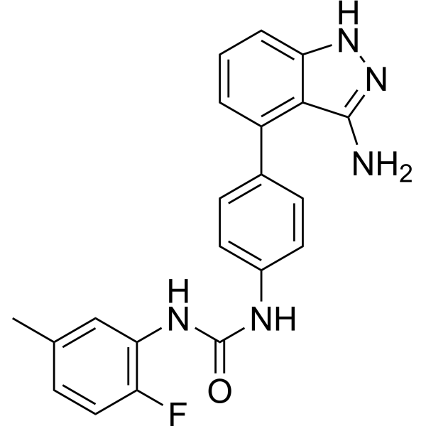
HY-50751
LinifanibMulti-Targeted RTK Inhibitor -
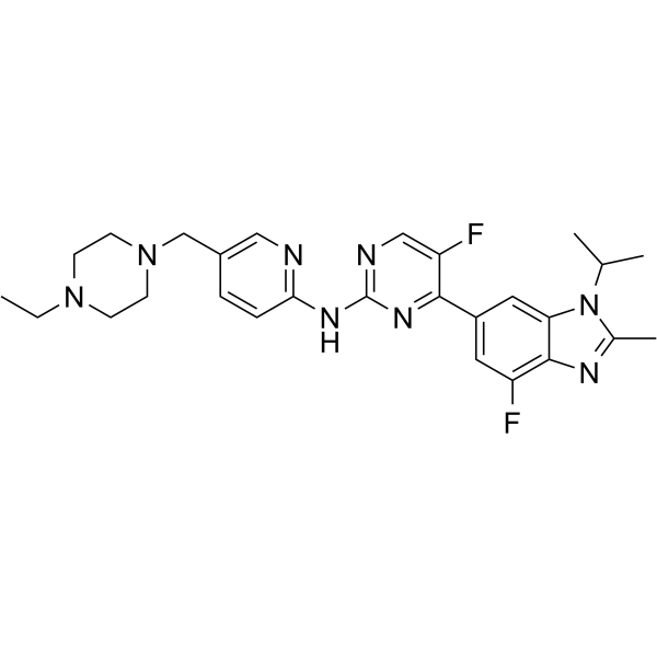
HY-16297A
AbemaciclibCDK4/6 Inhibitor -
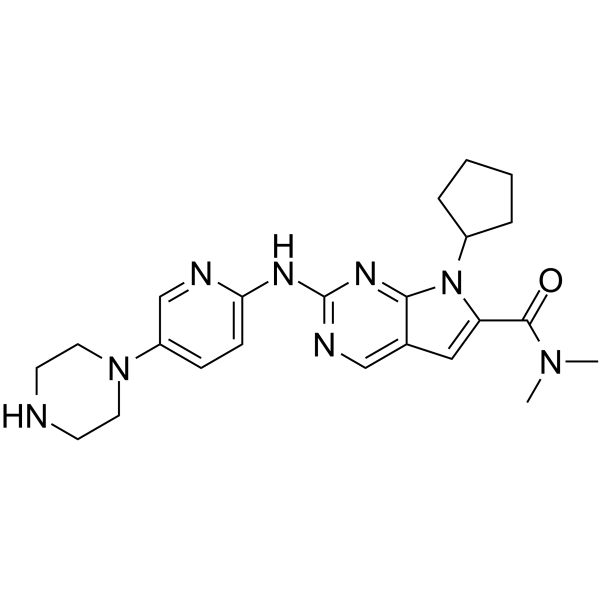
HY-15777
RibociclibCDK4/6 Inhibitor -
HY-10492
DinaciclibCDK2/5/9 Inhibitor -
HY-10987A
ENMD-2076 -
HY-103714
EnsartinibALK/MET Inhibitor -
HY-50856
RuxolitinibJAK1/2 Inhibitor -
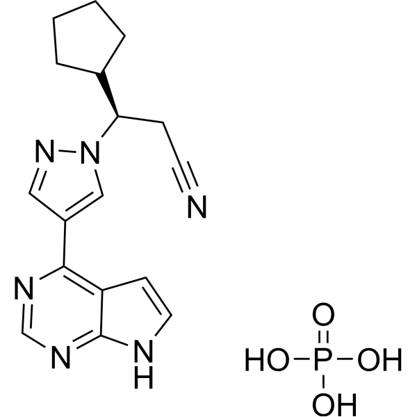
HY-50858
Ruxolitinib phosphateJAK1/2 Inhibitor -

HY-50856R
Ruxolitinib (Standard)JAK1/2 Inhibitor -
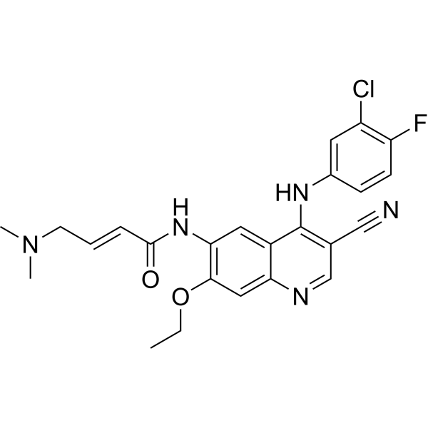
HY-32718
PelitinibSrc Inhibitor -
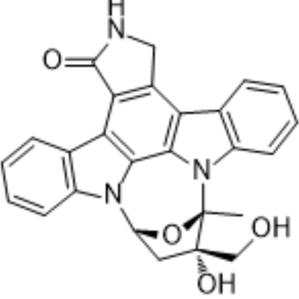
HY-50867
LestaurtinibRPTKs Inhibitor -
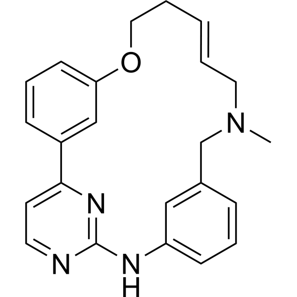
HY-15166
(E/Z)-ZotiraciclibCDK/JAK2/FLT3 Inhibitor
Publications Citing Use of MedChemExpress (MCE) Zotiraciclib
More

Activité biologique
Description
IC50 & Target
[1]|
CDK2 56 nM (IC50) |
JAK2 13 nM (IC50) |
Cellular Effect
|
Cell Line

|
Type | Value | Description | References |
|---|---|---|---|---|
| COLO 205 | IC50 |
0.072 μM
Compound: 26h, SB1317/TG02
|
Antiproliferative activity against human COLO205 cells after 48 hrs
Antiproliferative activity against human COLO205 cells after 48 hrs
|
[PMID: 22148278] |
| DU-145 | IC50 |
0.14 μM
Compound: 26h, SB1317/TG02
|
Antiproliferative activity against human DU145 cells after 48 hrs
Antiproliferative activity against human DU145 cells after 48 hrs
|
[PMID: 22148278] |
| HCT-116 | IC50 |
0.079 μM
Compound: 26h, SB1317/TG02
|
Antiproliferative activity against human HCT116 cells after 48 hrs
Antiproliferative activity against human HCT116 cells after 48 hrs
|
[PMID: 22148278] |
| HEL | GI50 |
0.79 μM
Compound: SB1317
|
Antiproliferative activity against human HEL cells assessed as growth inhibition after 48 hrs by CellTiter-Glo assay
Antiproliferative activity against human HEL cells assessed as growth inhibition after 48 hrs by CellTiter-Glo assay
|
[PMID: 25800646] |
| HL-60 | GI50 |
1.13 μM
Compound: SB1317
|
Antiproliferative activity against human HL60 cells assessed as growth inhibition after 48 hrs by CellTiter-Glo assay
Antiproliferative activity against human HL60 cells assessed as growth inhibition after 48 hrs by CellTiter-Glo assay
|
[PMID: 25800646] |
| HL-60 | IC50 |
0.059 μM
Compound: 26h, SB1317/TG02
|
Antiproliferative activity against human HL60 cells after 48 hrs
Antiproliferative activity against human HL60 cells after 48 hrs
|
[PMID: 22148278] |
| K562 | GI50 |
1.23 μM
Compound: SB1317
|
Antiproliferative activity against human K562 cells assessed as growth inhibition after 48 hrs by CellTiter-Glo assay
Antiproliferative activity against human K562 cells assessed as growth inhibition after 48 hrs by CellTiter-Glo assay
|
[PMID: 25800646] |
| MV4-11 | GI50 |
0.66 μM
Compound: SB1317
|
Antiproliferative activity against human MV4-11 cells assessed as growth inhibition after 48 hrs by CellTiter-Glo assay
Antiproliferative activity against human MV4-11 cells assessed as growth inhibition after 48 hrs by CellTiter-Glo assay
|
[PMID: 25800646] |
| MV4-11 | IC50 |
0.13 μM
Compound: 26h, SB1317/TG02
|
Inhibition of CDK2 in human MV411 cells assessed as Rb phosphorylation after 24 hrs by Western blot analysis
Inhibition of CDK2 in human MV411 cells assessed as Rb phosphorylation after 24 hrs by Western blot analysis
|
[PMID: 22148278] |
| Ramos | IC50 |
0.033 μM
Compound: 26h, SB1317/TG02
|
Antiproliferative activity against human Ramos cells after 48 hrs
Antiproliferative activity against human Ramos cells after 48 hrs
|
[PMID: 22148278] |
| Sf21 | IC50 |
37 nM
Compound: TG02
|
Inhibition of recombinant human C-terminal His6-tagged full length CDK7/untagged recombinant full length human Cyclin H/N-terminal GST-tagged recombinant full length human MAT1 expressed in baculovirus infected Sf21 insect cells using cdk7 peptide as subs
Inhibition of recombinant human C-terminal His6-tagged full length CDK7/untagged recombinant full length human Cyclin H/N-terminal GST-tagged recombinant full length human MAT1 expressed in baculovirus infected Sf21 insect cells using cdk7 peptide as subs
|
[PMID: 30543440] |
| Sf21 | IC50 |
8 nM
Compound: TG02
|
Inhibition of recombinant human full-length C-terminal His6-tagged CDK3/full-length human N-terminal GST-tagged Cyclin E expressed in baculovirus infected Sf21 insect cells using histone H1 as substrate
Inhibition of recombinant human full-length C-terminal His6-tagged CDK3/full-length human N-terminal GST-tagged Cyclin E expressed in baculovirus infected Sf21 insect cells using histone H1 as substrate
|
[PMID: 30543440] |
In Vitro
Zotiraciclib (up to 10 μM; 48 h) potently inhibits proliferation of HL-60, HCT-116, Ramos, COLO205, and DU145 cancer cell lines with IC50 values ranging from 0.033 μM to 0.14 μM[1].
Zotiraciclib (8-1000 nM; 24 h) inhibits phospho-Rb in HCT-116 cells, with detectable effects at 40 nM and complete inhibition at 200 nM after 24 h of treatment[1].
Zotiraciclib (0.05-25 μM; 5-60 min depending on isoform) inhibits human CYP2D6 with an IC50 of 0.95 μM, but does not inhibit CYP1A2, 2C9, 2C19, or 3A4 at concentrations up to 25 μM[1].
Zotiraciclib (5 μM; 120 min) exhibits high permeability with low efflux across Caco-2 cell monolayers, with Papp values of 28.0 × 10⁻⁶ cm/s (apical-to-basolateral) and 27.4 × 10⁻⁶ cm/s (basolateral-to-apical) at 5 μM for 120 min[2].
MedChemExpress (MCE) has not independently confirmed the accuracy of these methods. They are for reference only.
-
Cell Line:HL-60, HCT-116, Ramos, COLO205, DU145
-
Concentration:0-10 μM (IC50 values); up to 10 μM
-
Incubation Time:48 h
-
Result:Inhibited HL-60 cell proliferation with an IC50 of 0.059 μM.
Inhibited HCT-116 cell proliferation with an IC50 of 0.079 μM.
Inhibited Ramos cell proliferation with an IC50 of 0.033 μM.
Inhibited COLO205 cell proliferation with an IC50 of 0.072 μM.
Inhibited DU145 cell proliferation with an IC50 of 0.14 μM.
-
Cell Line:HCT-116
-
Concentration:8, 40, 200, 1000 nM
-
Incubation Time:24 h
-
Result:Inhibited phospho-Rb detectably at 40 nM.
Achieved complete inhibition of phospho-Rb at 200 nM.
Parmacokinetics
| Species | Dose | Route | CL | Vss | T1/2 | AUC0-t | AUC0-∞ | Bioavailability | Tmax | Cmax | CL/F | Vd/F |
|---|---|---|---|---|---|---|---|---|---|---|---|---|
| Mice[1] | 75 mg/kg | p.o. | / | / | 6.1 h | / | 2523 ng·h/mL | 24 % | 0.5 h | 1029 ng/mL | / | / |
| Dog[1] | 10 mg/kg | p.o. | / | / | / | / | / | 4 % | / | / | / | / |
| Rat[1] | 10 mg/kg | p.o. | / | / | / | / | / | 37 % | / | / | / | / |
| Mice[2] | 5 mg/kg | i.v. | 6.6 L/h/kg | 23 L/kg | 4.6 h | 732 ng·h/mL | 752 ng·h/mL | / | / | / | / | / |
| Mice[2] | 75 mg/kg | p.o. | / | / | 6.1 h | 2523 ng·h/mL | 2700 ng·h/mL | 24 % | 0.5 h | 1029 ng/mL | 28 L/h/kg | 245 L/kg |
| Rat[2] | 2 mg/kg | i.v. | 2.4 L/h/kg | 1.80 L/kg | 1.4 h | 897 ng·h/mL | 916 ng·h/mL | / | / | / | / | / |
| Rat[2] | 10 mg/kg | p.o. | / | / | / | 172 ± 38 ng·h/mL | / | 3.8 % | 0.08 ± 0.0 h | 62 ± 25 ng/mL | / | / |
| Dog[2] | 1 mg/kg | i.v. | 1.1 L/h/kg | 3.1 L/kg | 2.9 h | 819 ng·h/mL | 900 ng·h/mL | / | / | / | / | / |
| Dog[2] | 5 mg/kg | p.o. | / | / | 2.9 h | 1659 ng·h/mL | 1663 ng·h/mL | 37 % | 1.0 h | 493 ng/mL | 3.0 L/h/kg | 12.6 L/kg |
In Vivo
Zotiraciclib (15-75 mg/kg; p.o., i.p.; once daily, 2 days on/5 days off, 5 days on/5 days off; 14 days) administered orally at 75 mg/kg on a 2 days on/5 days off schedule achieves 42% TGI, and intraperitoneally at 15 mg/kg on a 5 days on/5 days off schedule achieves 63% TGI in Ramos B-cell lymphoma xenografts in nude mice[1].
MedChemExpress (MCE) has not independently confirmed the accuracy of these methods. They are for reference only.
-
Animal Model:BALB/c nude mice (female, 10−12 weeks of age)[1]
-
Dosage:50 mg/kg; 75 mg/kg
-
Administration:p.o.; 3 times per week (Monday, Wednesday, Friday); 15 days
-
Result:Achieved a mean tumor growth inhibition (TGI) of 82% (75 mg/kg).
Was marginally effective (50 mg/kg).
-
Animal Model:BALB/c nude mice (female, 10−12 weeks of age)[1]
-
Dosage:75 mg/kg (TGI 42%); 15 mg/kg (TGI 63%)
-
Administration:p.o.; once daily, 2 days on and 5 days off schedule; 14 days; i.p.; once daily, 5 days on and 5 days off schedule
-
Result:Achieved a mean TGI of 42% (75 mg/kg, oral, 2 days on/5 days off schedule).
Achieved a mean TGI of 63% (15 mg/kg, intraperitoneal, 5 days on/5 days off schedule).
- BrdU Incorporation Assay
- Immunoprecipitation
- Somatic Cell Culture
- CFSE Dye Dilution Proliferation Assay
- Dye-dilution cell tracking and proliferation staining
- Patient-Derived Orthotopic Xenograft (PDOX)
- CCK-8/WST-8 Cell Proliferation Assay
- Protocol for Cell Cycle
- Cell Counting-Based Growth Curve Assay
- Colony Formation (Clonogenic) Assay
- Cell Viability Determination by MTT Colorimetric Assay
- EdU Incorporation Assay (Click Chemistry-Based DNA Synthesis Measurement)
- Ki-67 Immunostaining Proliferation Assay
- PCNA Immunodetection Proliferation Assay
- Protocol for Cell Counting and Cell Density Analysis
- Patient-Derived Xenograft (PDX)
- Transepithelial/transendothelial electrical resistance assay
- MTT Cell Proliferation Assay
- Subcutaneous Cell-Line-Derived Xenograft
- Orthotopic Cell-Line Xenograft
Essai clinique
| NCT Number | Sponsor | Condition | Start Date |
Phase
|
|---|---|---|---|---|
| NCT01329991 | 2011-05 | PHASE1 |


Chemical Information
-
CAS No. 1204918-72-8
-
Appearance Solid
-
Masse moléculaire 372.46
-
Formule C23H24N4O
-
Color White to off-white
-
SMILES
CN1CC2=CC(NC3=NC(C4=CC(OCC/C=C/C1)=CC=C4)=CC=N3)=CC=C2
-
Synonyms
TG02; SB1317
-
Livraison
Room temperature in continental US; may vary elsewhere.
-
Stockage
Powder -20°C 3 years 4°C 2 years In solvent -80°C 6 months -20°C 1 month


Publications (3)
-
Journal Impact Factor
-
Most Recent
-
Mol Cancer Ther
2025 Dec 4. PMID: 41340469 -
-


Solvant et solubilité
In Vitro:
DMSO : 100 mg/mL (268.49 mM; Need ultrasonic; Hygroscopic DMSO has a significant impact on the solubility of product, please use newly opened DMSO)
Please refer to the solubility information to select the appropriate solvent. Once prepared, please aliquot and store the solution to prevent product inactivation from repeated freeze-thaw cycles.
Storage method and period of stock solution: -80°C, 6 months; -20°C, 1 month. When stored at -80°C, please use it within 6 months. When stored at -20°C, please use it within 1 month.
Please refer to the solubility information to select the appropriate solvent. Once prepared, please aliquot and store the solution to prevent product inactivation from repeated freeze-thaw cycles.
Storage method and period of stock solution: -80°C, 6 months; -20°C, 1 month. When stored at -80°C, please use it within 6 months. When stored at -20°C, please use it within 1 month.
Concentration (start) × Volume (start) = Concentration (final) × Volume (final)
In Vivo:
Select the appropriate dissolution method based on your experimental animal and administration route.
- For the following dissolution methods, please ensure to first prepare a clear stock solution using an In Vitro approach and then sequentially add co-solvents:
- To ensure reliable experimental results, the clarified stock solution can be appropriately stored based on storage conditions. As for the working solution for In Vivo experiments, it is recommended to prepare freshly and use it on the same day.
- The percentages shown for the solvents indicate their volumetric ratio in the final prepared solution. If precipitation or phase separation occurs during preparation, heat and/or sonication can be used to aid dissolution.
Add each solvent one by one: 10% DMSO 40% PEG300 5% Tween-80 45% Saline
Solubility: ≥ 2.5 mg/mL (6.71 mM); Clear solution
This protocol yields a clear solution of ≥ 2.5 mg/mL (saturation unknown).
Taking 1 mL working solution as an example, add 100 μL DMSO stock solution (25.0 mg/mL) to 400 μL PEG300, and mix evenly; then add 50 μL Tween-80 and mix evenly; then add 450 μL Saline to adjust the volume to 1 mL.
Preparation of Saline: Dissolve 0.9 g sodium chloride in ddH₂O and dilute to 100 mL to obtain a clear Saline solution.
In Vivo Dissolution Calculator
Please enter the basic information of animal experiments:
-
-
-
-
Recommended: Prepare an additional quantity of animals to account for potential losses during experiments.
Please enter your animal formula composition:
-
%DMSO +
Recommended: Keep the proportion of DMSO in working solution below 2% if your animal is weak.
-
%+
-
+%Tween-80 + +
-
%Saline +
The co-solvents required include: DMSO, . All of co-solvents are available by MedChemExpress (MCE). , Tween 80. All of co-solvents are available by MedChemExpress (MCE).
Working solution concentration: 0.22 mg/mL
Method for preparing stock solution: mg drug dissolved in μL DMSO. Stock solution concentration: mg/mL.
1. Take μL DMSO stock solution;
2. Add μL .
μL , mix evenly;
3. Then add μL Tween 80, mix evenly;
4. Then add μL
Please ensure that the stock solution in the first step is dissolved to a clear state, and add co-solvents in sequence. You can use ultrasonic heating (ultrasonic cleaner, recommended frequency 20-40 kHz), vortexing, etc. to assist dissolution.


Pureté et documentation
-
Fiche technique (284 KB)
-
SDS (396 KB)
- English - EN (396 KB)
- Français - FR (396 KB)
- Deutsch - DE (396 KB)
- Norwegian - NO (396 KB)
- Español - ES (396 KB)
- Swedish - SV (396 KB)
- Italian - IT (396 KB)
- Korean - KR (396 KB)
- Portuguese - PT (396 KB)
-
Instruction de manipulation (2659 KB)
Références
[1]. William AD, et al. Discovery of kinase spectrum selective macrocycle (16E)-14-methyl-20-oxa-5,7,14,26-tetraazatetracyclo[19.3.1.1(2,6).1(8,12)]heptacosa-1(25),2(26),3,5,8(27),9,11,16,21,23-decaene (SB1317/TG02), a potent inhibitor of cyclin dependent kinases (CDKs), Janus kinase 2 (JAK2), and fms-like tyrosine kinase-3 (FLT3) for the treatment of cancer. J Med Chem. 2012 Jan 12;55(1):169-96. [Content Brief]
[2]. Pasha MK, et al. Preclinical metabolism and pharmacokinetics of SB1317 (TG02), a potent CDK/JAK2/FLT3 inhibitor. Drug Metab Lett. 2012;6(1):33-42. [Content Brief]


Complete Stock Solution Preparation Table
Please refer to the solubility information to select the appropriate solvent. Once prepared, please aliquot and store the solution to prevent product inactivation from repeated freeze-thaw cycles.
Storage method and period of stock solution: -80°C, 6 months; -20°C, 1 month. When stored at -80°C, please use it within 6 months. When stored at -20°C, please use it within 1 month.
| Optional Solvent | Concentration Solvent Mass | 1 mg | 5 mg | 10 mg | 25 mg |
|---|---|---|---|---|---|
| DMSO | 1 mM | 2.6849 mL | 13.4243 mL | 26.8485 mL | 67.1213 mL |
| 5 mM | 0.5370 mL | 2.6849 mL | 5.3697 mL | 13.4243 mL | |
| 10 mM | 0.2685 mL | 1.3424 mL | 2.6849 mL | 6.7121 mL | |
| 15 mM | 0.1790 mL | 0.8950 mL | 1.7899 mL | 4.4748 mL | |
| 20 mM | 0.1342 mL | 0.6712 mL | 1.3424 mL | 3.3561 mL | |
| 25 mM | 0.1074 mL | 0.5370 mL | 1.0739 mL | 2.6849 mL | |
| 30 mM | 0.0895 mL | 0.4475 mL | 0.8950 mL | 2.2374 mL | |
| 40 mM | 0.0671 mL | 0.3356 mL | 0.6712 mL | 1.6780 mL | |
| 50 mM | 0.0537 mL | 0.2685 mL | 0.5370 mL | 1.3424 mL | |
| 60 mM | 0.0447 mL | 0.2237 mL | 0.4475 mL | 1.1187 mL | |
| 80 mM | 0.0336 mL | 0.1678 mL | 0.3356 mL | 0.8390 mL | |
| 100 mM | 0.0268 mL | 0.1342 mL | 0.2685 mL | 0.6712 mL |


Zotiraciclib Related Classifications
HY-15163 Related Classifications
 Powered by Bioz