



Monoethyl phthalate

Based on 1 Customer Validation
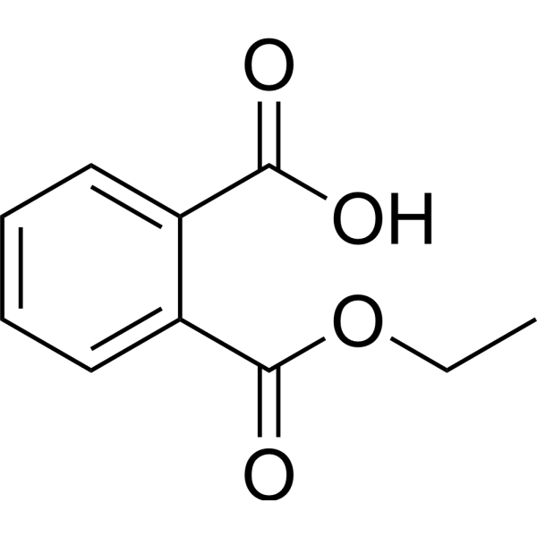
Monoethyl phthalate is an orally active PDX-1 activator and the major hydrolytic metabolite of Diethyl phthalate (HY-Y0284) in vivo, with reproductive toxicity. Monoethyl phthalate targets aromatase (aromatase/CYP19A1) and PPAR to induce cell proliferation. The plasma protein binding rate of Monoethyl phthalate in rats and humans is lower than that of Diethyl phthalate. It exhibits significant enterohepatic circulation in rats and mainly accumulates in liver tissues. Monoethyl phthalate shows no estrogenic activity in estrogen-dependent human breast cancer cells. Monoethyl phthalate can be used in studies of reproductive toxicity and related environmental endocrine disruption mechanisms.
For research use only. We do not sell to patients.
- Purity : 98.58%
- CAS No.: 2306-33-4
- Formula: C10H10O4
- Molecular Weight:194.19
-
Storage:Pure form -20°C, 3 years , 4°C, 2 years ; In solvent -80°C, 6 months , -20°C, 1 month

To place orders, for customer services and technical support, please contact: MedChemExpress USA
Tel: 609-228-6898 E-mail: [email protected] [email protected]


-
 Biological Activity
-
Chemical Information
-
Solvent & Solubility
- Protocols
- Purity & Documentation
- References
-
 Help & FAQs




Biological Activity
Description
IC50 & Target
|
Aromatase |
In Vitro
Monoethyl phthalate (1000 ng/mL; 20 min equilibration at 37 °C) exhibits low plasma protein binding, with 31.22% binding in rat plasma and 40.20% binding in human plasma[1].
Monoethyl phthalate (10-5-10-3 M; 6 d) does not exhibit estrogenic activity in estrogen-sensitive MCF-7 human breast cancer cells[2].
Monoethyl phthalate (10-5-10-3 M; 6 d) does not exhibit anti-estrogenic activity in estrogen-sensitive MCF-7 human breast cancer cells in vitro in the presence of 10-11 M 17β-estradiol[2].
Monoethyl phthalate (10-5-10-3 M; 24 h) does not exhibit cytotoxicity in MCF-7 human breast cancer cells[2].
MedChemExpress (MCE) has not independently confirmed the accuracy of these methods. They are for reference only.
-
Cell Line:MCF-7 human breast cancer cell
-
Concentration:10-5 M, 10-4 M, 10-3 M
-
Incubation Time:6 d
-
Result:Did not stimulate MCF-7 cell proliferation at any tested concentration; cell proliferation remained near the 100% vehicle control level across all doses.
-
Cell Line:MCF-7 human breast cancer cell
-
Concentration:10-5 M, 10-4 M, 10-3 M (in presence of 10-11 M 17β-estradiol)
-
Incubation Time:6 d
-
Result:Did not suppress 17β-estradiol-induced MCF-7 cell proliferation at any tested concentration; cell proliferation remained near the 100% baseline set by 17β-estradiol alone across all doses.
-
Cell Line:MCF-7 human breast cancer cell
-
Concentration:10-5 M, 10-4 M, 10-3 M
-
Incubation Time:24 h
-
Result:Did not inhibit neutral red uptake (a marker of cell viability) at any tested concentration; uptake remained near the 100% vehicle control level across all doses.
Parmacokinetics
| Species | Dose | Route | AUC0-24 | AUC0-∞ | T1/2 | Cmax | Tmax |
|---|---|---|---|---|---|---|---|
| Rat[1] | 0.1 mg/kg | i.v. | 15.48 ng·h/mL | 16.87 ng·h/mL | 1.18 h | 6.99 ng/mL | 0.75 h |
| Rat[1] | 0.5 mg/kg | i.v. | 78.92 ng·h/mL | 80.45 ng·h/mL | 1.27 h | 37.98 ng/mL | 0.92 h |
| Rat[1] | 2 mg/kg | i.v. | 558.27 ng·h/mL | 559.85 ng·h/mL | 1.29 h | 1025.74 ng/mL | / |
| Rat[1] | 10 mg/kg | i.v. | 2890.83 ng·h/mL | 2898.63 ng·h/mL | 2.08 h | 4568.39 ng/mL | / |
| Rat[1] | 0.1 mg/kg | p.o. | 15.48 ng·h/mL | 16.87 ng·h/mL | 1.18 h | 6.99 ng/mL | 0.75 h |
| Rat[1] | 0.5 mg/kg | p.o. | 78.92 ng·h/mL | 80.45 ng·h/mL | 1.27 h | 37.98 ng/mL | 0.92 h |
| Rat[1] | 2 mg/kg | p.o. | 319.46 ng·h/mL | 322.00 ng·h/mL | 1.51 h | 175.00 ng/mL | 0.75 h |
| Rat[1] | 10 mg/kg | p.o. | 1547.16 ng·h/mL | 1551.77 ng·h/mL | 1.96 h | 819.53 ng/mL | 0.75 h |
| Rat[1] | 2 mg/kg | i.v. | 507.52 ng·h/mL | 509.11 ng·h/mL | 1.34 h | 1073.45 ng/mL | / |
In Vivo
Monoethyl phthalate, a metabolite of diethyl phthalate (DEP) in male Sprague-Dawley rats, is generated following a single intravenous injection of diethyl phthalate (HY-Y0284) at doses of 0.1-10 mg/kg. It exhibits dose-linear pharmacokinetic characteristics within the range of 0.1-10 mg/kg, preferentially distributes to the liver, and is mainly excreted in urine[1].
Diethyl phthalate (DEP) (0.1-10 mg/kg; p.o.; single administration) exhibits dose-linear pharmacokinetic characteristics in rats. The plasma peak concentration of monoethyl phthalate is achieved within 0.75-0.92 h, and monoethyl phthalate is primarily excreted via urine[1].
MedChemExpress (MCE) has not independently confirmed the accuracy of these methods. They are for reference only.
-
Animal Model:Sprague-Dawley (male, 8-9 weeks old, 240-300 g)[1]
-
Dosage:0.1-10 mg/kg (parent diethyl phthalate dose)
-
Administration:i.v.; single dose
-
Result:From 0.1 mg/kg parent dose:
Reached an AUC0-∞ of 57.68 ng·h/mL, half-life of 0.96 hours, Cmax of 70.45 ng/mL, and cumulative urinary excretion of 15.14 μg.
From 0.5 mg/kg parent dose:
Reached an AUC0-∞ of 293.20 ng·h/mL, half-life of 1.11 hours, Cmax of 244.10 ng/mL, and cumulative urinary excretion of 82.77 μg.
From 2 mg/kg parent dose:
Reached an AUC0-∞ of 559.85 ng·h/mL, half-life of 1.29 hours, Cmax of 1025.74 ng/mL, and cumulative urinary excretion of 318.56 μg.
From 10 mg/kg parent dose:
Reached an AUC0-∞ of 2898.63 ng·h/mL, half-life of 2.08 hours, Cmax of 4568.39 ng/mL, cumulative urinary excretion of 1582.19 μg, and cumulative fecal excretion of 4.81 μg.
Exhibited highest tissue-to-plasma partition coefficients in liver (1.7-2.2) at 24 hours post-10 mg/kg parent dose administration.
- Reproductive and Developmental Toxicity Study
- BrdU Incorporation Assay
- Immunoprecipitation
- Somatic Cell Culture
- CFSE Dye Dilution Proliferation Assay
- Dye-dilution cell tracking and proliferation staining
- CCK-8/WST-8 Cell Proliferation Assay
- Protocol for Cell Cycle
- Cell Counting-Based Growth Curve Assay
- Colony Formation (Clonogenic) Assay
- Breast Cancer Modeling
- Cell Viability Determination by MTT Colorimetric Assay
- EdU Incorporation Assay (Click Chemistry-Based DNA Synthesis Measurement)
- Ki-67 Immunostaining Proliferation Assay
- PCNA Immunodetection Proliferation Assay
- Protocol for Cell Counting and Cell Density Analysis
- Research Protocol for Endocrine Diseases
- Patient-Derived Xenograft (PDX)
- MTT Cell Proliferation Assay


Chemical Information
-
CAS No. 2306-33-4
-
Appearance Liquid (Density: 1.0583 g/cm3)
-
Molecular Weight 194.19
-
Formula C10H10O4
-
Color Colorless to light yellow
-
SMILES
O=C(C1=CC=CC=C1C(O)=O)OCC
-
Structure Classification
-
Initial Source
-
Shipping
Room temperature in continental US; may vary elsewhere.
-
Storage
Pure form -20°C 3 years 4°C 2 years In solvent -80°C 6 months -20°C 1 month


Solvent & Solubility
In Vitro:
DMSO : 100 mg/mL (514.96 mM; Need ultrasonic; Hygroscopic DMSO has a significant impact on the solubility of product, please use newly opened DMSO)
Please refer to the solubility information to select the appropriate solvent. Once prepared, please aliquot and store the solution to prevent product inactivation from repeated freeze-thaw cycles.
Storage method and period of stock solution: -80°C, 6 months; -20°C, 1 month. When stored at -80°C, please use it within 6 months. When stored at -20°C, please use it within 1 month.
Please refer to the solubility information to select the appropriate solvent. Once prepared, please aliquot and store the solution to prevent product inactivation from repeated freeze-thaw cycles.
Storage method and period of stock solution: -80°C, 6 months; -20°C, 1 month. When stored at -80°C, please use it within 6 months. When stored at -20°C, please use it within 1 month.
Concentration (start) × Volume (start) = Concentration (final) × Volume (final)
In Vivo:
Select the appropriate dissolution method based on your experimental animal and administration route.
- For the following dissolution methods, please ensure to first prepare a clear stock solution using an In Vitro approach and then sequentially add co-solvents:
- To ensure reliable experimental results, the clarified stock solution can be appropriately stored based on storage conditions. As for the working solution for In Vivo experiments, it is recommended to prepare freshly and use it on the same day.
- The percentages shown for the solvents indicate their volumetric ratio in the final prepared solution. If precipitation or phase separation occurs during preparation, heat and/or sonication can be used to aid dissolution.
Add each solvent one by one: 10% DMSO 40% PEG300 5% Tween-80 45% Saline
Solubility: ≥ 2.5 mg/mL (12.87 mM); Clear solution
This protocol yields a clear solution of ≥ 2.5 mg/mL (saturation unknown).
Taking 1 mL working solution as an example, add 100 μL DMSO stock solution (25.0 mg/mL) to 400 μL PEG300, and mix evenly; then add 50 μL Tween-80 and mix evenly; then add 450 μL Saline to adjust the volume to 1 mL.
Preparation of Saline: Dissolve 0.9 g sodium chloride in ddH₂O and dilute to 100 mL to obtain a clear Saline solution.
Add each solvent one by one: 10% DMSO 90% (20% SBE-β-CD in Saline)
Solubility: ≥ 2.5 mg/mL (12.87 mM); Clear solution
This protocol yields a clear solution of ≥ 2.5 mg/mL (saturation unknown).
Taking 1 mL working solution as an example, add 100 μL DMSO stock solution (25.0 mg/mL) to 900 μL 20% SBE-β-CD in Saline, and mix evenly.
Preparation of 20% SBE-β-CD in Saline (4°C, storage for one week): 2 g SBE-β-CD powder is dissolved in 10 mL Saline, completely dissolve until clear.
In Vivo Dissolution Calculator
Please enter the basic information of animal experiments:
-
-
-
-
Recommended: Prepare an additional quantity of animals to account for potential losses during experiments.
Please enter your animal formula composition:
-
%DMSO +
Recommended: Keep the proportion of DMSO in working solution below 2% if your animal is weak.
-
%+
-
+%Tween-80 + +
-
%Saline +
The co-solvents required include: DMSO, . All of co-solvents are available by MedChemExpress (MCE). , Tween 80. All of co-solvents are available by MedChemExpress (MCE).
Working solution concentration: 0.22 mg/mL
Method for preparing stock solution: mg drug dissolved in μL DMSO. Stock solution concentration: mg/mL.
1. Take μL DMSO stock solution;
2. Add μL .
μL , mix evenly;
3. Then add μL Tween 80, mix evenly;
4. Then add μL
Please ensure that the stock solution in the first step is dissolved to a clear state, and add co-solvents in sequence. You can use ultrasonic heating (ultrasonic cleaner, recommended frequency 20-40 kHz), vortexing, etc. to assist dissolution.


Purity & Documentation
-
Data Sheet (311 KB)
-
SDS (394 KB)
- English - EN (394 KB)
- Français - FR (394 KB)
- Deutsch - DE (394 KB)
- Norwegian - NO (394 KB)
- Español - ES (394 KB)
- Swedish - SV (394 KB)
- Italian - IT (394 KB)
- Korean - KR (394 KB)
- Portuguese - PT (394 KB)
-
Handling Instructions (2659 KB)
References
[1]. Jeong SH, et al. Risk assessment for humans using physiologically based pharmacokinetic model of diethyl phthalate and its major metabolite, monoethyl phthalate. Arch Toxicol. 2020;94(7):2377-2400. [Content Brief]
[2]. Okubo T, et al. Estimation of estrogenic and anti-estrogenic activities of some phthalate diesters and monoesters by MCF-7 cell proliferation assay in vitro. Biol Pharm Bull. 2003;26(8):1219-1224. [Content Brief]


Complete Stock Solution Preparation Table
Please refer to the solubility information to select the appropriate solvent. Once prepared, please aliquot and store the solution to prevent product inactivation from repeated freeze-thaw cycles.
Storage method and period of stock solution: -80°C, 6 months; -20°C, 1 month. When stored at -80°C, please use it within 6 months. When stored at -20°C, please use it within 1 month.
| Optional Solvent | Concentration Solvent Mass | 1 mg | 5 mg | 10 mg | 25 mg |
|---|---|---|---|---|---|
| DMSO | 1 mM | 5.1496 mL | 25.7480 mL | 51.4960 mL | 128.7399 mL |
| 5 mM | 1.0299 mL | 5.1496 mL | 10.2992 mL | 25.7480 mL | |
| 10 mM | 0.5150 mL | 2.5748 mL | 5.1496 mL | 12.8740 mL | |
| 15 mM | 0.3433 mL | 1.7165 mL | 3.4331 mL | 8.5827 mL | |
| 20 mM | 0.2575 mL | 1.2874 mL | 2.5748 mL | 6.4370 mL | |
| 25 mM | 0.2060 mL | 1.0299 mL | 2.0598 mL | 5.1496 mL | |
| 30 mM | 0.1717 mL | 0.8583 mL | 1.7165 mL | 4.2913 mL | |
| 40 mM | 0.1287 mL | 0.6437 mL | 1.2874 mL | 3.2185 mL | |
| 50 mM | 0.1030 mL | 0.5150 mL | 1.0299 mL | 2.5748 mL | |
| 60 mM | 0.0858 mL | 0.4291 mL | 0.8583 mL | 2.1457 mL | |
| 80 mM | 0.0644 mL | 0.3218 mL | 0.6437 mL | 1.6092 mL | |
| 100 mM | 0.0515 mL | 0.2575 mL | 0.5150 mL | 1.2874 mL |


Monoethyl phthalate Related Classifications
HY-133668 Related Classifications
 Powered by Bioz
Keywords
- Monoethyl phthalate
- 2306-33-4
- Drug Metabolite
- Cytochrome P450
- PPAR
- peroxisome proliferator-activated receptor
- PDX-1
- human plasma
- female Sprague-Dawley rats
- aromatase enzyme
- MCF-7 human breast cancer cells
- male Sprague-Dawley rats
- estrogen-dependent human breast cancer cells
- rat liver
- rat plasma
- Inhibitor
- inhibitor
- inhibit