



Lonafarnib
Based on 15 publication(s) in Google Scholar
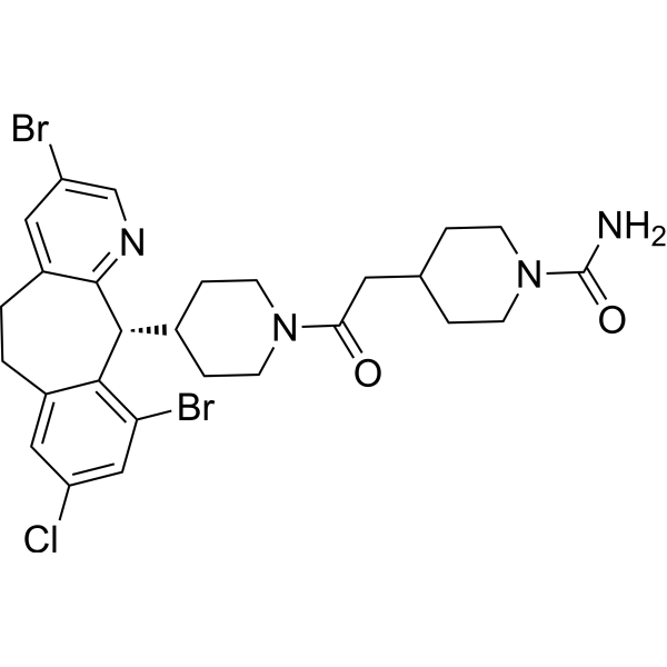
Lonafarnib (Sch66336) is an orally active, blood-brain barrier penetrant farnesyltransferase (FPTase) inhibitor. Lonafarnib increases the phosphorylation levels of Akt, CaMKII and CREB, upregulates BDNF expression in the hippocampus, elevates the content of α7nAChR on cell membranes, and blocks the isoprenylation of H-Ras. Lonafarnib repairs synapses and reverses spatial memory deficits in Aβ1-42 model mice. Lonafarnib inhibits RSV fusion and replication, and alleviates virus-induced lung injury. Lonafarnib activates the lysosomal and autophagic pathways, and reduces the phosphorylation and aggregation of tau. Lonafarnib acts synergistically with Sorafenib (HY-10201) to promote apoptosis, and inhibits the proliferation and invasion of melanoma cells. Lonafarnib inhibits the farnesylation of progerin, repairs nuclear membrane defects, and activates the cGAS-STING-STAT1 signaling pathway. Lonafarnib can be used in research related to diseases including Alzheimer's disease, viral infections, melanoma, and progeria.
For research use only. We do not sell to patients.
- Purity : 99.85%
- CAS No.: 193275-84-2
- Formula: C27H31Br2ClN4O2
- Molecular Weight:638.82
-
Storage:Powder -20°C, 3 years , 4°C, 2 years ; In solvent -80°C, 2 years , -20°C, 1 year

To place orders, for customer services and technical support, please contact: MedChemExpress USA
Tel: 609-228-6898 E-mail: [email protected] [email protected]


-
 Biological Activity
-
Chemical Information
-
Solvent & Solubility
- Protocols
- Purity & Documentation
- References
-
 Help & FAQs


-
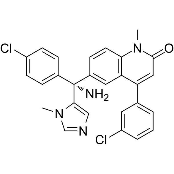
HY-10502
TipifarnibFTase Inhibitor -
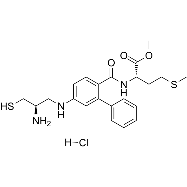
HY-15872A
FTI-277 hydrochlorideFTase Inhibitor -
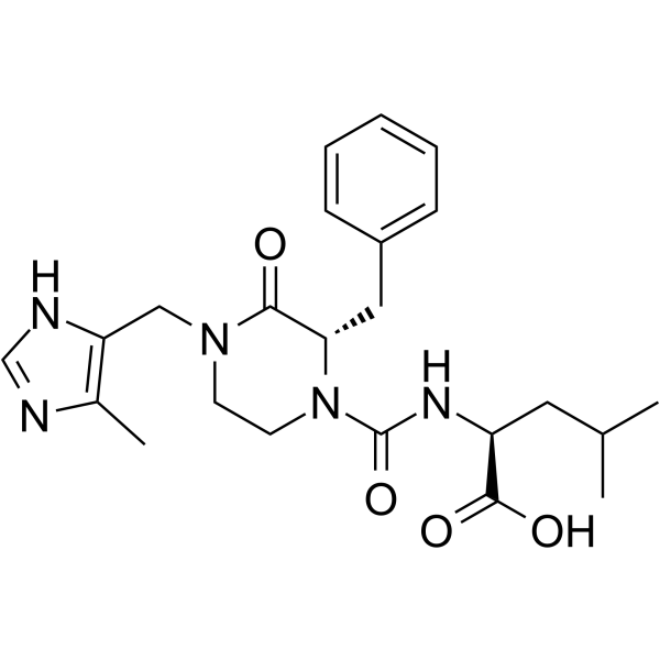
HY-16231
CamprenstatGGTase I Inhibitor -
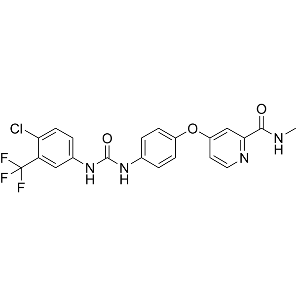
HY-10201
SorafenibMultikinase Inhibitor -
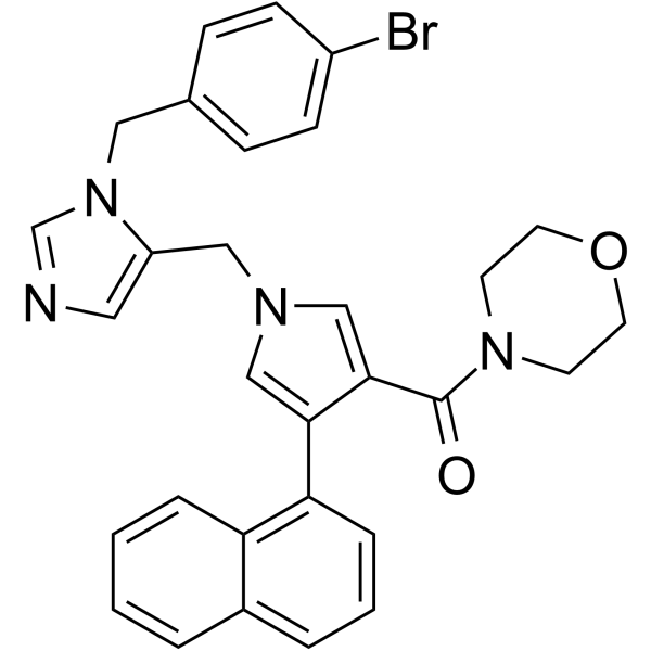
HY-15879
LB42708Farnesyltransferase Inhibitor -

HY-B0015
PaclitaxelAntitumor Agent -
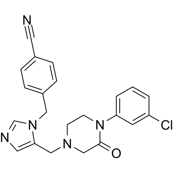
HY-16273
L-778123FPTase/GGPTase-I Iinhibitor -
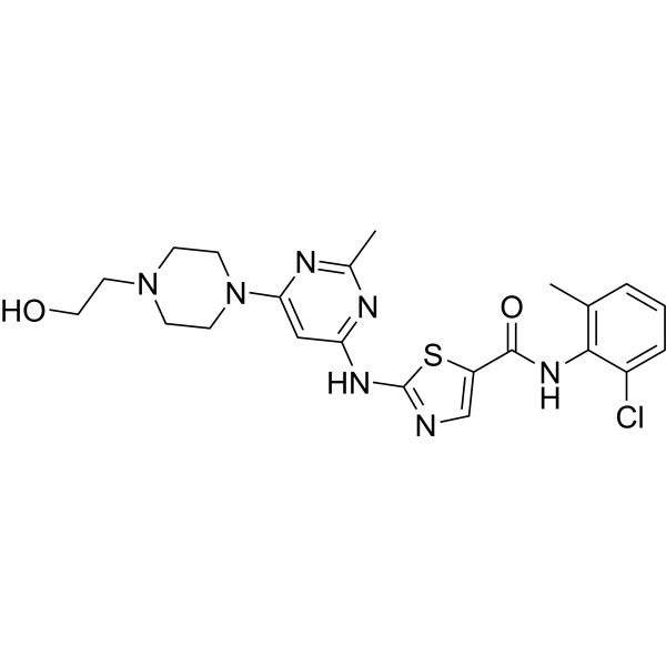
HY-10181
DasatinibSrc/Bcr-Abl Inhibitor -
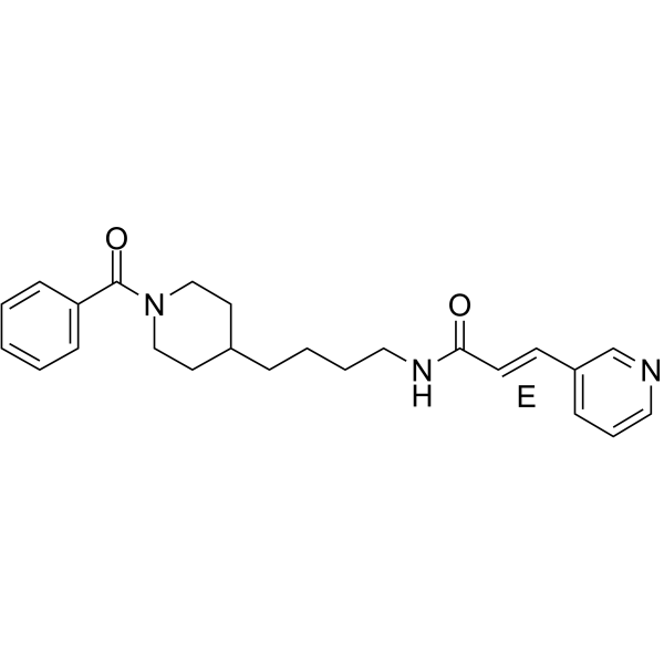
HY-50876
DaporinadNMPRT Inhibitor -
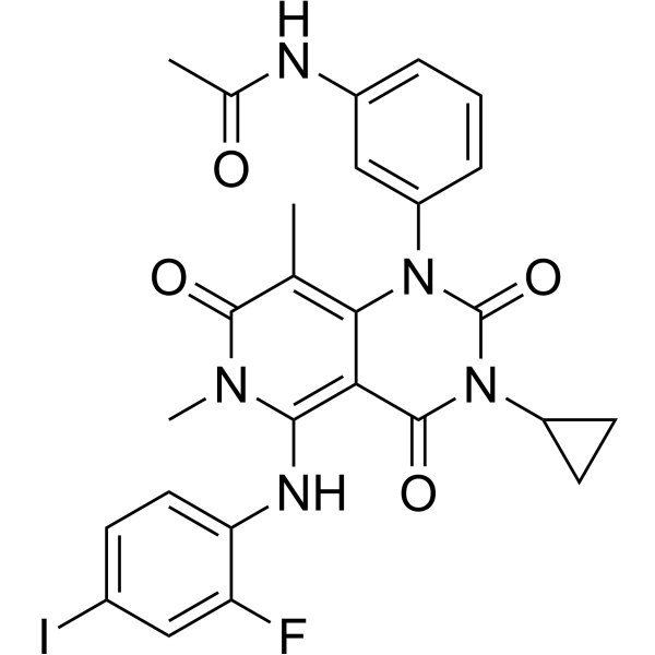
HY-10999
TrametinibMEK Inhibitor -
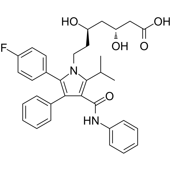
HY-B0589
AtorvastatinHMG-CoA Reductase Inhibitor -
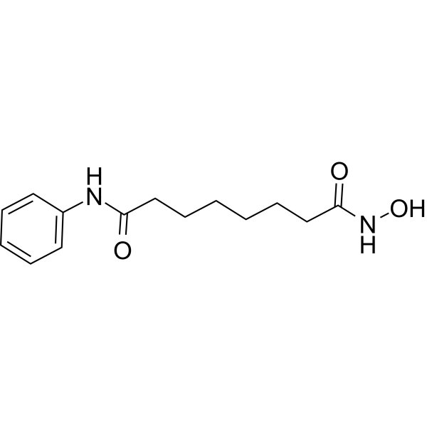
HY-10221
VorinostatHDAC Inhibitor
Publications Citing Use of MedChemExpress (MCE) Lonafarnib
More- Nat Commun. 2024 Apr 23;15(1):3422. [Abstract]
- Nat Commun. 2019 May 22;10(1):2265. [Abstract]
- J Clin Invest. 2025 Jul 15;135(14):e189048. [Abstract]
- Adv Sci (Weinh). 2025 Sep;12(33):e15176. [Abstract]
- Cell Death Dis. 2025 Aug 7;16(1):595. [Abstract]
- Int J Biol Sci. 2025 Jan 1;21(2):758-771. [Abstract]
- Phytomedicine. 2023 Nov:120:155066. [Abstract]
- Aging Cell. 2019 Aug;18(4):e12979. [Abstract]
- Inflamm Res. 2026 Aug 18;75(1):188.
- Eur J Pharmacol. 2026 Mar 28:1019:178723. [Abstract]
- Sci Rep. 2019 Jul 10;9(1):10021. [Abstract]
- Virol Sin. 2023 Oct;38(5):778-786. [Abstract]
- J Neurosci. 2022 Aug 3;42(31):6090-6107. [Abstract]
- J Virol. 2025 Dec 9:e0148725. [Abstract]
- Med Mycol. 2018 Jun 1;56(4):452-457. [Abstract]
 Customer Validation & Images

-
Histological Imaging/Staining
-
IF
-
WB
-
RT-PCR
-
RT-PCR


Biological Activity
Description
IC50 & Target
|
STAT1 |
Bcl-2 |
H-Ras |
Akt |
Cellular Effect
|
Cell Line

|
Type | Value | Description | References |
|---|---|---|---|---|
| Caco-2 | CC50 |
10.71 μM
Compound: LONAFARNIB
|
Toxicity against Caco-2 cells determined at 48 hours by intracellular ATP concentration using the CellTiter-Glo Luminescent Cell Viability Assay
Toxicity against Caco-2 cells determined at 48 hours by intracellular ATP concentration using the CellTiter-Glo Luminescent Cell Viability Assay
|
10.21203/rs.3.rs-23951/v1 |
| Caco-2 | IC50 |
5.68 μM
Compound: LONAFARNIB
|
Determination of IC50 values for inhibition of SARS-CoV-2 induced cytotoxicity of Caco-2 cells after 48 hours by high content imaging
Determination of IC50 values for inhibition of SARS-CoV-2 induced cytotoxicity of Caco-2 cells after 48 hours by high content imaging
|
10.21203/rs.3.rs-23951/v1 |
| COS-1 | IC50 |
75 nM
Compound: 5
|
inhibition of tumor colony formation in soft agar
inhibition of tumor colony formation in soft agar
|
[PMID: 10411485] |
| COS-7 | IC50 |
10 nM
Compound: 15
|
Inhibiting the farnesylation of H-ras proteins in COS-7 monkey cells transiently expressing H-ras[Val12]-CVLS in the whole cell assay.
Inhibiting the farnesylation of H-ras proteins in COS-7 monkey cells transiently expressing H-ras[Val12]-CVLS in the whole cell assay.
|
[PMID: 9822558] |
| COS-7 | IC50 |
10 nM
Compound: 9b
|
Inhibition of FTase in human COS7 cells
Inhibition of FTase in human COS7 cells
|
[PMID: 20925433] |
| HCT-116 | EC50 |
74 nM
Compound: SCH66336
|
Inhibition of anchorage-independent growth in human HCT-116 cells assessed as inhibition of colony formation in presence of 10% FBS
Inhibition of anchorage-independent growth in human HCT-116 cells assessed as inhibition of colony formation in presence of 10% FBS
|
[PMID: 16222147] |
| HCT-116 | EC50 |
94 nM
Compound: SCH66336
|
Inhibition of anchorage-independent growth in human HCT-116 cells assessed as inhibition of colony formation incubated for 14 days in presence of 10% FBS by imaging based soft agar colony formation assay
Inhibition of anchorage-independent growth in human HCT-116 cells assessed as inhibition of colony formation incubated for 14 days in presence of 10% FBS by imaging based soft agar colony formation assay
|
[PMID: 16222147] |
| HCT-116 | IC50 |
0.07 μM
Compound: 15
|
Compound was measured for inhibition of HCT116 tumor cell line in colon under soft agar assay.
Compound was measured for inhibition of HCT116 tumor cell line in colon under soft agar assay.
|
[PMID: 9822558] |
| MCF7 | IC50 |
0.05 μM
Compound: 15
|
Compound was measured for inhibition of MCF-7 tumor cell line in breast under soft agar assay.
Compound was measured for inhibition of MCF-7 tumor cell line in breast under soft agar assay.
|
[PMID: 9822558] |
| NIH3T3 | EC50 |
0.16 μM
Compound: 2, SCH-66336
|
Effective concentration against Ha-RAS processing in NIH3T3 ras-transformed cells
Effective concentration against Ha-RAS processing in NIH3T3 ras-transformed cells
|
[PMID: 12657284] |
| NIH3T3 | EC50 |
100 nM
Compound: Lonafarnib
|
Inhibition of Ras farnesylation in H-Ras transformed NIH3T3 cells
Inhibition of Ras farnesylation in H-Ras transformed NIH3T3 cells
|
[PMID: 15454228] |
| NIH3T3 | EC50 |
50 nM
Compound: SCH66336
|
Inhibition of anchorage-independent growth in H-Ras transformed mouse NIH3T3 cells assessed as inhibition of colony formation in presence of 10% bovine serum
Inhibition of anchorage-independent growth in H-Ras transformed mouse NIH3T3 cells assessed as inhibition of colony formation in presence of 10% bovine serum
|
[PMID: 16222147] |
| NIH3T3 | EC50 |
56 nM
Compound: SCH66336
|
Inhibition of anchorage-independent growth in H-Ras transformed mouse NIH3T3 cells assessed as inhibition of colony formation incubated for 14 days in presence of 10% bovine serum by imaging based soft agar colony formation assay
Inhibition of anchorage-independent growth in H-Ras transformed mouse NIH3T3 cells assessed as inhibition of colony formation incubated for 14 days in presence of 10% bovine serum by imaging based soft agar colony formation assay
|
[PMID: 16222147] |
| NIH3T3 | IC50 |
2.7 μM
Compound: SCH66336
|
TP_TRANSPORTER: inhibition of DNR efflux (DNR: ? uM) in MDR1-expressing NIH3T3 cells
TP_TRANSPORTER: inhibition of DNR efflux (DNR: ? uM) in MDR1-expressing NIH3T3 cells
|
[PMID: 11606389] |
| NIH3T3 | IC50 |
500 μM
Compound: SCH-66336
|
Inhibition of K-Ras transformed NIH3T3 cell proliferation
Inhibition of K-Ras transformed NIH3T3 cell proliferation
|
[PMID: 15501065] |
| NIH3T3 | IC50 |
72 μM
Compound: SCH-66336
|
Inhibition of H-Ras transformed NIH3T3 cell proliferation
Inhibition of H-Ras transformed NIH3T3 cell proliferation
|
[PMID: 15501065] |
| NIH-H | IC50 |
72 nM
Compound: 15
|
Compound ability to inhibit anchorage-independent growth of NIH-H tumor cell lines in soft agar.
Compound ability to inhibit anchorage-independent growth of NIH-H tumor cell lines in soft agar.
|
[PMID: 9822558] |
| NIH-K | IC50 |
500 nM
Compound: 15
|
Compound ability to inhibit anchorage-independent growth of NIH-K tumor cell lines in soft agar.
Compound ability to inhibit anchorage-independent growth of NIH-K tumor cell lines in soft agar.
|
[PMID: 9822558] |
In Vitro
Lonafarnib (72 h) potently inhibits RSV A2 infection in HEp-2 cells (EC50 = 57.7 nM, selectivity index > 168.2) and RSV B01 infection in HEp-2 cells (EC50 = 75.5 nM, selectivity index > 168.2), while it also suppresses RSV A2 replication in human primary bronchial epithelial cells (EC50 = 599.9 nM, selectivity index = 31.0) and RSV B01 replication in human primary bronchial epithelial cells (EC50 = 515.0 nM, selectivity index = 36.1)[2].
Lonafarnib inhibits RSV A2 infection in HEp-2 cells via a farnesyltransferase-independent mechanism[2].
Lonafarnib (3.3 μM; pre (-2-0 h), during (0-2 h), and post (2-16 h) infection) blocks RSV A2 infection in HEp-2 cells primarily by inhibiting the viral entry process, with significant inhibitory effects observed during the 0-2 h entry window or over the full −2-22 h period[2].
Lonafarnib inhibits RSV entry into HEp-2 cells and suppresses RSV F-induced intercellular fusion in HEK293T cells, confirming that it targets the RSV membrane fusion process[2].
Lonafarnib binds to the pre-fusion RSV F trimer (KD = 20.1 μM) and stabilizes its native conformation, while mutations M396A, S398A, D486A, F488L, and D489A in the RSV F protein abolish its fusion inhibitory effect[2].
Lonafarnib (0.5-1.0 μM; 48 h) dose-dependently enhances macroautophagic flux in NIH3T3 mouse fibroblasts, primarily by accelerating autophagosome clearance via lysosomes; it also stimulates basal chaperone-mediated autophagy in NIH3T3 mouse fibroblasts[3].
Lonafarnib (0.1-0.5 μM; 48 h) stimulates endosomal microautophagy in NIH3T3 mouse fibroblasts, promoting the transport of substrates to late endosomal compartments and their degradation[3].
Lonafarnib (0.5-1.0 μM; up to 24 h) increases total proteolysis in NIH3T3 mouse fibroblasts in a dose-dependent manner, and this effect is completely mediated by the lysosomal pathway[3].
Lonafarnib potently inhibits Rhes-mediated phosphorylated tau protein (PHF-1) accumulation in primary mouse hippocampal neurons, with an IC50 of 61.00 nM[3].
Lonafarnib (10 μM; 9 days) reduces the level of phosphorylated tau (Thr181) in wild-type and MAPTR406W mutant human induced pluripotent stem cell-derived neurons[3].
Lonafarnib (0.1-5 μM; 24 h) inhibits farnesyltransferase activity in BLM, MV3, MEWO and SKMel19 human metastatic melanoma cell lines[4].
Lonafarnib (5 μM; 48-72 h) combined with 4 μM Sorafenib (HY-10201) synergistically suppresses the proliferation of BLM, MV3, MEWO and SKMel1 human metastatic melanoma cell lines, strengthens growth inhibition on primary melanoma cells derived from human skin metastases and reduces BLM cell viability[4].
Lonafarnib (5 μM; 24 h) inhibits the mTOR pathway in human metastatic melanoma cells. When combined with Sorafenib, it significantly downregulates intracellular Bcl-2, completely eliminates Mcl-1, and only slightly alters Bcl-xL expression[4].
Lonafarnib (3 μM; 14 days) reduces the invasion and proliferation of BL melanoma cells in organotypic skin models, and the combination with 2 μM Sorafenib completely blocks the invasion and proliferation of these cells[4].
Lonafarnib (5 μM; 48 h) slightly increases the proportion of sub-G1 apoptotic cells in various melanoma cells, and its combination with Sorafenib greatly amplifies this pro-apoptotic effect without damaging human fibroblasts[4].
Lonafarnib (5 μM; 24 h) combined with Sorafenib triggers typical apoptotic morphological features such as cell fragmentation in human metastatic melanoma BLM cells[4].
Lonafarnib (5 μM; 12 h) combined with Sorafenib further elevates p8 mRNA in BLM, MV3, MEWO, SKMel19 human metastatic melanoma cell lines and increases CHOP mRNA in all detected melanoma cell lines[4].
Lonafarnib (0.025 µM; 9 days) upregulates the proportion of p21-positive cells and phosphorylated STAT1 in normal and progeria fibroblasts, increases the formation of ring-shaped nuclei and micronuclei, elevates IFN-β mRNA levels, glycolysis and ATP content, downregulates the mRNA levels of multiple pro-inflammatory cytokines, reduces progerin levels and proteasome activity, and upregulates prelamin A levels and autophagic activity[5].
MedChemExpress (MCE) has not independently confirmed the accuracy of these methods. They are for reference only. Further protocols information, click here.
-
Cell Line:HEp-2 cells infected with RSV A2
-
Concentration:3.3 μM
-
Incubation Time:-2-22 h, 0-2 h, 2-22 h, 4-22 h, 6-22 h, 8-22 h, 16-22 h
-
Result:Significantly reduced RSV infection when added during full-time (-2-22 h) and entry (0-2 h) periods.
Showed weak inhibition at post-entry stages.
-
Cell Line:NIH3T3 mouse fibroblasts
-
Concentration:0.5 μM, 1.0 μM
-
Incubation Time:48 h
-
Result:Increased the number of autophagic vacuoles to ~30 puncta/cell, autophagosomes to ~30 puncta/cell, and autolysosomes to ~30 puncta/cell at 0.5 μM compared to untreated cells.
Further increased autophagic vacuoles to ~70 puncta/cell, autophagosomes to ~20 puncta/cell, and autolysosomes to ~50 puncta/cell at 1.0 μM, with majority of increase driven by enhanced autolysosome abundance.\nIncreased CMA activity to ~30 puncta/cell at 0.5 μM.
Reduced CMA activity to ~15 puncta/cell at 1.0 μM, which was still significantly higher than untreated cells at ~5 puncta/cell.
-
Cell Line:human induced pluripotent stem cell (hiPSC)-derived neurons (MAPT-WT, MAPT-R406W)
-
Concentration:10 μM
-
Incubation Time:9 days
-
Result:Significantly reduced intracellular phospho-tau (Thr181) levels in both MAPT-WT and MAPT-R406W hiPSC-derived neurons.
-
Cell Line:BLM, MV3, MEWO, SKMel19 human metastatic melanoma cell lines
-
Concentration:0.1 μM, 1 μM, 5 μM
-
Incubation Time:24 h
-
Result:Caused HDJ2 to migrate to a higher apparent molecular weight in all tested melanoma cell lines, with increasing concentrations associated with this shift.
-
Cell Line:BLM, MV3, MEWO, SKMel19 human metastatic melanoma cell lines
-
Concentration:5 μM (lonafarnib)
4 μM (Sorafenib) -
Incubation Time:72 h
-
Result:Synergistically enhanced growth inhibition compared to either agent alone, with absolute enhancement of growth inhibition rates ranging from 53% (MEWO) to 90% (MV3).
-
Cell Line:BLM human metastatic melanoma cell line
-
Concentration:5 μM (lonafarnib)
4 μM (Sorafenib) -
Incubation Time:48 h, 72 h
-
Result:Reduced the percentage of detectable cells to 50% after 48 hours and 30% after 72 hours relative to the start of treatment, significantly inhibiting cell proliferation compared to monotreatment.
-
Cell Line:BLM, MV3, MEWO, SKMel19 human metastatic melanoma cell lines
-
Concentration:5 μM
-
Incubation Time:48 h
-
Result:Increased the sub-G1 apoptotic cell fraction, with percentages ranging from 6% (BLM) to 18% (MV3).
-
Cell Line:BLM, MV3, MEWO, SKMel19 human metastatic melanoma cell lines; human fibroblasts
-
Concentration:5 μM (lonafarnib)
4 μM (Sorafenib) -
Incubation Time:48 h
-
Result:Significantly increased the sub-G1 apoptotic cell fraction compared to either agent alone, with percentages ranging from 27% (BLM) to 75% (SKMel19).
Did not increase the sub-G1 fraction in human fibroblasts.
-
Cell Line:BLM, MV3, MEWO, SKMel19 human metastatic melanoma cell lines
-
Concentration:5 μM
-
Incubation Time:24 h
-
Result:Marginally affected expression levels of Bcl-2, Bcl-xL, and Mcl-1 across all tested cell lines.\nDid not affect phosphorylation of ERK or AKT in any of the tested melanoma cell lines.\nDecreased phosphorylation of S6 kinase and ribosomal S6 protein in all tested melanoma cell lines.
-
Cell Line:BLM, MV3, MEWO, SKMel19 human metastatic melanoma cell lines
-
Concentration:5 μM (lonafarnib)
4 μM (Sorafenib) -
Incubation Time:24 h
-
Result:Significantly reduced Bcl-2 expression, slightly affected Bcl-xL expression, and completely abolished Mcl-1 expression in all tested melanoma cell lines.
-
Cell Line:BLM, MV3, MEWO, SKMel19 human metastatic melanoma cell lines
-
Concentration:5 μM
-
Incubation Time:12 h
-
Result:Slightly affected p8 mRNA levels across all tested melanoma cell lines.
-
Cell Line:BLM, MV3, MEWO, SKMel19 human metastatic melanoma cell lines
-
Concentration:5 μM (lonafarnib)
2 μM, 4 μM (Sorafenib) -
Incubation Time:12 h
-
Result:Further upregulated p8 mRNA in most tested cell lines, and increased CHOP mRNA levels in all tested cell lines, compared to either agent alone.
-
Cell Line:GM01651C, GM01652C (control primary fibroblasts); HGADFN003, HGADFN127 (HGPS primary fibroblasts)
-
Concentration:0.025 µM
-
Incubation Time:9 days
-
Result:Maintained cell growth rates similar to mock-treated control and HGPS fibroblasts.
Increased the percentage of p21-positive cells by 9% in control fibroblasts relative to mock-treated cells.
Increased the percentage of p21-positive cells by 12% in HGPS fibroblasts relative to mock-treated cells.
-
Cell Line:GM01651C, GM01652C (control primary fibroblasts); HGADFN003, HGADFN127 (HGPS primary fibroblasts)
-
Concentration:0.025 µM
-
Incubation Time:9 days
-
Result:Elevated P-STAT1 levels by 48% in control fibroblasts and 51% in HGPS fibroblasts, while leaving total STAT1 unaltered in both cell lines versus mock groups
-
Cell Line:GM01651C (control primary fibroblasts); HGADFN127 (HGPS primary fibroblasts)
-
Concentration:0.025 µM
-
Incubation Time:9 days
-
Result:Elevated the proportion of cells carrying cGAS-positive cytosolic foci by 3% in control fibroblasts and 4% in HGPS fibroblasts versus mock groups
-
Cell Line:GM01651C, GM01652C (control primary fibroblasts); HGADFN003, HGADFN127 (HGPS primary fibroblasts)
-
Concentration:0.025 µM
-
Incubation Time:9 days
-
Result:Elevated IFN-β mRNA by 58% in control fibroblasts and 67% in HGPS fibroblasts, and downregulated IL-1α, CCL2, IL-6, CXCL8 mRNA by 30%/11%, 41%/26%, 14%/10%, 43%/42% in two cell types separately compared with mock groups
In Vivo
Lonafarnib (17-34 mg/kg; p.o.; twice daily; for 4 consecutive days) dose-dependently reduces RSV replication levels in BALB/c mice, and improves body weight and pulmonary pathological conditions of mice when administered via early intervention or post-infection oral delivery[2].
Lonafarnib (80 mg/kg per day; p.o.; intermittent administration/acute administration) reduces tau protein inclusions, brain atrophy, glial reactivity and behavioral deficits in rTg4510 mice, whereas acute administration has no effect on established tau protein pathology[3].
MedChemExpress (MCE) has not independently confirmed the accuracy of these methods. They are for reference only.
-
Animal Model:C57BL/6J (male, 2 months old, intracerebroventricular injection of soluble Aβ1-42 to induce pathology)[1]
-
Dosage:10 mg/kg; 30 mg/kg; 50 mg/kg; 80 mg/kg
-
Administration:i.p.; daily; 14 days
-
Result:Reduced MWM escape latency and prolonged target quadrant residence time, thereby improved spatial learning and memory in Aβ1-42 mice at 50 mg/kg and 80 mg/kg.
Rescued impaired hippocampal synaptic electrophysiology including EPSP slopes, paired-pulse ratios, PTP and L-LTP at 50 mg/kg. It also restored damaged dendritic architecture, elevated synaptic PSD-95 and synaptophysin levels, upregulated surface α7nAChR expression, activated Akt/CaMKII/CREB phosphorylation, and increased mature hippocampal BDNF content.
Demonstrated that the BDNF upregulation relied on α7nAChR and CaMKII signaling rather than ERK or PI3K pathways. H-Ras overexpression reversed the elevations of surface α7nAChR and BDNF, while TrkB/Fc chimera completely abolished all synaptic and cognitive improvements.
-
Animal Model:BALB/c (female, 8 weeks old, intranasally inoculated with 1 × 10⁶ PFU RSV A2)[2]
-
Dosage:17 mg/kg; 34 mg/kg
-
Administration:p.o.; twice daily; 4 days
-
Result:Improved body weight from day 1 post-infection at 34 mg/kg early intervention, while post-infection treatment only slightly recovered weight at day 4.
Reduced lung viral titers dose-dependently: early 17/34 mg/kg downregulated titers 3.8/7.4-fold; post-infection 17/34 mg/kg downregulated titers 3.7/4.1-fold.
Attenuated RSV-mediated lung inflammation in all early-intervention groups and relieved lung injury at 34 mg/kg post-infection, with quantitative pathology confirming alleviated alveolitis and lesions in early groups.
-
Animal Model:rTg4510 (tauopathy transgenic, 10 weeks old at initiation, evaluated at 20 weeks old); wild-type littermate controls[3]
-
Dosage:80 mg/kg
-
Administration:p.o.; intermittent schedule (5 days on, 5 days off); 10 weeks (chronic intermittent treatment)
p.o.; daily; 2 weeks (acute treatment) -
Result:Lowered cortical/hippocampal MC1-tau inclusions and cortical/hippocampal PHF-1 phosphorylated tau; cut cortical/hippocampal Rhes-positive cell density.
Enlarged coronal brain area, mitigated hippocampal microgliosis and cortical astrogliosis.
Rescued nest-building impairment and suppressed circling behavior.
Decreased sumoylation- and ubiquitination-positive cells, changed co-labeling likelihood of MC1-tau with SUMO/ubiquitin.
Lessened pathological tau in cortical lysates without altering total tau, and erased insoluble high-molecular-weight tau aggregates.
Exerted no shift in MC1 tau pathology distribution or range after acute dosing.
Clinical Trial
| NCT Number | Sponsor | Condition | Start Date |
Phase
|
|---|---|---|---|---|
| NCT01329991 | Plexxikon| | 2011-05 | PHASE1 |


Chemical Information
-
CAS No. 193275-84-2
-
Appearance Solid
-
Molecular Weight 638.82
-
Formula C27H31Br2ClN4O2
-
Color White to off-white
-
SMILES
O=C(N)N1CCC(CC1)CC(N2CCC(CC2)[C@@H]3C4=C(C=C(C=C4CCC5=CC(Br)=CN=C53)Cl)Br)=O
-
Synonyms
Sch66336
-
Shipping
Room temperature in continental US; may vary elsewhere.
-
Storage
Powder -20°C 3 years 4°C 2 years In solvent -80°C 2 years -20°C 1 year


Publications (15)
-
Journal Impact Factor
-
Most Recent
-
Nat Commun
Targeting NRAS via miR-1304-5p or farnesyltransferase inhibition confers sensitivity to ALK inhibitors in ALK-mutant neuroblastoma. [Abstract]2024 Apr 23;15(1):3422. PMID: 38653965
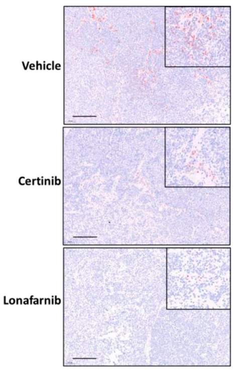
Lonafarnib purchased from MedChemExpress. Usage Cited in: Nat Commun. 2024 Apr 23;15(1):3422. [Abstract]
Immunohistochemistry (pERK) in tumours from NSG mice treated with Lonafarnib (10 μL/g; p.o.; once daily). Lonafarnib led to a profound decrease in pERK expression levels.
-
Nat Commun
Recapitulation of HDV infection in a fully permissive hepatoma cell line allows efficient drug evaluation. [Abstract]2019 May 22;10(1):2265. PMID: 31118422
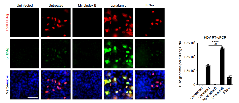
Lonafarnib purchased from MedChemExpress. Usage Cited in: Nat Commun. 2019 May 22;10(1):2265. [Abstract]
Lonafarnib (1.8 μM) treatment resulted in significant and selective increase in intracellular L-HDAg of HepNB2.7 cells.
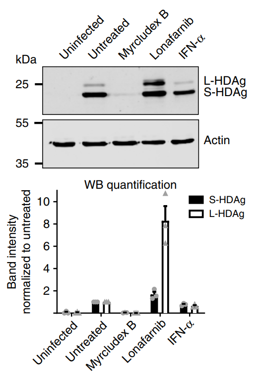
Lonafarnib purchased from MedChemExpress. Usage Cited in: Nat Commun. 2019 May 22;10(1):2265. [Abstract]
Lonafarnib (1.8 μM) treatment caused an eightfold increase of intracellular L-HDAg in HepNB2.7 cells compared with the untreated controls, while the levels of S-HDAg only marginally (1.6-fold) increased.
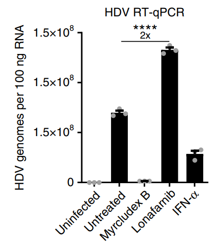
Lonafarnib purchased from MedChemExpress. Usage Cited in: Nat Commun. 2019 May 22;10(1):2265. [Abstract]
Lonafarnib (1.8 μM) treatment led to a twofold increase in the total intracellular HDV genomes.
Lonafarnib purchased from MedChemExpress. Usage Cited in: Nat Commun. 2019 May 22;10(1):2265. [Abstract]
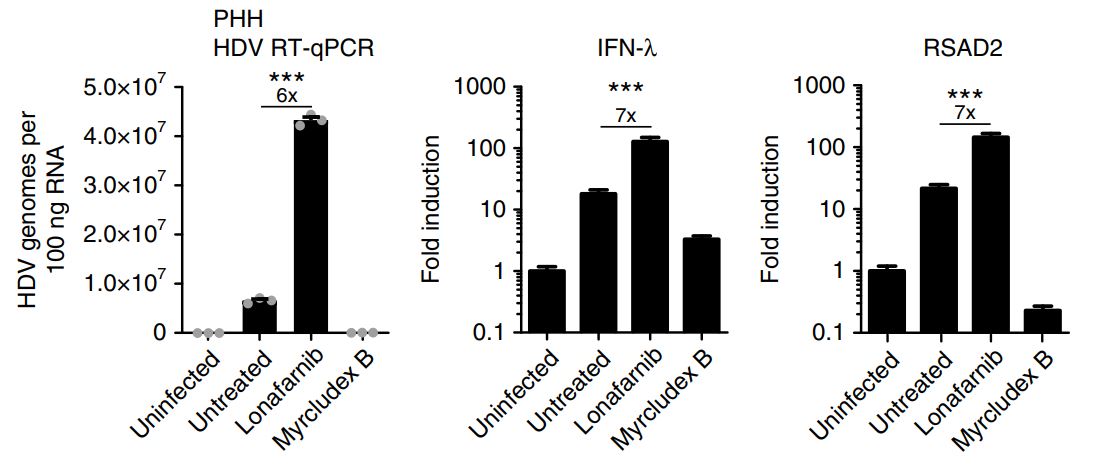
Quantitative PCR on intracellular HDV replicative intermediates and the two markers for IFN induction (IFN-λ and RSAD2) was performed. Lonafarnib (1.8 μM) profoundly increased the intracellular HDV replication and the induction of IFN responses in PHH co-infected with HDV and HBV.
-
J Clin Invest
Cancer-associated SPOP mutations enlarge nuclear size and facilitate nuclear envelope rupture upon farnesyltransferase inhibitor treatment. [Abstract]2025 Jul 15;135(14):e189048. PMID: 40662365 -
Adv Sci (Weinh)
Multimodal Investigation of Angiogenesis and Its Prevention by Small Compounds in a Zebrafish Cancer Model. [Abstract]2025 Sep;12(33):e15176. PMID: 40557748 -
Cell Death Dis
Activation of AKT via a dual mechanism enhances the susceptibility of melanoma cells to glucose deprivation. [Abstract]2025 Aug 7;16(1):595. PMID: 40774947 -
Int J Biol Sci
2025 Jan 1;21(2):758-771. PMID: 39781460 -
Phytomedicine
Astragaloside IV protects against lung injury and pulmonary fibrosis in COPD by targeting GTP-GDP domain of RAS and downregulating the RAS/RAF/FoxO signaling pathway. [Abstract]2023 Nov:120:155066. PMID: 37690229 -
Aging Cell
2019 Aug;18(4):e12979. PMID: 31152494 -
-
Eur J Pharmacol
Heat stroke-induced structural and functional impairments of cognition-relevant brain areas in mice is associated with alpha-7 nicotinic acetylcholine receptors downregulation. [Abstract]2026 Mar 28:1019:178723. PMID: 41786068 -
Sci Rep
Generation and characterization of a stable cell line persistently replicating and secreting the human hepatitis delta virus. [Abstract]2019 Jul 10;9(1):10021. PMID: 31292511 -
Virol Sin
Identification of a receptor tyrosine kinase inhibitor CP-724714 inhibits SADS-CoV related swine diarrhea coronaviruses infection in vitro. [Abstract]2023 Oct;38(5):778-786. PMID: 37406816 -
J Neurosci
Ras Inhibitor Lonafarnib Rescues Structural and Functional Impairments of Synapses of Aβ1-42 Mice via α7nAChR-Dependent BDNF Upregulation. [Abstract]2022 Aug 3;42(31):6090-6107. PMID: 35760529 -
J Virol
Molecular mechanism of resistance to lonafarnib conferred by mutations in the cysteine-rich region of respiratory syncytial virus fusion glycoprotein and discovery of a lonafarnib-derived antiviral PROTAC. [Abstract]2025 Dec 9:e0148725. PMID: 41364004 -
Med Mycol
2018 Jun 1;56(4):452-457. PMID: 29420769


Solvent & Solubility
In Vitro:
DMSO : 25 mg/mL (39.13 mM; ultrasonic and warming and heat to 60°C; Hygroscopic DMSO has a significant impact on the solubility of product, please use newly opened DMSO)
Please refer to the solubility information to select the appropriate solvent. Once prepared, please aliquot and store the solution to prevent product inactivation from repeated freeze-thaw cycles.
Storage method and period of stock solution: -80°C, 2 years; -20°C, 1 year. When stored at -80°C, please use it within 2 years. When stored at -20°C, please use it within 1 year.
Please refer to the solubility information to select the appropriate solvent. Once prepared, please aliquot and store the solution to prevent product inactivation from repeated freeze-thaw cycles.
Storage method and period of stock solution: -80°C, 2 years; -20°C, 1 year. When stored at -80°C, please use it within 2 years. When stored at -20°C, please use it within 1 year.
Concentration (start) × Volume (start) = Concentration (final) × Volume (final)
In Vivo:
Select the appropriate dissolution method based on your experimental animal and administration route.
- For the following dissolution methods, please ensure to first prepare a clear stock solution using an In Vitro approach and then sequentially add co-solvents:
- To ensure reliable experimental results, the clarified stock solution can be appropriately stored based on storage conditions. As for the working solution for In Vivo experiments, it is recommended to prepare freshly and use it on the same day.
- The percentages shown for the solvents indicate their volumetric ratio in the final prepared solution. If precipitation or phase separation occurs during preparation, heat and/or sonication can be used to aid dissolution.
Add each solvent one by one: 10% DMSO 40% PEG300 5% Tween-80 45% Saline
Solubility: ≥ 2.5 mg/mL (3.91 mM); Clear solution
This protocol yields a clear solution of ≥ 2.5 mg/mL (saturation unknown).
Taking 1 mL working solution as an example, add 100 μL DMSO stock solution (25.0 mg/mL) to 400 μL PEG300, and mix evenly; then add 50 μL Tween-80 and mix evenly; then add 450 μL Saline to adjust the volume to 1 mL.
Preparation of Saline: Dissolve 0.9 g sodium chloride in ddH₂O and dilute to 100 mL to obtain a clear Saline solution.
Add each solvent one by one: 10% DMSO 90% (20% SBE-β-CD in Saline)
Solubility: ≥ 2.5 mg/mL (3.91 mM); Clear solution
This protocol yields a clear solution of ≥ 2.5 mg/mL (saturation unknown).
Taking 1 mL working solution as an example, add 100 μL DMSO stock solution (25.0 mg/mL) to 900 μL 20% SBE-β-CD in Saline, and mix evenly.
Preparation of 20% SBE-β-CD in Saline (4°C, storage for one week): 2 g SBE-β-CD powder is dissolved in 10 mL Saline, completely dissolve until clear.
In Vivo Dissolution Calculator
Please enter the basic information of animal experiments:
-
-
-
-
Recommended: Prepare an additional quantity of animals to account for potential losses during experiments.
Please enter your animal formula composition:
-
%DMSO +
Recommended: Keep the proportion of DMSO in working solution below 2% if your animal is weak.
-
%+
-
+%Tween-80 + +
-
%Saline +
The co-solvents required include: DMSO, . All of co-solvents are available by MedChemExpress (MCE). , Tween 80. All of co-solvents are available by MedChemExpress (MCE).
Working solution concentration: 0.22 mg/mL
Method for preparing stock solution: mg drug dissolved in μL DMSO. Stock solution concentration: mg/mL.
1. Take μL DMSO stock solution;
2. Add μL .
μL , mix evenly;
3. Then add μL Tween 80, mix evenly;
4. Then add μL
Please ensure that the stock solution in the first step is dissolved to a clear state, and add co-solvents in sequence. You can use ultrasonic heating (ultrasonic cleaner, recommended frequency 20-40 kHz), vortexing, etc. to assist dissolution.

Protocols
-
Western Blot
Western blotting (WB) is a commonly used experimental method in molecular biology, biochemistry, and immunogenetics for identifying and quantifying target proteins. It combines gel electrophoresis with immunoassay, enabling researchers to analyze protein expression, post-translational modifications, and molecular weight.
-
RNA extraction experimental
By lysing cells, releasing RNA, and removing impurities such as proteins and DNA, high-purity RNA products are finally obtained. The commonly used traditional method is the guanidine isothiocyanate/phenol/chloroform method (Trizol), which is suitable for a variety of animal materials including animal tissues, microorganisms, cultured cells, etc., and most plant materials.
-
Annexin V plus membrane-impermeant dye apoptosis staining
Annexin V-based apoptosis assays rely on the detection of phosphatidylserine (PS) externalization from the inner leaflet of the plasma membrane to the outer leaflet, an early biochemical hallmark of apoptosis. Fluorescently labeled Annexin V binds PS in a calcium-dependent manner, enabling identification of early apoptotic cells by flow cytometry or fluorescence microscopy. When combined with a membrane-impermeant DNA-binding dye (e. g. , propidium iodide), this approach allows discrimination between viable (Annexin V−/dye−), early apoptotic (Annexin V+/dye−), and late apoptotic or necrotic (Annexin V+/dye+) cell populations by assessing membrane integrity and PS exposure.
-
Protocol for Water Maze
The Morris Water Maze is a rodent spatial learning and memory assay in which a mouse or rat swims in opaque water to find an escape platform; in the hidden-platform version, the animal cannot see the platform and must use distal extra-maze cues to learn its fixed spatial location. The assay primarily measures hippocampus-dependent spatial learning during acquisition trials and spatial reference memory during probe trials after platform removal; readouts include escape latency, swim path length, swim speed, quadrant occupancy, platform-site crossings, and proximity to the former platform location.
-
Cell Viability Determination by MTT Colorimetric Assay
The following protocol uses the MTT colorimetric assay as a classic literature-established method for assessing cell viability/metabolic activity in cultured mammalian cells. MTT[3-(4,5-dimethylthiazol-2-yl)-2,5-diphenyltetrazolium bromide] is reduced by metabolically active cells to a colored formazan product; the amount of formazan is quantified spectrophotometrically and provides an indirect measure of metabolically active viable cells. Importantly, MTT reduction reflects cellular oxidoreductase/metabolic activity rather than an absolute direct count of living cells, so changes in cellular metabolism can alter the signal independently of cell number.
-
Alzheimer’s Disease Modeling
Alzheimer’s Disease (AD) is a neurodegenerative disorder characterized by a progressive decline in cognitive functions and loss of specific types of neurons and synapses. Alzheimer's symptoms can be simulated in mice by injecting drugs (such as Aβ) or genetically modified.
-
Kinase activity and phosphorylation assays
Kinase activity assays measure the ability of kinases to transfer phosphate groups from ATP to specific substrates, while phosphorylation assays detect the presence and levels of phosphorylated proteins. Common methods include radiolabeled ATP incorporation (e. g. ,), ADP release detection via bioluminescence (e. g. ,[3]), enzyme-linked immunosorbent assays (ELISA) for phospho-specific epitopes (e. g. ,[6]), and microtiter-based formats for high-throughput screening (e. g. ,[8]). The ADP-Glo assay quantifies kinase activity by measuring ADP produced during phosphorylation using a luciferase-based system. Radiometric assays involve autoradiography or scintillation counting after incorporation of 32P-labeled ATP into substrate proteins. ELISA-based approaches rely on phospho-specific antibodies to detect activated kinases in cell lysates or purified samples.
-
Detection of Apoptosis
Apoptosis, also called programmed cell death, is generally characterized by distinct morphological characteristics.
-
TUNEL staining for apoptotic DNA fragmentation
TUNEL staining detects DNA strand breaks by using terminal deoxynucleotidyl transferase to add labeled nucleotides to exposed 3′-OH DNA termini, generating either microscopic staining in fixed cells or tissue sections, or fluorescence/cytometric signal in cell suspensions. TUNEL positivity reflects DNA fragmentation but should not be interpreted alone as definitive apoptosis, because TUNEL can also label necrotic, autolytic, mechanically damaged, or DNA-repair-associated DNA breaks.
-
Autophagy
Autophagy is a process in which eukaryotic cells use lysosomes to degrade their own cytoplasmic proteins and damaged organelles under the regulation of autophagy related gene (Atg). Microtubule-associated proteins light chain 3 (LC3) is recognized as autophagy marker, which transfers from cytoplasmic LC3 (LC3-I) to membrane type (LC3-II). LC3-II/I ratio could be detected by Western Blot and fluorescence microscopy.
-
Apoptosis Solutions
Apoptosis is a regulated, generally non-lytic cell-death pathway that removes unwanted, damaged, infected, or abnormal cells through coordinated morphological changes, caspase activation, DNA fragmentation, and membrane remodeling. The intrinsic apoptosis pathway is controlled mainly by mitochondrial outer membrane permeabilization, BCL-2 family proteins, cytochrome c release, apoptosome formation, caspase-9 activation, and downstream executioner caspase-3/7 activation. The extrinsic apoptosis pathway is initiated by death receptors such as Fas, TNFR, and TRAIL receptors, which recruit adaptor proteins and activate caspase-8 before engaging executioner caspases or mitochondrial amplification through BID cleavage. Apoptosis is linked to many phenotypes, including cancer cell killing, tissue homeostasis, immune regulation, neurodegeneration, infection response, and treatment-induced cytotoxicity; unresolved questions include how apoptosis interacts with necroptosis, pyroptosis, ferroptos
-
Cell invasion
Cell invasion is the ability of cells to migrate from one area to another via the extracellular matrix. Cell invasion is the response of normal and cancer cells to chemical and mechanical stimuli. Before migrating to a new region, the extracellular matrix is degraded by proteases within the cell. Cell invasion often occurs during wound repair, vascularization and inflammation, abnormal tissue invasion, and tumor cell metastasis.
-
Protocol for Kinase activity and phosphorylation assays
Kinase activity assays measure transfer of phosphate from ATP to a protein or peptide substrate, generating phosphorylated substrate, ADP, or incorporated radiolabeled phosphate as the readout; phosphorylation assays measure site-specific phosphorylation in cells or tissues as a proxy for kinase-pathway activation, inhibition, or substrate regulation. Phosphorylation can be detected by phospho-specific Western blot, immunoprecipitation kinase assay, phospho-immunofluorescence, phospho-flow cytometry, luminescent ADP detection, radiolabeled ATP incorporation, or reporter-based pathway assays, and these readouts can be applied to cancer cells, primary neurons, mouse tumors, organoids, inflammatory macrophages, ferroptosis studies, and mitophagy studies when the kinase target is biologically relevant.
-
Lysosome and acidic-vesicle live-cell staining
Lysosome and acidic-vesicle live-cell staining detects acidic intracellular compartments by using membrane-permeant acidotropic probes that accumulate in low-pH vesicles, including lysosomes, late endosomes, autolysosomes, and acidic phagosomes. LysoTracker staining is commonly used as an intensity-based readout of acidic lysosomal compartment abundance or enlargement, while acridine orange produces green fluorescence in less concentrated compartments and red fluorescence after concentration-dependent accumulation in acidic vesicular organelles. Loss or reduction of acridine-orange red signal can be used as a readout of lysosomal membrane permeabilization or reduced acidic-vesicle integrity. This protocol is designed for live cultured cells and can be adapted for fluorescence microscopy, high-content imaging, plate-reader readout, or flow cytometry when the selected literature supports the readout. Because these dyes report acidotropic accumulation rather than lysosome identity alone,
-
Inhalation Toxicity Study
Inhalation toxicity studies expose rodents to a controlled aerosol, vapor, gas, or smoke atmosphere and assess respiratory and systemic toxicity using exposure-atmosphere characterization, clinical observations, body and organ weights, bronchoalveolar lavage fluid, histopathology, blood chemistry, hematology, and, when included, molecular endpoints such as transcriptomics, proteomics, lipidomics, or tissue burden analysis. The primary biological readouts are airway irritation, pulmonary inflammation, cytotoxicity, altered surfactant or lipid homeostasis, impaired particle clearance, and tissue remodeling, reflected by BALF cell differentials, BALF protein, LDH, phosphatase activities, cytokines, lung weight, microscopic respiratory-tract lesions, and retained lung burden.


Purity & Documentation
-
Data Sheet (320 KB)
-
SDS (396 KB)
- English - EN (396 KB)
- Français - FR (396 KB)
- Deutsch - DE (396 KB)
- Norwegian - NO (396 KB)
- Español - ES (396 KB)
- Swedish - SV (396 KB)
- Italian - IT (396 KB)
- Korean - KR (396 KB)
- Portuguese - PT (396 KB)
-
Handling Instructions (2659 KB)
References
[1]. Cai C, et al. Ras Inhibitor Lonafarnib Rescues Structural and Functional Impairments of Synapses of Aβ Mice via α7nAChR-Dependent BDNF Upregulation. The Journal of neuroscience : the official journal of the Society for Neuroscience. 2022 Aug 03;42(31):6090-6107. [Content Brief]
[2]. Yang Q, et al. Farnesyltransferase inhibitor lonafarnib suppresses respiratory syncytial virus infection by blocking conformational change of fusion glycoprotein. Signal transduction and targeted therapy. 2024 Jun 10;9(1):144. [Content Brief]
[3]. Hernandez I, et al. A farnesyltransferase inhibitor activates lysosomes and reduces tau pathology in mice with tauopathy. Science translational medicine. 2019 Mar 27;11(485):eaat3005. [Content Brief]
[4]. Niessner H, et al. The farnesyl transferase inhibitor lonafarnib inhibits mTOR signaling and enforces sorafenib-induced apoptosis in melanoma cells. The Journal of investigative dermatology. 2011 Feb;131(2):468-79. [Content Brief]
[5]. Arnold R, et al. Baricitinib, a JAK-STAT Inhibitor, Reduces the Cellular Toxicity of the Farnesyltransferase Inhibitor Lonafarnib in Progeria Cells. International journal of molecular sciences. 2021 Jul 12;22(14):7474. [Content Brief]


Complete Stock Solution Preparation Table
Please refer to the solubility information to select the appropriate solvent. Once prepared, please aliquot and store the solution to prevent product inactivation from repeated freeze-thaw cycles.
Storage method and period of stock solution: -80°C, 2 years; -20°C, 1 year. When stored at -80°C, please use it within 2 years. When stored at -20°C, please use it within 1 year.
| Optional Solvent | Concentration Solvent Mass | 1 mg | 5 mg | 10 mg | 25 mg |
|---|---|---|---|---|---|
| DMSO | 1 mM | 1.5654 mL | 7.8269 mL | 15.6539 mL | 39.1347 mL |
| 5 mM | 0.3131 mL | 1.5654 mL | 3.1308 mL | 7.8269 mL | |
| 10 mM | 0.1565 mL | 0.7827 mL | 1.5654 mL | 3.9135 mL | |
| 15 mM | 0.1044 mL | 0.5218 mL | 1.0436 mL | 2.6090 mL | |
| 20 mM | 0.0783 mL | 0.3913 mL | 0.7827 mL | 1.9567 mL | |
| 25 mM | 0.0626 mL | 0.3131 mL | 0.6262 mL | 1.5654 mL | |
| 30 mM | 0.0522 mL | 0.2609 mL | 0.5218 mL | 1.3045 mL |


Lonafarnib Related Classifications
HY-15136 Related Classifications
 Powered by Bioz
Keywords
- Lonafarnib
- 193275-84-2
- Sch66336
- Sch 66336
- Sch-66336
- Farnesyl Transferase
- Tau Protein
- RSV
- Akt
- Ras
- Bcl-2 Family
- STAT
- nAChR
- Apoptosis
- Autophagy
- α7nAChR
- tau inclusions
- Hutchinson-Gilford progeria syndrome
- Alzheimer’s disease
- Aβ1-42 mice
- farnesyltransferase
- respiratory syncytial virus
- melanoma
- HEp-2 cells
- hippocampal BDNF
- Inhibitor
- inhibitor
- inhibit