



Garcinone C
Based on 3 publication(s) in Google Scholar
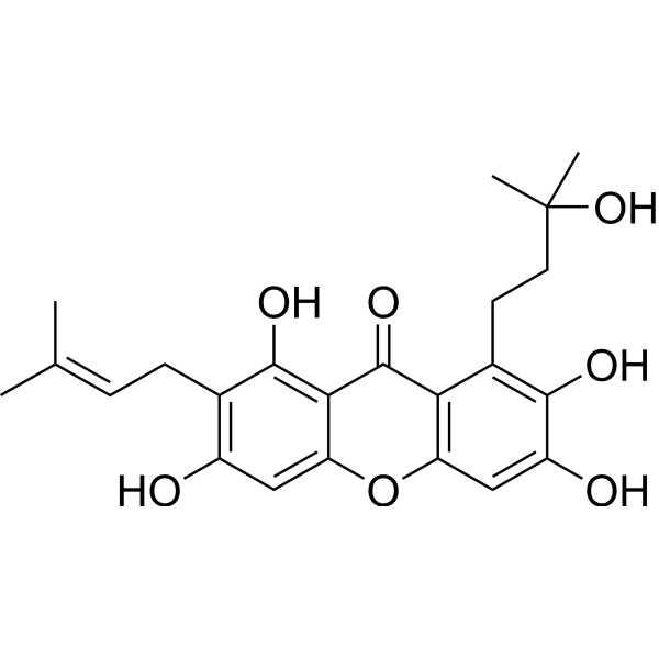
Garcinone C, a xanthone derivative, is a natural compound extracted from Garcinia oblongifolia that is used as an anti-inflammatory, astringency and granulation-promoting medicine, and has potential cytotoxic effects on certain cancers. Garcinone C stimulates the expression levels of ATR and 4E-BP1, arrests the cell cycle, inhibits cell viability of the human Nasopharyngeal carcinoma (NPC) cell lines CNE1, CNE2, HK1 and HONE1 in a time‑ and dose‑dependent manner through inhibition of Hedgehog signaling pathway. Garcinone C is orally active.
연구목적의 판매만을 진행합니다. 환자를 대상으로 한 판매는 하지 않습니다.
- Purity : 99.51%
- CAS No.: 76996-27-5
- 화학식: C23H26O7
- 분자량:414.45
-
보관:
4°C, protect from light
* In solvent : -80°C, 6 months; -20°C, 1 month (protect from light)

To place orders, for customer services and technical support, please contact: MedChemExpress USA
Tel: 609-228-6898 E-mail: [email protected] [email protected]


-
 Biological Activity
-
Chemical Information
-
용액&용해도
- Protocol
- 순도&문서
- References
-
 Help & FAQs


-

HY-N6954R
Garcinone C (Standard)CDK Inhibitor -
HY-139039
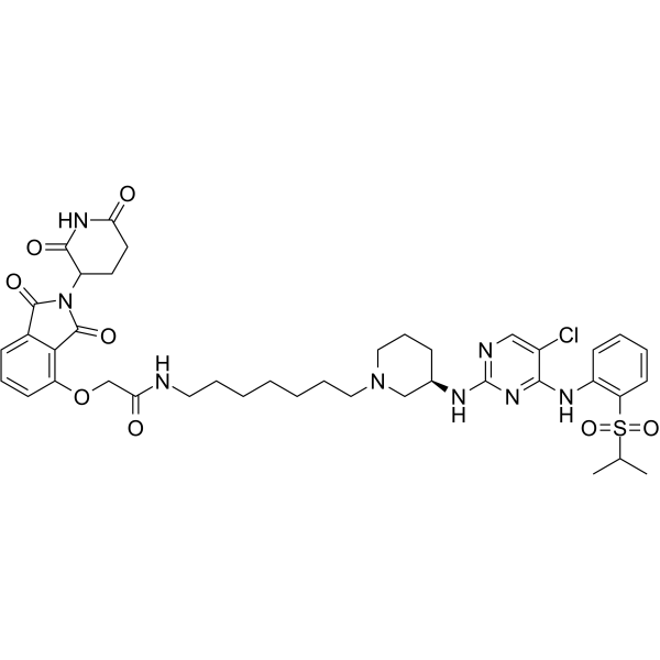
BSJ-4-116PROTAC CDK12 Degrader -
HY-138946
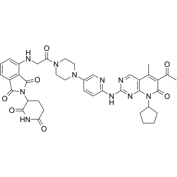
XY028-140CDK4/6 PROTAC -
HY-130709
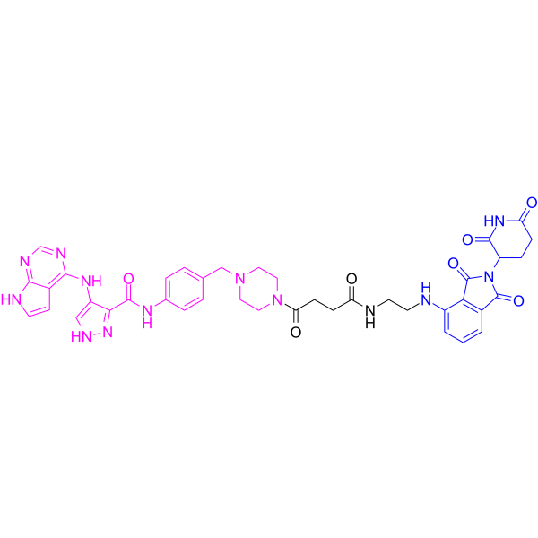
PROTAC CDK2/9 Degrader-1PROTAC CDK2/9 Degrader -
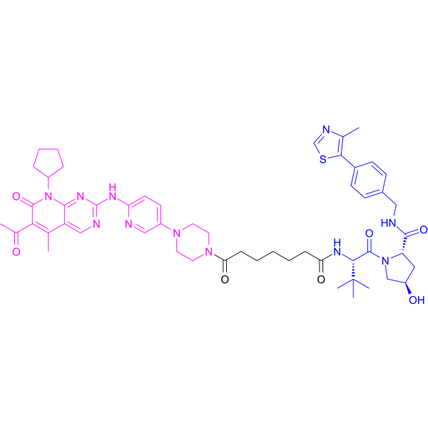
HY-129180
XY028-133CDK4/6 PROTAC Degrader -
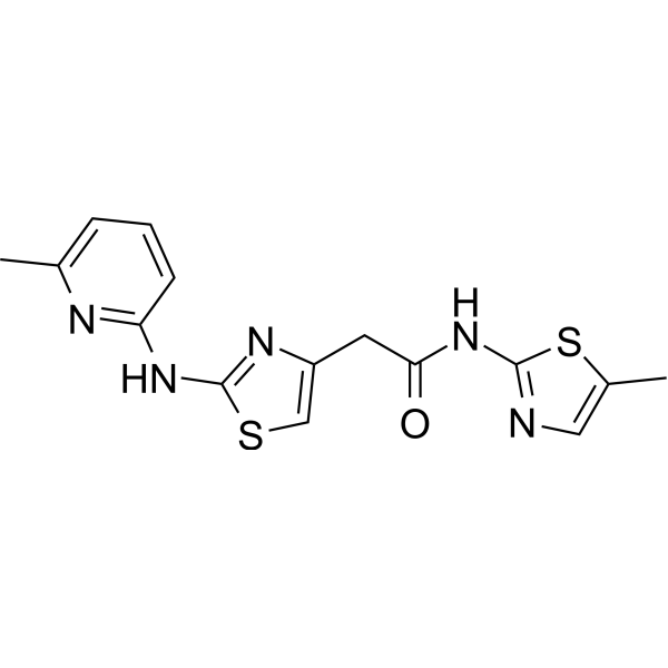
HY-144981
HQ461Cyclin K Degrader -
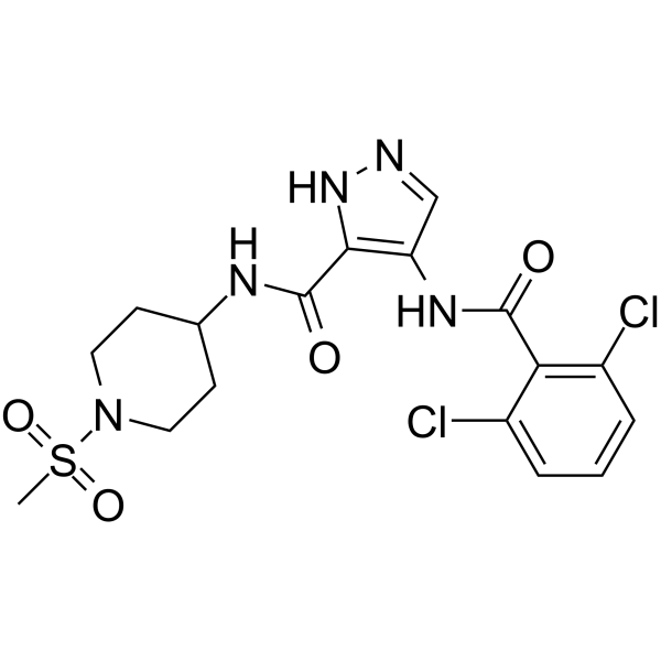
HY-15241
NVP-LCQ195CDK Inhibitor -
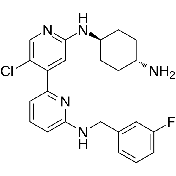
HY-16462
CDK9-IN-2CDK9 Inhibitor -
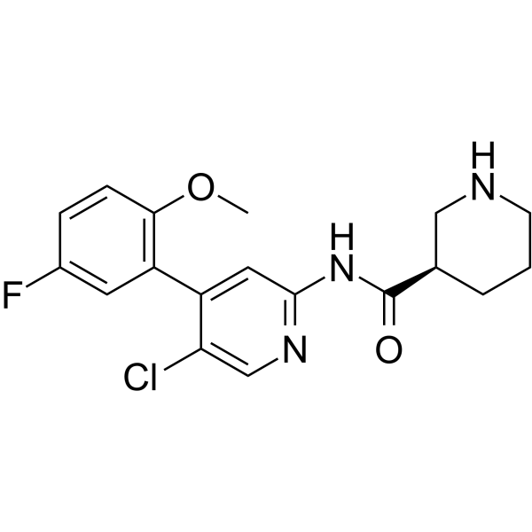
HY-13033
CDK-IN-2CDK Inhibitor -
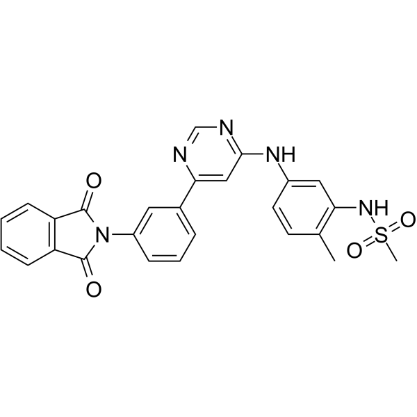
HY-13231
CDK9-IN-1CDK9 Inhibitor -

HY-13231R
CDK9-IN-1 (Standard)CDK9 Inhibitor
Publications Citing Use of MedChemExpress (MCE) Garcinone C
More Customer Validation & Images

-
WB

-
ELISA

-
RT-PCR


Biological Activity
제품 설명
IC50 & Target
[1]|
ATR |
Stat-3 |
CDK7 |
In Vitro
Garcinone C (1-5 μg/mL; 24 h-2 weeks) inhibits the the expression of cyclin D1, cyclin E and CDK6, increases the expression of p21, arrests the cell cycle at G0/G1 phase, inhibits the colony formation of HT29[2].
MedChemExpress (MCE) has not independently confirmed the accuracy of these methods. They are for reference only.
-
Cell Line:HT29
-
Concentration:0.3-3 μg/mL
-
Incubation Time:24 h
-
Result:Reduced the expression of cyclin D1/E and CDK6, increased the expression of p21.
-
Cell Line:HT29
-
Concentration:1-5 μg/mL
-
Incubation Time:24 h
-
Result:Arrested the cell cycle at G0/G1 phase.
In Vivo
MedChemExpress (MCE) has not independently confirmed the accuracy of these methods. They are for reference only.
-
Animal Model:AOM/DSS-induced colon cancer mouse models[2]
-
Dosage:10-50 mg/kg
-
Administration:po, 5 days per week for 9 weeks
-
Result:Reduced the number of colon tumors.
- RNA extraction experimental
- Flow cytometric DNA-content cell-cycle staining
- Cell Cytotoxicity Assay
- Mammalian live/dead viability and cytotoxicity staining
- BrdU Incorporation Assay
- Human pluripotent stem cell neural induction and neuron differentiation
- Protocol for Cell Cycle
- CCK-8/WST-8 Cell Proliferation Assay
- Research Protocol for Inflammation-related Diseases
- MTT Cell Proliferation Assay
- Cell Viability Determination by MTT Colorimetric Assay


Chemical Information
-
CAS No. 76996-27-5
-
Appearance Solid
-
분자량 414.45
-
화학식 C23H26O7
-
Color Light yellow to yellow
-
SMILES
CC(C)(O)CCC1=C(O)C(O)=CC(OC2=CC(O)=C(C/C=C(C)/C)C(O)=C23)=C1C3=O
-
Structure Classification
-
선적
Room temperature in continental US; may vary elsewhere.
-
보관
4°C, protect from light
* In solvent : -80°C, 6 months; -20°C, 1 month (protect from light)


Publications (3)
-
Journal Impact Factor
-
Most Recent
-
-
EMBO Rep
2025 Feb;26(3):635-655. PMID: 39747659
Garcinone C purchased from MedChemExpress. Usage Cited in: EMBO Rep. 2025 Feb;26(3):635-655. [Abstract]
PD-L1 KO MDA-MB-231 cells were untreated (day 0), treated with NCS, and cultured with vehicle DMSO or Garcinone C for the indicated times, then subjected to western blotting.
Garcinone C purchased from MedChemExpress. Usage Cited in: EMBO Rep. 2025 Feb;26(3):635-655. [Abstract]
WT and PD-L1 KO MDA-MB-231 cells were untreated (control) or treated with NCS and cultured for 5 days with or without Garcinone C, and concentrations of cellular cGAMP were analyzed using ELISA.
Garcinone C purchased from MedChemExpress. Usage Cited in: EMBO Rep. 2025 Feb;26(3):635-655. [Abstract]
WT and PD-L1 KO MDA-MB-231 cells were untreated (control) or treated with NCS, cultured for 5 days with or without the indicated doses of Garcinone C (5, 10, 25 μM), and analyzed for the mRNA levels of IL-6 and GM-CSF using qRT-PCR.
-


용액&용해도
In Vitro:
DMSO : 100 mg/mL (241.28 mM; Need ultrasonic; Hygroscopic DMSO has a significant impact on the solubility of product, please use newly opened DMSO)
Please refer to the solubility information to select the appropriate solvent. Once prepared, please aliquot and store the solution to prevent product inactivation from repeated freeze-thaw cycles.
Storage method and period of stock solution: -80°C, 6 months; -20°C, 1 month (protect from light). When stored at -80°C, please use it within 6 months. When stored at -20°C, please use it within 1 month.
Please refer to the solubility information to select the appropriate solvent. Once prepared, please aliquot and store the solution to prevent product inactivation from repeated freeze-thaw cycles.
Storage method and period of stock solution: -80°C, 6 months; -20°C, 1 month (protect from light). When stored at -80°C, please use it within 6 months. When stored at -20°C, please use it within 1 month.
Concentration (start) × Volume (start) = Concentration (final) × Volume (final)
In Vivo:
Select the appropriate dissolution method based on your experimental animal and administration route.
- For the following dissolution methods, please ensure to first prepare a clear stock solution using an In Vitro approach and then sequentially add co-solvents:
- To ensure reliable experimental results, the clarified stock solution can be appropriately stored based on storage conditions. As for the working solution for In Vivo experiments, it is recommended to prepare freshly and use it on the same day.
- The percentages shown for the solvents indicate their volumetric ratio in the final prepared solution. If precipitation or phase separation occurs during preparation, heat and/or sonication can be used to aid dissolution.
Add each solvent one by one: 10% DMSO 90% (20% SBE-β-CD in Saline)
Solubility: ≥ 2.5 mg/mL (6.03 mM); Clear solution
This protocol yields a clear solution of ≥ 2.5 mg/mL (saturation unknown).
Taking 1 mL working solution as an example, add 100 μL DMSO stock solution (25.0 mg/mL) to 900 μL 20% SBE-β-CD in Saline, and mix evenly.
Preparation of 20% SBE-β-CD in Saline (4°C, storage for one week): 2 g SBE-β-CD powder is dissolved in 10 mL Saline, completely dissolve until clear.
In Vivo Dissolution Calculator
Please enter the basic information of animal experiments:
-
-
-
-
Recommended: Prepare an additional quantity of animals to account for potential losses during experiments.
Please enter your animal formula composition:
-
%DMSO +
Recommended: Keep the proportion of DMSO in working solution below 2% if your animal is weak.
-
%+
-
+%Tween-80 + +
-
%Saline +
The co-solvents required include: DMSO, . All of co-solvents are available by MedChemExpress (MCE). , Tween 80. All of co-solvents are available by MedChemExpress (MCE).
Working solution concentration: 0.22 mg/mL
Method for preparing stock solution: mg drug dissolved in μL DMSO. Stock solution concentration: mg/mL. * In solvent : -80°C, 6 months; -20°C, 1 month (protect from light)
1. Take μL DMSO stock solution;
2. Add μL .
μL , mix evenly;
3. Then add μL Tween 80, mix evenly;
4. Then add μL
Please ensure that the stock solution in the first step is dissolved to a clear state, and add co-solvents in sequence. You can use ultrasonic heating (ultrasonic cleaner, recommended frequency 20-40 kHz), vortexing, etc. to assist dissolution.


순도&문서
-
Data Sheet (277 KB)
-
SDS (393 KB)
- English - EN (393 KB)
- Français - FR (393 KB)
- Deutsch - DE (393 KB)
- Norwegian - NO (393 KB)
- Español - ES (393 KB)
- Swedish - SV (393 KB)
- Italian - IT (393 KB)
- Korean - KR (393 KB)
- Portuguese - PT (393 KB)
-
Handling Instructions (2659 KB)
References
[1]. Xia Y, et al. Garcinone C exerts antitumor activity by modulating the expression of ATR/Stat3/4E BP1 in nasopharyngeal carcinoma cells. Oncol Rep. 2018 Mar;39(3):1485-1493. [Content Brief]
[2]. Chen J, et al., Garcinone C suppresses colon tumorigenesis through the Gli1-dependent hedgehog signaling pathway. Phytomedicine. 2020 Dec;79:153334. [Content Brief]


Complete Stock Solution Preparation Table
Please refer to the solubility information to select the appropriate solvent. Once prepared, please aliquot and store the solution to prevent product inactivation from repeated freeze-thaw cycles.
Storage method and period of stock solution: -80°C, 6 months; -20°C, 1 month (protect from light). When stored at -80°C, please use it within 6 months. When stored at -20°C, please use it within 1 month.
| Optional Solvent | Concentration Solvent Mass | 1 mg | 5 mg | 10 mg | 25 mg |
|---|---|---|---|---|---|
| DMSO | 1 mM | 2.4128 mL | 12.0642 mL | 24.1284 mL | 60.3209 mL |
| 5 mM | 0.4826 mL | 2.4128 mL | 4.8257 mL | 12.0642 mL | |
| 10 mM | 0.2413 mL | 1.2064 mL | 2.4128 mL | 6.0321 mL | |
| 15 mM | 0.1609 mL | 0.8043 mL | 1.6086 mL | 4.0214 mL | |
| 20 mM | 0.1206 mL | 0.6032 mL | 1.2064 mL | 3.0160 mL | |
| 25 mM | 0.0965 mL | 0.4826 mL | 0.9651 mL | 2.4128 mL | |
| 30 mM | 0.0804 mL | 0.4021 mL | 0.8043 mL | 2.0107 mL | |
| 40 mM | 0.0603 mL | 0.3016 mL | 0.6032 mL | 1.5080 mL | |
| 50 mM | 0.0483 mL | 0.2413 mL | 0.4826 mL | 1.2064 mL | |
| 60 mM | 0.0402 mL | 0.2011 mL | 0.4021 mL | 1.0053 mL | |
| 80 mM | 0.0302 mL | 0.1508 mL | 0.3016 mL | 0.7540 mL | |
| 100 mM | 0.0241 mL | 0.1206 mL | 0.2413 mL | 0.6032 mL |


Garcinone C Related Classifications
HY-N6954 Related Classifications
 Powered by Bioz