



1-Oleoyl lysophosphatidic acid sodium (GMP)
1-Oleoyl lysophosphatidic acid sodium (GMP) is the GMP-grade form of 1-Oleoyl lysophosphatidic acid sodium (HY-107614). GMP-grade small molecules serve as auxiliary reagents in cell therapy. 1-Oleoyl lysophosphatidic acid sodium is a bioactive lipid signaling molecule. 1-Oleoyl lysophosphatidic acid sodium inhibits lysoPLD-catalyzed hydrolysis of lysophosphatidylcholine and FS-3. 1-Oleoyl lysophosphatidic acid sodium activates LPA1 and LPA2, thereby triggering calcium mobilization, NFATc1 translocation, Rho/ROCK activation, Smad2/3 phosphorylation and c-Fos expression. 1-Oleoyl lysophosphatidic acid sodium induces anxiety-like, depression-like and hypoactivity phenotypes, regulates osteoclast cytoskeleton and viability, reduces osteoclast bone resorptive activity, and drives mesenchymal stem cell differentiation into myofibroblast-like cells. 1-Oleoyl lysophosphatidic acid sodium stimulates the secretion of transforming growth factor-β1 and stromal cell-derived factor-1. 1-Oleoyl lysophosphatidic acid sodium is applicable to research related to anxiety, depression and ovarian cancer.
For research use only. We do not sell to patients.
- CAS No.: 325465-93-8
- Formula: C21H40NaO7P
- Molecular Weight:458.50
-
Storage:
Please store the product under the recommended conditions in the Certificate of Analysis.


To place orders, for customer services and technical support, please contact: MedChemExpress USA
Tel: 609-228-6898 E-mail: [email protected] [email protected]


-
 Biological Activity
-
Chemical Information
- Protocols
- Purity & Documentation
- References
-
 Help & FAQs


-
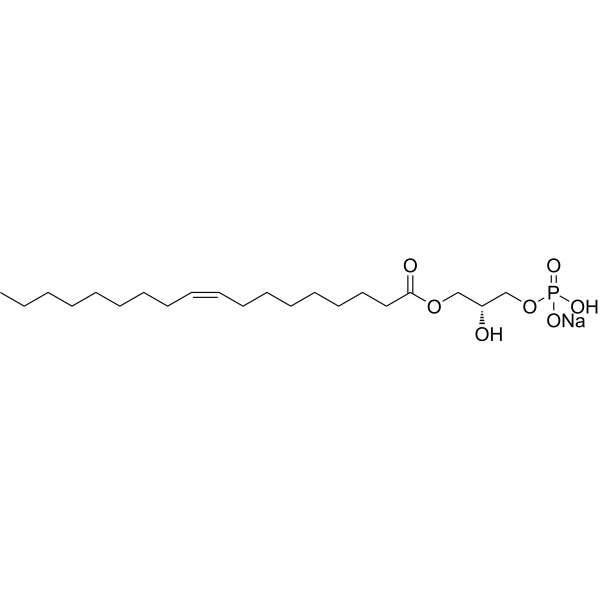
HY-107614
1-Oleoyl lysophosphatidic acid sodiumLPA Receptor Activator -
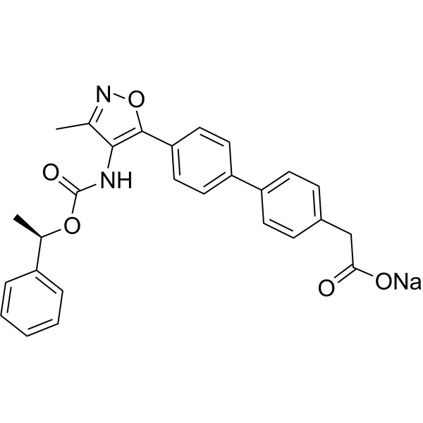
HY-16039
AM095LPA1 Antagonist -
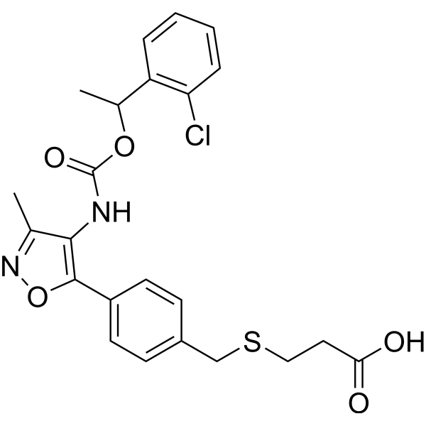
HY-13285
Ki16425LPA1/3 Antagonist -
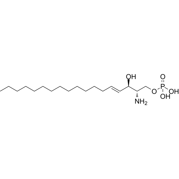
HY-108496
Sphingosine-1-phosphateS1PR Agonist -
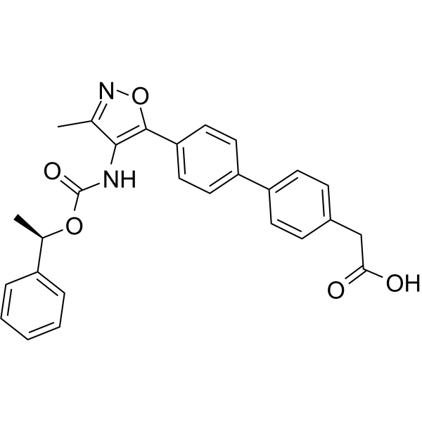
HY-16040
AM095 free acid -
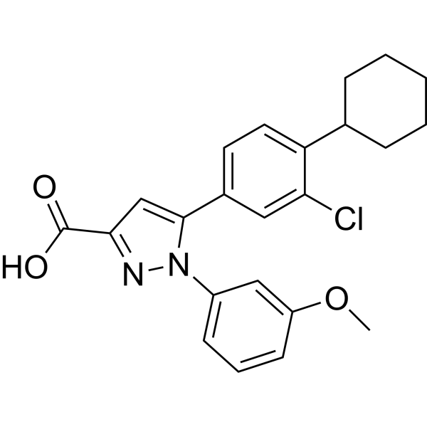
HY-107615
TC LPA5 4LPA5/GPR92 Antagonist -

HY-107614R
1-Oleoyl lysophosphatidic acid sodium (Standard)Reference Standard -
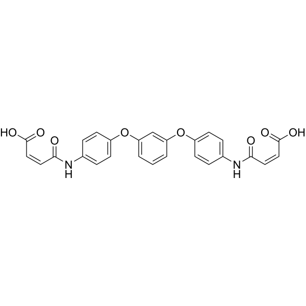
HY-107616
H2L5186303LPA2 Receptor Antagonist -
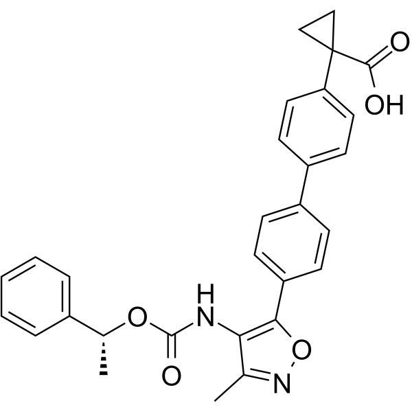
HY-100619
BMS-986020LPA1 Antagonist -
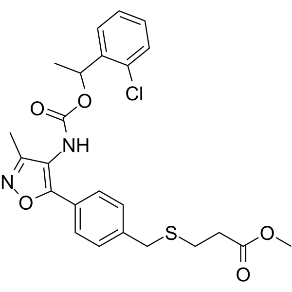
HY-18641
Ki16198LPA Receptor Antagonist -
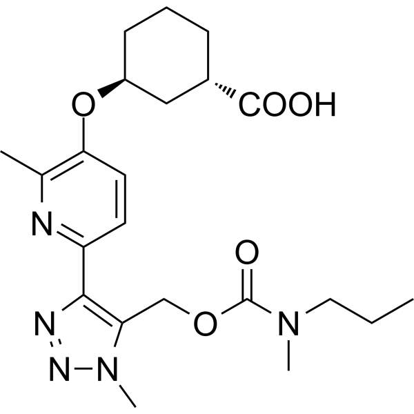
HY-139853
BMS-986278LPA1 Antagonist -
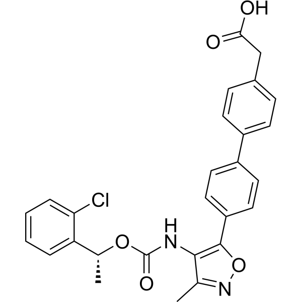
HY-15277
AM966LPA1 Receptor Antagonist


Biological Activity
Description
In Vitro
1-Oleoyl lysophosphatidic acid sodium competitively inhibits the activity of purified bovine serum lysophospholipase D (lysoPLD) in spectrophotometry and fluorescence analysis assays, with Ki values of 9.6 and 1.2 μM[1].
1-Oleoyl lysophosphatidic acid sodium (GMP) (400 nM-10 μM) induces concentration-dependent transient elevation of [Ca2+]i in primary rat and rabbit osteoclasts. Approximately 400 nM of the reagent elicits responses in 50% of the cells, and this response is mediated by Gi/o-coupled LPA1/3 receptors[3].
1-Oleoyl lysophosphatidic acid sodium (GMP) (5 μM) induces sustained retraction of lamellipodia and disrupts the peripheral actin belt in primary rat osteoclasts via a pathway involving Rho-associated kinases[3].
1-Oleoyl lysophosphatidic acid sodium (0-5 μM; 3-18 h) enhances the survival rate of primary rat osteoclasts, inhibits the apoptosis of primary rat osteoclasts, and induces the nuclear translocation of NFATc1 in primary rabbit osteoclasts via the Gi/o-coupled LPA1 receptor pathway[3].
1-Oleoyl lysophosphatidic acid sodium (5 μM; 5 min) induces mild ERK1/2 phosphorylation via a MEK1/2-dependent pathway in RAW 264.7-derived osteoclast-like cells[3].
1-Oleoyl lysophosphatidic acid sodium (0-10 μM; 0-96 h) induces delayed phosphorylation of Smad2 and Smad3 and subsequent expression of α-smooth muscle actin in human adipose tissue-derived mesenchymal stem cells via LPA receptor-dependent and Smad2/3-dependent signaling pathways[4].
1-Oleoyl lysophosphatidic acid sodium (5 μM; 0-48 hours, 4 days) induces the expression of α-smooth muscle actin in human adipose tissue-derived mesenchymal stem cells by stimulating autocrine TGF-β1 secretion and subsequent Smad2 activation[4].
1-Oleoyl lysophosphatidic acid sodium (0-20 μM; 0-48 h) induces the expression and secretion of stromal cell-derived factor-1 (SDF-1) in human adipose tissue-derived mesenchymal stem cells in a dose- and time-dependent manner via LPA receptor-dependent and TGF-β1-dependent signaling pathways[4].
1-Oleoyl lysophosphatidic acid sodium (5 μM; 4 days) induces the differentiation of human adipose tissue-derived mesenchymal stem cells into myofibroblast-like cells (which express α-smooth muscle actin and calmodulin, but not smooth muscle myosin heavy chain)[4].
MedChemExpress (MCE) has not independently confirmed the accuracy of these methods. They are for reference only. Further protocols information, click here.
-
Cell Line:RAW 264.7-derived osteoclast-like cells
-
Concentration:5 μM
-
Incubation Time:5 min
-
Result:Induced a small but significant increase in phospho-ERK1/2 levels.
Had effect blocked by 10 μM U0126.
-
Cell Line:human adipose tissue-derived mesenchymal stem cells (hADSCs)
-
Concentration:0-10 μM (dose-response); 5 μM (time-course); 5 μM (Ki16425 pre-incubation group); 10 μM (Ki16425)
-
Incubation Time:4 days (dose-response); 3-96 hours (time-course); 4 days (Ki16425 pre-incubation group)
-
Result:Increased α-SMA expression dose-dependently, with maximal increase at 5 μM after 4 days.
Increased α-SMA expression starting 48 hours after 5 μM treatment, with maximal levels at 96 hours.
Completely abolished the 5 μM-induced α-SMA expression when pre-incubated with 10 μM Ki16425.
-
Cell Line:human adipose tissue-derived mesenchymal stem cells (hADSCs)
-
Concentration:5 μM (time-course); 5 μM (Ki16425 pre-incubation group); 10 μM (Ki16425); 10 μM (SIS3); siRNA transfection (Smad2/3); adenoviral Smad7 expression
-
Incubation Time:0-96 hours (time-course); 4 days (inhibitor/siRNA/adenovirus groups)
-
Result:Induced delayed phosphorylation of Smad2 and Smad3, with maximal levels at 48 hours, sustained through 96 hours.
Completely abolished Smad2/3 phosphorylation when pre-incubated with 10 μM Ki16425.
Completely abrogated 5 μM-induced α-SMA expression and corresponding Smad phosphorylation after siRNA-mediated knockdown of Smad2 or Smad3.
Attenuated 5 μM-induced α-SMA expression and Smad3 phosphorylation (but not Smad2 phosphorylation) when pre-incubated with 10 μM SIS3.
Abrogated 5 μM-induced α-SMA expression and Smad2/3 phosphorylation after adenoviral overexpression of Smad7.
-
Cell Line:human adipose tissue-derived mesenchymal stem cells (hADSCs)
-
Concentration:5 μM
-
Incubation Time:4 days
-
Result:Increased expression of α-SMA and calponin in hADSCs.
Did not induce expression of smooth muscle myosin heavy chain (SM-MHC).
In Vivo
MedChemExpress (MCE) has not independently confirmed the accuracy of these methods. They are for reference only.
-
Animal Model:Wistar rats (9-week-old male, 250-300 g)[2]
-
Dosage:0.4 μg/5 μL; 2 μg/5 μL
-
Administration:i.c.v.; single injection
-
Result:Reduced time spent in open arms by approximately 89% (2 μg dose, elevated plus-maze novelty condition); showed no significant effect on time spent in open arms (0.4 μg dose, elevated plus-maze novelty condition).
Reduced locomotor activity (crossings) by approximately 40% (2 μg dose, open-field test novelty condition); showed no significant effect on locomotor activity (0.4 μg dose, open-field test novelty condition).
Increased locomotor activity by approximately 180% (2 μg dose, open-field test habituation condition); showed no significant effect on locomotor activity (0.4 μg dose, open-field test habituation condition).
Reduced the percentage of rats that first entered the novel arm to chance levels (both doses, Y maze test); significantly reduced total time spent in the novel arm (0.4 μg dose, Y maze test); showed a near-significant reduction in total time spent in the novel arm (p=0.0558, 2 μg dose, Y maze test).
Increased immobility time by 75% (0.4 μg dose, forced swimming test); increased immobility time by 120% (2 μg dose, forced swimming test).
Increased c-Fos immunoreactivity in the dorsal periaqueductal gray matter (DPAG) by 94% (2 μg dose).


Chemical Information
-
CAS No. 325465-93-8
-
Molecular Weight 458.50
-
Formula C21H40NaO7P
-
SMILES
CCCCCCCC/C=C\CCCCCCCC(OC[C@@H](O)COP(O)(O[Na])=O)=O
-
Synonyms
1-Oleoyl-sn-glycero-3-phosphate sodium (GMP); 1-Oleoyl-LPA sodium (GMP)
-
Shipping
Room temperature in continental US; may vary elsewhere.
-
Storage
Please store the product under the recommended conditions in the Certificate of Analysis.

Protocols
-
Kinase activity and phosphorylation assays
Kinase activity assays measure the ability of kinases to transfer phosphate groups from ATP to specific substrates, while phosphorylation assays detect the presence and levels of phosphorylated proteins. Common methods include radiolabeled ATP incorporation (e. g. ,), ADP release detection via bioluminescence (e. g. ,[3]), enzyme-linked immunosorbent assays (ELISA) for phospho-specific epitopes (e. g. ,[6]), and microtiter-based formats for high-throughput screening (e. g. ,[8]). The ADP-Glo assay quantifies kinase activity by measuring ADP produced during phosphorylation using a luciferase-based system. Radiometric assays involve autoradiography or scintillation counting after incorporation of 32P-labeled ATP into substrate proteins. ELISA-based approaches rely on phospho-specific antibodies to detect activated kinases in cell lysates or purified samples.
-
Western Blot
Western blotting (WB) is a commonly used experimental method in molecular biology, biochemistry, and immunogenetics for identifying and quantifying target proteins. It combines gel electrophoresis with immunoassay, enabling researchers to analyze protein expression, post-translational modifications, and molecular weight.
-
RNA extraction experimental
By lysing cells, releasing RNA, and removing impurities such as proteins and DNA, high-purity RNA products are finally obtained. The commonly used traditional method is the guanidine isothiocyanate/phenol/chloroform method (Trizol), which is suitable for a variety of animal materials including animal tissues, microorganisms, cultured cells, etc., and most plant materials.
-
Protocol for Kinase activity and phosphorylation assays
Kinase activity assays measure transfer of phosphate from ATP to a protein or peptide substrate, generating phosphorylated substrate, ADP, or incorporated radiolabeled phosphate as the readout; phosphorylation assays measure site-specific phosphorylation in cells or tissues as a proxy for kinase-pathway activation, inhibition, or substrate regulation. Phosphorylation can be detected by phospho-specific Western blot, immunoprecipitation kinase assay, phospho-immunofluorescence, phospho-flow cytometry, luminescent ADP detection, radiolabeled ATP incorporation, or reporter-based pathway assays, and these readouts can be applied to cancer cells, primary neurons, mouse tumors, organoids, inflammatory macrophages, ferroptosis studies, and mitophagy studies when the kinase target is biologically relevant.
-
iPSC cell differentiation
Induced pluripotent stem cells (iPSCs) are a type of cell that has similar properties to embryonic stem cells through somatic cell reprogramming.
-
Directly Induced Neuron Culture
Directly induced neuron culture converts somatic cells, most commonly fibroblasts, into induced neurons without passing through a pluripotent or neural progenitor stage; classic evidence shows that mouse fibroblasts can be converted by Ascl1, Brn2/Pou3f2, and Myt1l, human fibroblasts can be converted by defined neuronal transcription factors, and human fibroblasts can also be converted by miR-9/9-124 with neurogenic or subtype-specifying transcription factors. The readout is acquisition of neuronal identity and function, assessed by neuronal morphology, neuronal markers such as Tuj1/βIII-tubulin, MAP2, synapsin, and subtype markers when relevant, together with functional assays such as action-potential firing, synaptic activity, and electrophysiology.
-
Osteoclast differentiation from monocyte/macrophage precursors
Osteoclast differentiation is an in vitro induction assay in which monocyte/macrophage-lineage precursors are exposed to macrophage colony-stimulating factor (M-CSF) and receptor activator of NF-κB ligand (RANKL), generating multinucleated osteoclasts that are commonly identified by tartrate-resistant acid phosphatase (TRAP) staining and functionally confirmed by resorption pits on dentin, bone, or mineralized substrates. M-CSF supports survival and expansion of osteoclast precursors, while RANKL binding to RANK drives osteoclast commitment, fusion, maturation, and resorptive function; osteoprotegerin inhibits this pathway by binding RANKL and preventing RANK activation. The main readouts are the number of TRAP-positive multinucleated cells, formation of F-actin rings, and resorbed surface area; TRAP-positive multinucleated cells indicate osteoclast differentiation, whereas pit formation on dentin, bone, or mineralized coating indicates functional bone-resorbing activity.
-
Phalloidin F-actin cytoskeleton staining
Phalloidin F-actin staining detects polymerized filamentous actin in fixed and permeabilized specimens by using fluorescent phalloidin or phalloidin-derived phallotoxins that bind actin filaments and generate a fluorescence microscopy readout corresponding to F-actin organization, including stress fibers, cortical actin, filament bundles, and tissue-specific actin networks. Phalloidin stabilizes F-actin by reducing actin subunit dissociation from filament ends, and fluorescent phallotoxins were established as tools for visualizing actin-containing structures in eukaryotic cells.
-
Cell Viability Determination by MTT Colorimetric Assay
The following protocol uses the MTT colorimetric assay as a classic literature-established method for assessing cell viability/metabolic activity in cultured mammalian cells. MTT[3-(4,5-dimethylthiazol-2-yl)-2,5-diphenyltetrazolium bromide] is reduced by metabolically active cells to a colored formazan product; the amount of formazan is quantified spectrophotometrically and provides an indirect measure of metabolically active viable cells. Importantly, MTT reduction reflects cellular oxidoreductase/metabolic activity rather than an absolute direct count of living cells, so changes in cellular metabolism can alter the signal independently of cell number.


Purity & Documentation
References
[1]. Liu XW, et al. Inhibition of lysophospholipase D activity by unsaturated lysophosphatidic acids or seed extracts containing 1-linoleoyl and 1-oleoyl lysophosphatidic acid. J Agric Food Chem. 2007;55(21):8717-8722. [Content Brief]
[2]. Castilla-Ortega E, et al. 1-Oleoyl lysophosphatidic acid: a new mediator of emotional behavior in rats. PLoS One. 2014;9(1):e85348. Published 2014 Jan 7. [Content Brief]
[3]. Lapierre DM, et al. Lysophosphatidic acid signals through multiple receptors in osteoclasts to elevate cytosolic calcium concentration, evoke retraction, and promote cell survival. J Biol Chem. 2010;285(33):25792-25801. [Content Brief]
[4]. Jeon ES, et al. Cancer-derived lysophosphatidic acid stimulates differentiation of human mesenchymal stem cells to myofibroblast-like cells. Stem Cells. 2008;26(3):789-797. [Content Brief]

Calculators
Concentration (start) × Volume (start) = Concentration (final) × Volume (final)


1-Oleoyl lysophosphatidic acid sodium (GMP) Related Classifications
HY-107614G Related Classifications
 Powered by Bioz
Keywords
- 1-Oleoyl lysophosphatidic acid sodium (GMP)
- 325465-93-8
- 1-Oleoyl-sn-glycero-3-phosphate sodium (GMP)
- 1-Oleoyl-LPA sodium (GMP)
- LPL Receptor
- ROCK
- TGF-beta/Smad
- TGF-β Receptor
- bioactive lipid signaling molecule
- anxiety
- depression
- ovarian cancer
- osteoclasts
- mesenchymal stem cells
- Wistar rats
- Inhibitor
- inhibitor
- inhibit