Delivery of temperature sensitive items including proteins and kits will be paused on 6/19 for the Juneteenth holiday. For urgent orders please contact customer service.
ODN 2216 is a type A CpG oligodeoxynucleotide vaccine adjuvant and a TLR9 agonist. ODN 2216 interacts with TLR9 in the lysosomes of CD4 + T cells and activates feedback-dependent signaling pathways. ODN 2216 induces the production of type I interferons, IL-6 and TGF-β via the IRAK4/IRF7 axis, while increasing intracellular ATP levels. ODN 2216 not only induces the differentiation of CD4 + T cells into anti-inflammatory Th3-like regulatory phenotypes to inhibit autologous proliferation, but also enhances the specific CD8+ T cell-mediated cytotoxicity against Mammaglobin-A in breast cancer cells. ODN 2216 is widely used in studies related to breast cancer and systemic lupus erythematosus .
Anti-Mouse CD8beta Antibody (53-5.8) is an anti-mouse CD8beta IgG2a monoclonal antibody. Anti-Mouse CD8beta Antibody (53-5.8) can deplete CD8+ T cells and enhance cytotoxicity. Anti-Mouse CD8beta Antibody (53-5.8) can be used for research on immunology .
diABZI-V/C-DBCO is a STING agonist with an EC50 of 1.47 nM. diABZI-V/C-DBCO activates the STING pathway, induces the production of IFN-I, and stimulates the secretion of IFN-β. diABZI-V/C-DBCO serves as a substrate for cathepsin B, and releases active diABZI-amine via cathepsin B-mediated cleavage. In an orthotopic mouse model of breast cancer, diABZI-V/C-DBCO increases serum IFN-β levels and the frequency of granzyme B +CD8+ T cells. diABZI-V/C-DBCO is applicable to research related to triple-negative breast cancer .
ODN 2216 sodium is a type A CpG oligodeoxynucleotide vaccine adjuvant and a TLR9 agonist. ODN 2216 sodium interacts with TLR9 in the lysosomes of CD4 + T cells and activates feedback-dependent signaling pathways. ODN 2216 sodium induces the production of type I interferons, IL-6 and TGF-β via the IRAK4/IRF7 axis, while increasing intracellular ATP levels. ODN 2216 sodium not only induces the differentiation of CD4 + T cells into anti-inflammatory Th3-like regulatory phenotypes to inhibit autologous proliferation, but also enhances the specific CD8+ T cell-mediated cytotoxicity against Mammaglobin-A in breast cancer cells. ODN 2216 sodium is widely used in studies related to breast cancer and systemic lupus erythematosus .
Anti-Mouse LPAM-1/Integrin α4β7 Antibody (DATK32) is a rat-derived anti-LPAM-1/Integrin α4β7 IgG2a, κ type antibody inhibitor. Anti-Mouse LPAM-1/Integrin α4β7 Antibody (DATK32) specifically reacts with both chains of the α4β7 heterodimer and blocks the adhesion to immobilized mucosal addressin cell adhesion molecule-1 (MAdCAM-1). Anti-Mouse LPAM-1/Integrin α4β7 Antibody (DATK32) suppresses the proliferation and cytokine secretion of CD8+ T cells. Anti-Mouse LPAM-1/Integrin α4β7 Antibody (DATK32) decreases Peyer’s patches and follicular B cells in mice. Anti-Mouse LPAM-1/Integrin α4β7 Antibody (DATK32) can be used for the researches of inflammation, such as ulcerative colitis .
Kp7-6 is a Fas mimetic peptide and also a Fas/FasL antagonist. Kp7-6 specifically binds to Fas and FasL, disrupts receptor complexes, and blocks downstream apoptosis signaling pathways. Kp7-6 inhibits the phosphorylation of ERK1-2, induces the phosphorylation of IκBα, and activates NF-κB. Kp7-6 inhibits the activation of caspase-8, caspase-3 and JNK, and suppresses human amylin-induced β-cell apoptosis. Kp7-6 inhibits FasL-induced lymphoid cytotoxicity and apoptosis. Kp7-6 reduces local tumor FasL expression, increases CD8+Fas + T cell infiltration, and decreases tumor volume in pancreatic neuroendocrine tumor models. Kp7-6 prevents concanavalin A-induced liver injury in mice. Kp7-6 is applicable to research related to type 2 diabetes, concanavalin A-induced hepatitis and pancreatic neuroendocrine tumors .
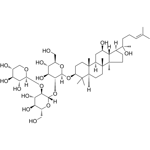
Pinocembrin 7-O-[3''-O-galloyl-4'',6''-hexahydroxydiphenoyl]-β-D-glucoside is a TRPV1 antagonist and HDAC7 inhibitor. Pinocembrin 7-O-[3''-O-galloyl-4'',6''-hexahydroxydiphenoyl]-β-D-glucoside blocks TRPV1-mediated calcium influx, suppresses phosphorylation of p65, IκBα, p38, JNK, and ERK1/2, inhibiting NF-κB and MAPK signaling cascades.Pinocembrin 7-O-[3''-O-galloyl-4'',6''-hexahydroxydiphenoyl]-β-D-glucoside reduces production and gene expression of pro-inflammatory cytokines IL-1β, IL-6, and TNF-α.Pinocembrin 7-O-[3''-O-galloyl-4'',6''-hexahydroxydiphenoyl]-β-D-glucoside exhibits potent analgesic activity, elevates thermal pain threshold and mechanical pain threshold in murine models.Pinocembrin 7-O-[3''-O-galloyl-4'',6''-hexahydroxydiphenoyl]-β-D-glucoside restores CD8+ T cell infiltration into bladder cancer tumors and improves bladder cancer immunotherapy efficacy.Pinocembrin 7-O-[3''-O-galloyl-4'',6''-hexahydroxydiphenoyl]-β-D-glucoside can be used for the researches of painand bladder cancer .
STING agonist-45 is a selective STING agonist (EC50 = 0.28 μM). STING agonist-45 activates the innate immune response through the cGAS-STING pathway, upregulating key markers such as p-TBK1 and IRF3. STING agonist-45 exhibits robust STING activation in human peripheral blood mononuclear cells (PBMCs), inducing the production of type I interferons (such as IFN-β) and downstream cytokines (such as TNF-α and IL-6). STING agonist-45 enhances anti-tumor immunity, inhibits tumor growth, and increases CD8+ T cell infiltration in mouse models. STING agonist-45 is promising for the study of STING-related diseases .
Notoginsenoside Ft1 is an orally active bioactive saponin. Notoginsenoside Ft1 inhibits the PI3K/AKT/mTOR signaling pathway, activates the p38 MAPK and ERK1/2 signaling pathways, and increases the proportion of CD8+ T cells, thereby inducing apoptosis and lysosomal cell death in various cancer cells, and promoting angiogenesis. Notoginsenoside Ft1 causes vasodilation by activating glucocorticoid receptors (GR) and estrogen receptor beta (ERβ) in endothelial cells. Notoginsenoside Ft1 increases intracellular Ca 2+ accumulation, reduces cAMP levels by activating a signaling network mediated through P2Y12 receptors, and promotes platelet aggregation, thereby exerting a procoagulant effect. Notoginsenoside Ft1 inhibits ferroptosis (ferroptosis) in renal tubular epithelial cells by activating the TGR5 receptor, thereby demonstrating a renal protective effect. Notoginsenoside Ft1 acts as a TGR5 agonist and an FXR antagonist to combat obesity and insulin resistance .
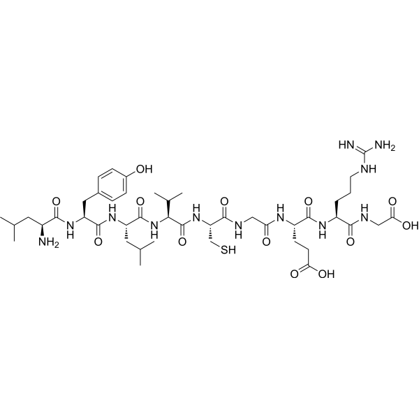
His-D-beta-Nal-Ala-Trp-D-Phe-Lys-NH2 TFA, is a growth hormone releasing peptide, as well as a metabolite of GHRP-1. GHRP-1, or Ala-His-D-beta Nal-Ala-Trp-D-Phe-Lys-NH2, has the effect of promoting the release of growth hormone (GH). GHRP-1 increases GH release and increases [Ca2+]i levels in static monolayer cells of rat pituitary gland, but does not affect cAMP levels .
SMU-L11-R is a selective TLR7 agonist with an EC50 of 0.012 μM for human TLR7. SMU-L11-R specifically activates TLR7, recruits MyD88, and triggers MAPK/NF-κB pathways, leading to TNF-α/IL-1β/IL-6 secretion in both mouse and human peripheral blood mononuclear cells. SMU-L11-R promotes M1-like macrophage polarization. SMU-L11-R exhibits excellent synergistic anti-tumor effects with PD-L1 inhibitors by upregulating CD8+T cells. SMU-L11-R shows potential in colorectal cancer studies .
EP4 receptor antagonist 7 (Compound 14) is an antagonist of the prostaglandin E2 (PGE2) receptor subtype EP4 with an IC50 value of 1.1 nM. EP4 receptor antagonist 7 inhibits PGE2-induced β-arrestin recruitment in HEK293 cells with an IC50 value of 0.9 nM. EP4 receptor antagonist 7 decreases PGE2-induced expression of mRNA encoding IL-4, macrophage mannose receptor 1 (Mrc1), chitinase-like protein 3 (Chil3), chemokine (C-X-C) motif ligand 1 (Cxcl1), triggering receptor expressed on myeloid cells 2 (Trem2), and arginase-1 (Arg1), in RAW 264.7 macrophages. EP4 receptor antagonist 7 combined with an anti-PD-1 antibody inhibits tumor growth and increases infiltration of CD 8+ T cells into tumors in a CT26 murine colon cancer model .
FGT-4 is a folate receptor β (FR-β) targeting chimeric molecule. FGT-4 is a TLR7 agonist. FGT-4 facilitates the secretion of iNOS and proinflammatory cytokine IL-6 associated with M1 macrophages and enhances the proliferation of cytotoxic CD8+ T cells. FGT-4 has anti-tumor activity in the 4T1 breast cancer mouse model. FGT-4 can be used for the study of cancer immunity. (Pink: target protein TLR7/8 agonist 1 ligand (HY-103698); black: linker (HY-172936); blue: FR-β ligand (HY-172935)) .
PARP1-IN-44, an Olaparib (HY-10162) derivative, is an orally active PARP1 inhibitor (IC50 = 0.6 nM), and also inhibits PARP2 (IC50 = 1.0 nM) and PARP7 (IC50 = 7.5 nM). PARP1-IN-44 has selective antiproliferative activity against BRCA-deficient cancer cells with minimal toxicity to normal cells. PARP1-IN-44 induces G2/M phase arrest, promotes apoptosis, elevates ROS levels, disrupts mitochondrial membrane potential. PARP1-IN-44 suppresses PARylation while increasing γH2AX accumulation. PARP1-IN-44 activates the cGAS-STING pathway, upregulating IFN-β and CXCL10 expression. PARP1-IN-44 enhancing CD8+ T cell infiltration in a CT26 tumor mouse model, demonstrating robust in vivo antitumor efficacy .
AB-3PRGD2 is a radiotherapeutic agent targeting integrin αvβ3. AB-3PRGD2 shows improved tumor uptake and prolonged tumor retention, leading to significantly enhanced tumor growth suppression. AB-3PRGD2 can remodel the tumor immune microenvironment by upregulating PD-L1 expression and increasing tumor-infiltrating CD8+ T cells .
CD8B Human Pre-designed siRNA Set A contains three designed siRNAs for CD8B gene (Human), as well as a negative control, a positive control, and a FAM-labeled negative control.
HM-279 is a potent and orally active ALK5 inhibitor with an IC50 of 4.7 nM. HM-279 shows cross-reactivity with ALK7 (IC50 = 6.8 nM), but HM-279 has fair to good selectivity against other TGF-β receptor family kinases, with 4.5-693-fold selectivity. HM-279 demonstrates antitumor activity in vivo through CD8+ T cell immunity. HM-279 can be used for the research of colon cancer .
FB102 is an anti-human CD122 (IL-2Rβ) monoclonal antibody with selective activity. FB102 blocks the proliferation and activation of pathogenic NK cells and specific T cell subsets induced by IL-2 and IL-15, without affecting the proliferation of regulatory T cells. FB102 inhibits IL-2/IL-15-induced activation of CD4+ and CD8+ T cells in in vitro disease models. FB102 is applicable to research related to celiac disease .
SMU-Z1 is a TLR1/2 heterodimer agonist with an EC50 of 4.88 nM. SMU-Z1 activates the NF-κB pathway, triggers pro-inflammatory cytokine production, and induces the generation of TNF-α, IL-1β, IL-6 and NO. SMU-Z1 promotes splenocyte proliferation and upregulates the expression of CD8+T cells, NK cells and dendritic cells. SMU-Z1 exhibits significant anti-tumor effects in mouse leukemia models. SMU-Z1 can be used for leukemia-related research .
XMU-MP-10 is a selective NEDD4 inhibitor with a KD of 43.92 nM. XMU-MP-10 selectively inhibits NEDD4 auto-ubiquitination without affecting other ubiquitination activity, upregulates of β-TrCP and results YAP degradation without affecting NEDD4 protein expression. XMU-MP-10 exhibits significant in vivo efficacy in inhibiting TNBC tumor growth by enhancing CD8+ T cell infiltration. XMU-MP-10 enhances antitumor immune responses through the β-TrCP/YAP/ECM axis. XMU-MP-10 can be used for Triple-Negative Breast Cancer (TNBC) research .
Kp7-6 is a Fas mimetic peptide and also a Fas/FasL antagonist. Kp7-6 specifically binds to Fas and FasL, disrupts receptor complexes, and blocks downstream apoptosis signaling pathways. Kp7-6 inhibits the phosphorylation of ERK1-2, induces the phosphorylation of IκBα, and activates NF-κB. Kp7-6 inhibits the activation of caspase-8, caspase-3 and JNK, and suppresses human amylin-induced β-cell apoptosis. Kp7-6 inhibits FasL-induced lymphoid cytotoxicity and apoptosis. Kp7-6 reduces local tumor FasL expression, increases CD8+Fas + T cell infiltration, and decreases tumor volume in pancreatic neuroendocrine tumor models. Kp7-6 prevents concanavalin A-induced liver injury in mice. Kp7-6 is applicable to research related to type 2 diabetes, concanavalin A-induced hepatitis and pancreatic neuroendocrine tumors .
His-D-beta-Nal-Ala-Trp-D-Phe-Lys-NH2 TFA, is a growth hormone releasing peptide, as well as a metabolite of GHRP-1. GHRP-1, or Ala-His-D-beta Nal-Ala-Trp-D-Phe-Lys-NH2, has the effect of promoting the release of growth hormone (GH). GHRP-1 increases GH release and increases [Ca2+]i levels in static monolayer cells of rat pituitary gland, but does not affect cAMP levels .
Anti-Mouse CD8beta Antibody (53-5.8) is an anti-mouse CD8beta IgG2a monoclonal antibody. Anti-Mouse CD8beta Antibody (53-5.8) can deplete CD8+ T cells and enhance cytotoxicity. Anti-Mouse CD8beta Antibody (53-5.8) can be used for research on immunology .
Anti-Mouse LPAM-1/Integrin α4β7 Antibody (DATK32) is a rat-derived anti-LPAM-1/Integrin α4β7 IgG2a, κ type antibody inhibitor. Anti-Mouse LPAM-1/Integrin α4β7 Antibody (DATK32) specifically reacts with both chains of the α4β7 heterodimer and blocks the adhesion to immobilized mucosal addressin cell adhesion molecule-1 (MAdCAM-1). Anti-Mouse LPAM-1/Integrin α4β7 Antibody (DATK32) suppresses the proliferation and cytokine secretion of CD8+ T cells. Anti-Mouse LPAM-1/Integrin α4β7 Antibody (DATK32) decreases Peyer’s patches and follicular B cells in mice. Anti-Mouse LPAM-1/Integrin α4β7 Antibody (DATK32) can be used for the researches of inflammation, such as ulcerative colitis .
FB102 is an anti-human CD122 (IL-2Rβ) monoclonal antibody with selective activity. FB102 blocks the proliferation and activation of pathogenic NK cells and specific T cell subsets induced by IL-2 and IL-15, without affecting the proliferation of regulatory T cells. FB102 inhibits IL-2/IL-15-induced activation of CD4+ and CD8+ T cells in in vitro disease models. FB102 is applicable to research related to celiac disease .
Pinocembrin 7-O-[3''-O-galloyl-4'',6''-hexahydroxydiphenoyl]-β-D-glucoside is a TRPV1 antagonist and HDAC7 inhibitor. Pinocembrin 7-O-[3''-O-galloyl-4'',6''-hexahydroxydiphenoyl]-β-D-glucoside blocks TRPV1-mediated calcium influx, suppresses phosphorylation of p65, IκBα, p38, JNK, and ERK1/2, inhibiting NF-κB and MAPK signaling cascades.Pinocembrin 7-O-[3''-O-galloyl-4'',6''-hexahydroxydiphenoyl]-β-D-glucoside reduces production and gene expression of pro-inflammatory cytokines IL-1β, IL-6, and TNF-α.Pinocembrin 7-O-[3''-O-galloyl-4'',6''-hexahydroxydiphenoyl]-β-D-glucoside exhibits potent analgesic activity, elevates thermal pain threshold and mechanical pain threshold in murine models.Pinocembrin 7-O-[3''-O-galloyl-4'',6''-hexahydroxydiphenoyl]-β-D-glucoside restores CD8+ T cell infiltration into bladder cancer tumors and improves bladder cancer immunotherapy efficacy.Pinocembrin 7-O-[3''-O-galloyl-4'',6''-hexahydroxydiphenoyl]-β-D-glucoside can be used for the researches of painand bladder cancer .
Notoginsenoside Ft1 is an orally active bioactive saponin. Notoginsenoside Ft1 inhibits the PI3K/AKT/mTOR signaling pathway, activates the p38 MAPK and ERK1/2 signaling pathways, and increases the proportion of CD8+ T cells, thereby inducing apoptosis and lysosomal cell death in various cancer cells, and promoting angiogenesis. Notoginsenoside Ft1 causes vasodilation by activating glucocorticoid receptors (GR) and estrogen receptor beta (ERβ) in endothelial cells. Notoginsenoside Ft1 increases intracellular Ca 2+ accumulation, reduces cAMP levels by activating a signaling network mediated through P2Y12 receptors, and promotes platelet aggregation, thereby exerting a procoagulant effect. Notoginsenoside Ft1 inhibits ferroptosis (ferroptosis) in renal tubular epithelial cells by activating the TGR5 receptor, thereby demonstrating a renal protective effect. Notoginsenoside Ft1 acts as a TGR5 agonist and an FXR antagonist to combat obesity and insulin resistance .
CD8 beta is an important immune glycoprotein that serves as a coreceptor for MHC class I:peptide complexes, linking T cells to antigens presented by APCs. It interacts with TCR and MHC class I, recruits LCK kinase, and initiates multiple signaling pathways. CD8 beta Protein, Human (HEK293) is the recombinant human-derived CD8 beta protein, expressed by HEK293 , with tag free.
CD8 beta is an important immune glycoprotein that serves as a coreceptor for MHC class I:peptide complexes, linking T cells to antigens presented by APCs. It interacts with TCR and MHC class I, recruits LCK kinase, and initiates multiple signaling pathways. CD8 beta Protein, Human (HEK293, Fc) is the recombinant human-derived CD8 beta protein, expressed by HEK293 , with C-hFc labeled tag.
CD8 beta is an important immune glycoprotein that serves as a coreceptor for MHC class I:peptide complexes, linking T cells to antigens presented by APCs. It interacts with TCR and MHC class I, recruits LCK kinase, and initiates multiple signaling pathways. CD8 beta Protein, Human (HEK293, His) is the recombinant human-derived CD8 beta protein, expressed by HEK293 , with C-6*His labeled tag.
CD8 beta is an important immune glycoprotein that serves as a coreceptor for MHC class I:peptide complexes, linking T cells to antigens presented by APCs. It interacts with TCR and MHC class I, recruits LCK kinase, and initiates multiple signaling pathways. CD8 beta Protein, Human (Biotinylated, HEK293) is the recombinant human-derived CD8 beta protein, expressed by HEK293 , with tag free.
BOP; CD8 beta opposite; CD8b opposite; Histone lysine N methyltransferase SMYD1; KMT3D; SET and MYND domain-containing protein 1; SMYD1; SMYD1_HUMAN; Zinc finger MYND domain containing 18; ZMYND18; ZMYND22; zinc finger, MYND domain containing 18.
WB
Rat
SMYD1 Antibody (YA5193) is a Mouse-derived and non-conjugated monoclonal antibody, targeting to SMYD1.
diABZI-V/C-DBCO is a STING agonist with an EC50 of 1.47 nM. diABZI-V/C-DBCO activates the STING pathway, induces the production of IFN-I, and stimulates the secretion of IFN-β. diABZI-V/C-DBCO serves as a substrate for cathepsin B, and releases active diABZI-amine via cathepsin B-mediated cleavage. In an orthotopic mouse model of breast cancer, diABZI-V/C-DBCO increases serum IFN-β levels and the frequency of granzyme B +CD8+ T cells. diABZI-V/C-DBCO is applicable to research related to triple-negative breast cancer .
ODN 2216 is a type A CpG oligodeoxynucleotide vaccine adjuvant and a TLR9 agonist. ODN 2216 interacts with TLR9 in the lysosomes of CD4 + T cells and activates feedback-dependent signaling pathways. ODN 2216 induces the production of type I interferons, IL-6 and TGF-β via the IRAK4/IRF7 axis, while increasing intracellular ATP levels. ODN 2216 not only induces the differentiation of CD4 + T cells into anti-inflammatory Th3-like regulatory phenotypes to inhibit autologous proliferation, but also enhances the specific CD8+ T cell-mediated cytotoxicity against Mammaglobin-A in breast cancer cells. ODN 2216 is widely used in studies related to breast cancer and systemic lupus erythematosus .
ODN 2216 sodium is a type A CpG oligodeoxynucleotide vaccine adjuvant and a TLR9 agonist. ODN 2216 sodium interacts with TLR9 in the lysosomes of CD4 + T cells and activates feedback-dependent signaling pathways. ODN 2216 sodium induces the production of type I interferons, IL-6 and TGF-β via the IRAK4/IRF7 axis, while increasing intracellular ATP levels. ODN 2216 sodium not only induces the differentiation of CD4 + T cells into anti-inflammatory Th3-like regulatory phenotypes to inhibit autologous proliferation, but also enhances the specific CD8+ T cell-mediated cytotoxicity against Mammaglobin-A in breast cancer cells. ODN 2216 sodium is widely used in studies related to breast cancer and systemic lupus erythematosus .
CD8B Human Pre-designed siRNA Set A contains three designed siRNAs for CD8B gene (Human), as well as a negative control, a positive control, and a FAM-labeled negative control.
Inquiry Online
Your information is safe with us. * Required Fields.
Western blot analysis of extracts from THP-1(lane 2(20μg), Jurkat (lane 3(20μg) and NIH3T3(lane 4(20μg) using FOXO1A (HY-P80132) Rabbit mAb. Proteins were transferred
to a PVDF membrane and blocked with 5% non-fat milk in TBST for 2 hour at room temperature. The primary antibody (1/1000) and Loading control antibody (Beta Actin, HY-P80438, 1/10000) was
used in 5% non-fat milk in TBST at 4°C overnight. Goat Anti-Mouse/Rabbit IgG-HRP Secondary Antibody (1/10000) was used for 1 hour at room temperature.
Western blot analysis of extracts from THP-1(lane 2(20μg), Jurkat (lane 3(20μg) and NIH3T3(lane 4(20μg) using FOXO1A (HY-P80132) Rabbit mAb. Proteins were transferred
to a PVDF membrane and blocked with 5% non-fat milk in TBST for 2 hour at room temperature. The primary antibody (1/1000) and Loading control antibody (Beta Actin, HY-P80438, 1/10000) was
MedchemExpress Validation 03
Western blot analysis of extracts from THP-1(lane 2(20μg), Jurkat (lane 3(20μg) and NIH3T3(lane 4(20μg) using FOXO1A (HY-P80132) Rabbit mAb. Proteins were transferred
MedchemExpress Validation 04
Western blot analysis of extracts from THP-1(lane 2(20μg), Jurkat (lane 3(20μg) and NIH3T3(lane 4(20μg) using FOXO1A (HY-P80132) Rabbit mAb. Proteins were transferred
to a PVDF membrane and blocked with 5% non-fat milk in TBST for 2 hour at room temperature. The primary antibody (1/1000) and Loading control antibody (Beta Actin, HY-P80438, 1/10000) was
used in 5% non-fat milk in TBST at 4°C overnight. Goat Anti-Mouse/Rabbit IgG-HRP Secondary Antibody (1/10000) was used for 1 hour at room temperature.
MedchemExpress Validation
Western blot analysis of extracts from THP-1(lane 2(20μg), Jurkat (lane 3(20μg) and NIH3T3(lane 4(20μg) using FOXO1A (HY-P80132) Rabbit mAb. Proteins were transferred
to a PVDF membrane and blocked with 5% non-fat milk in TBST for 2 hour at room temperature. The primary antibody (1/1000) and Loading control antibody (Beta Actin, HY-P80438, 1/10000) was
used in 5% non-fat milk in TBST at 4°C overnight. Goat Anti-Mouse/Rabbit IgG-HRP Secondary Antibody (1/10000) was used for 1 hour at room temperature.
MedchemExpress Validation
Western blot analysis of extracts from THP-1(lane 2(20μg), Jurkat (lane 3(20μg) and NIH3T3(lane 4(20μg) using FOXO1A (HY-P80132) Rabbit mAb. Proteins were transferred
to a PVDF membrane and blocked with 5% non-fat milk in TBST for 2 hour at room temperature. The primary antibody (1/1000) and Loading control antibody (Beta Actin, HY-P80438, 1/10000) was
used in 5% non-fat milk in TBST at 4°C overnight. Goat Anti-Mouse/Rabbit IgG-HRP Secondary Antibody (1/10000) was used for 1 hour at room temperature.
MedChemExpress values your privacy and your trust is important to us. We use cookies to enhance your website experience. Some cookies are necessary to run the website.
Privacy and Cookie Policy


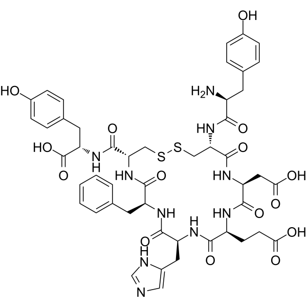
![Pinocembrin 7-O-[3''-O-galloyl-4'',6''-hexahydroxydiphenoyl]-β-D-glucoside](http://file.medchemexpress.com/product_pic/hy-n5084.gif)