



Kp7-6
Based on 3 publication(s) in Google Scholar
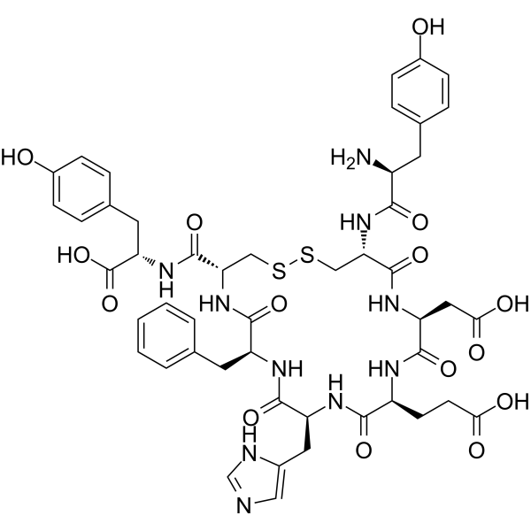
Kp7-6 is a Fas mimetic peptide and also a Fas/FasL antagonist. Kp7-6 specifically binds to Fas and FasL, disrupts receptor complexes, and blocks downstream apoptosis signaling pathways. Kp7-6 inhibits the phosphorylation of ERK1-2, induces the phosphorylation of IκBα, and activates NF-κB. Kp7-6 inhibits the activation of caspase-8, caspase-3 and JNK, and suppresses human amylin-induced β-cell apoptosis. Kp7-6 inhibits FasL-induced lymphoid cytotoxicity and apoptosis. Kp7-6 reduces local tumor FasL expression, increases CD8+Fas+ T cell infiltration, and decreases tumor volume in pancreatic neuroendocrine tumor models. Kp7-6 prevents concanavalin A-induced liver injury in mice. Kp7-6 is applicable to research related to type 2 diabetes, concanavalin A-induced hepatitis and pancreatic neuroendocrine tumors.
For research use only. We do not sell to patients.
- Purity : 99.28%
- CAS No.: 629628-53-1
- Formula: C48H56N10O15S2
- Molecular Weight:1077.15
-
Storage:
Sealed storage, away from moisture.
Powder -80°C, 2 years , -20°C, 1 year* In solvent : -80°C, 6 months; -20°C, 1 month (sealed storage, away from moisture)

To place orders, for customer services and technical support, please contact: MedChemExpress USA
Tel: 609-228-6898 E-mail: [email protected] [email protected]


-
 Biological Activity
-
Chemical Information
-
Solvent & Solubility
- Protocols
- Purity & Documentation
- References
-
 Help & FAQs



Publications Citing Use of MedChemExpress (MCE) Kp7-6
MoreAll Caspase Isoforms
More

Biological Activity
Description
In Vitro
Kp7-6 (1 h, 0.1-5 mM) dose-dependently inhibits apoptosis of CM cells, RINm5F cells and isolated mouse islet β-cells induced by human islet amyloid polypeptide (hA), with stronger activity in cultured insulinoma cell lines[1].
Kp7-6 (5 mM) inhibits the activation of hA-induced active caspase-8, phosphorylated JNK1 and active caspase-3 in isolated mouse islets when used as a pre-treatment or post-treatment[1].
Kp7-6 (300 μM; 2 h) potently inhibits the binding of FasL to Fas receptors in solid-phase ELISA assays[2].
Kp7-6 (12.5-100 μM; 300 s) binds specifically to FasL (Kd = 11.2 μM) and Fas (Kd = 13.2 μM) with comparable affinity[2].
Kp7-6 (0-1000 μM; 25 h) dose-dependently protects Jurkat cells against FasL-induced cytotoxicity, with a cell survival rate of >90% at the concentration of 1000 μM, and exhibits no toxicity when applied alone to Jurkat cells[2].
Kp7-6 (0-1000 μg/mL; 3 h) dose-dependently inhibits FasL-induced apoptosis in Jurkat cells[2].
Kp7-6 (1 mM; 2 h) activates the NF-κB signaling pathway (via IκBα phosphorylation) in FasL-stimulated Jurkat cells, inhibits ERK1/2 phosphorylation, and does not alter the activation level of JNK[2].
MedChemExpress (MCE) has not independently confirmed the accuracy of these methods. They are for reference only.
-
Cell Line:CM insulinoma cells, RINm5F insulinoma cells, isolated murine islet β-cells
-
Concentration:0.1 mM, 0.5 mM, 1 mM, 5 mM, 10 mM
-
Incubation Time:1 h
-
Result:Dose-dependently suppressed hA-evoked apoptosis.
Partially but significantly suppressed apoptosis in CM and RINm5F cells at 0.5-1 mM.
Completely suppressed apoptosis in both cell lines at 5 mM.
Exhibited more potent protective effect in cultured cell lines than in isolated islets.
Effectively inhibited apoptosis in all three β-cell systems.
Showed similar rescue effect between cultured cell lines and islet β-cells.
-
Cell Line:Jurkat cells
-
Concentration:10, 100, 1000 μM
-
Incubation Time:1 h (preincubation with FasL); 24 h (incubation with cells before [3H]thymidine pulsing)
-
Result:Showed dose-dependent inhibitory activity against FasL-induced cytotoxicity.
Protected >90% of Jurkat cells from Fas-mediated cytotoxicity at the highest tested concentration.
Did not mediate cytotoxicity to Jurkat cells at the tested concentrations.
-
Cell Line:Jurkat cells
-
Concentration:300, 1000 μg/mL
-
Incubation Time:3 h
-
Result:Dose-dependently reduced the percentage of annexin V-positive apoptotic Jurkat cells treated with FasL.
Reduced apoptotic cells to 31.5% at 300 μg/mL, and to 21.5% at 1000 μg/mL, compared to 49.6% apoptotic cells with FasL alone.
-
Cell Line:Jurkat cells
-
Concentration:1 mM
-
Incubation Time:2 h (preincubation with cells before FasL treatment)
-
Result:Enhanced phosphorylation of IκBα in the presence of FasL at 5 min.
Significantly inhibited phosphorylation of ERK1/2 at 5-30 min compared to treatment with FasL alone.
Had no effect on JNK activation.
In Vivo
Kp7-6 (100 mg/kg; i.p.; daily) reduces tumor FasL levels, increases CD8+Fas+ T cell infiltration, and significantly decreases tumor weight in Rip1-Tag2 pancreatic neuroendocrine tumor mice[3].
MedChemExpress (MCE) has not independently confirmed the accuracy of these methods. They are for reference only.
-
Animal Model:C57BL-6 (8-week-old)[2]
-
Dosage:3 mg per mouse
-
Administration:i.p.; single dose
-
Result:Reduced serum alanine aminotransferase (ALT) and aspartate aminotransferase (AST) activities significantly (P < 0.01 vs.
Con A-treated mice).
-
Animal Model:Rip1-Tag2 (C57BL/6 background; 12 weeks old; spontaneous pancreatic neuroendocrine tumor model)[3]
-
Dosage:100 mg/kg
-
Administration:i.p.; daily
-
Result:Significantly reduced tumor FasL levels compared to vehicle controls.
Increased infiltration of CD8+Fas+ T cells in tumor tissues compared to vehicle controls.
Significantly decreased tumor weight compared to vehicle controls.
Left ACSS2 expression in tumor tissues unchanged compared to vehicle controls.
- Western Blot
- RNA extraction experimental
- Mammalian live/dead viability and cytotoxicity staining
- Annexin V plus membrane-impermeant dye apoptosis staining
- Research Protocol for Inflammation-related Diseases
- Research Protocol for Metabolic Diseases
- Research Protocol for Cancer Immunology
- Cell Viability Determination by MTT Colorimetric Assay
- Kinase activity and phosphorylation assays
- Detection of Apoptosis
- TUNEL staining for apoptotic DNA fragmentation
- Cell Cytotoxicity Assay
- Apoptosis Solutions
- Protocol for Kinase activity and phosphorylation assays


Chemical Information
-
CAS No. 629628-53-1
-
Appearance Solid
-
Molecular Weight 1077.15
-
Formula C48H56N10O15S2
-
Color White to off-white
-
Sequence
Tyr-Cys-Asp-Glu-His-Phe-Cys-Tyr (Disulfide bridge:Cys2-Cys7)
-
Sequence Shortening
YCDEHFCY (Disulfide bridge:Cys2-Cys7)
-
Shipping
Room temperature in continental US; may vary elsewhere.
-
Storage
Sealed storage, away from moisture
Powder -80°C 2 years -20°C 1 year * In solvent : -80°C, 6 months; -20°C, 1 month (sealed storage, away from moisture)


Publications (3)
-
Journal Impact Factor
-
Most Recent
-
-
Adv Sci (Weinh)
ACSS2/AATF Drives Soluble FasL-Mediated CD8+ T Cell Apoptosis in Pancreatic Neuroendocrine Tumors. [Abstract]2025 Aug 12:e06883. PMID: 40791180 -
Cell Rep Med
Tumor-infiltrated double-negative regulatory T cells predict outcome of T cell-based immunotherapy in nasopharyngeal carcinoma. [Abstract]2025 May 20;6(5):102096. PMID: 40315843


Solvent & Solubility
In Vitro:
DMSO : 100 mg/mL (92.84 mM; Need ultrasonic; Hygroscopic DMSO has a significant impact on the solubility of product, please use newly opened DMSO)
Please refer to the solubility information to select the appropriate solvent. Once prepared, please aliquot and store the solution to prevent product inactivation from repeated freeze-thaw cycles.
Storage method and period of stock solution: -80°C, 6 months; -20°C, 1 month (sealed storage, away from moisture). When stored at -80°C, please use it within 6 months. When stored at -20°C, please use it within 1 month.
Please refer to the solubility information to select the appropriate solvent. Once prepared, please aliquot and store the solution to prevent product inactivation from repeated freeze-thaw cycles.
Storage method and period of stock solution: -80°C, 6 months; -20°C, 1 month (sealed storage, away from moisture). When stored at -80°C, please use it within 6 months. When stored at -20°C, please use it within 1 month.
Concentration (start) × Volume (start) = Concentration (final) × Volume (final)
In Vivo:
Select the appropriate dissolution method based on your experimental animal and administration route.
- For the following dissolution methods, please ensure to first prepare a clear stock solution using an In Vitro approach and then sequentially add co-solvents:
- To ensure reliable experimental results, the clarified stock solution can be appropriately stored based on storage conditions. As for the working solution for In Vivo experiments, it is recommended to prepare freshly and use it on the same day.
- The percentages shown for the solvents indicate their volumetric ratio in the final prepared solution. If precipitation or phase separation occurs during preparation, heat and/or sonication can be used to aid dissolution.
Add each solvent one by one: 10% DMSO 40% PEG300 5% Tween-80 45% Saline
Solubility: ≥ 2.5 mg/mL (2.32 mM); Clear solution
This protocol yields a clear solution of ≥ 2.5 mg/mL (saturation unknown).
Taking 1 mL working solution as an example, add 100 μL DMSO stock solution (25.0 mg/mL) to 400 μL PEG300, and mix evenly; then add 50 μL Tween-80 and mix evenly; then add 450 μL Saline to adjust the volume to 1 mL.
Preparation of Saline: Dissolve 0.9 g sodium chloride in ddH₂O and dilute to 100 mL to obtain a clear Saline solution.
Add each solvent one by one: 10% DMSO 90% (20% SBE-β-CD in Saline)
Solubility: ≥ 2.5 mg/mL (2.32 mM); Clear solution
This protocol yields a clear solution of ≥ 2.5 mg/mL (saturation unknown).
Taking 1 mL working solution as an example, add 100 μL DMSO stock solution (25.0 mg/mL) to 900 μL 20% SBE-β-CD in Saline, and mix evenly.
Preparation of 20% SBE-β-CD in Saline (4°C, storage for one week): 2 g SBE-β-CD powder is dissolved in 10 mL Saline, completely dissolve until clear.
In Vivo Dissolution Calculator
Please enter the basic information of animal experiments:
-
-
-
-
Recommended: Prepare an additional quantity of animals to account for potential losses during experiments.
Please enter your animal formula composition:
-
%DMSO +
Recommended: Keep the proportion of DMSO in working solution below 2% if your animal is weak.
-
%+
-
+%Tween-80 + +
-
%Saline +
The co-solvents required include: DMSO, . All of co-solvents are available by MedChemExpress (MCE). , Tween 80. All of co-solvents are available by MedChemExpress (MCE).
Working solution concentration: 0.22 mg/mL
Method for preparing stock solution: mg drug dissolved in μL DMSO. Stock solution concentration: mg/mL. * In solvent : -80°C, 6 months; -20°C, 1 month (sealed storage, away from moisture)
1. Take μL DMSO stock solution;
2. Add μL .
μL , mix evenly;
3. Then add μL Tween 80, mix evenly;
4. Then add μL
Please ensure that the stock solution in the first step is dissolved to a clear state, and add co-solvents in sequence. You can use ultrasonic heating (ultrasonic cleaner, recommended frequency 20-40 kHz), vortexing, etc. to assist dissolution.


Purity & Documentation
-
Data Sheet (293 KB)
-
SDS (252 KB)
- English - EN (252 KB)
- Français - FR (252 KB)
- Deutsch - DE (252 KB)
- Norwegian - NO (252 KB)
- Español - ES (252 KB)
- Swedish - SV (252 KB)
- Italian - IT (252 KB)
- Korean - KR (252 KB)
- Portuguese - PT (252 KB)
-
Handling Instructions (2659 KB)
References
[1]. Zhang S, et al. Fas-associated death receptor signaling evoked by human amylin in islet beta-cells. Diabetes. 2008;57(2):348-356. [Content Brief]
[2]. Hasegawa A, et al. Fas-disabling small exocyclic peptide mimetics limit apoptosis by an unexpected mechanism. Proc Natl Acad Sci U S A. 2004;101(17):6599-6604. [Content Brief]
[3]. Dang Q, et al. ACSS2/AATF Drives Soluble FasL-Mediated CD8+ T Cell Apoptosis in Pancreatic Neuroendocrine Tumors. Adv Sci (Weinh). 2025;12(40):e06883. [Content Brief]


Complete Stock Solution Preparation Table
Please refer to the solubility information to select the appropriate solvent. Once prepared, please aliquot and store the solution to prevent product inactivation from repeated freeze-thaw cycles.
Storage method and period of stock solution: -80°C, 6 months; -20°C, 1 month (sealed storage, away from moisture). When stored at -80°C, please use it within 6 months. When stored at -20°C, please use it within 1 month.
| Optional Solvent | Concentration Solvent Mass | 1 mg | 5 mg | 10 mg | 25 mg |
|---|---|---|---|---|---|
| DMSO | 1 mM | 0.9284 mL | 4.6419 mL | 9.2838 mL | 23.2094 mL |
| 5 mM | 0.1857 mL | 0.9284 mL | 1.8568 mL | 4.6419 mL | |
| 10 mM | 0.0928 mL | 0.4642 mL | 0.9284 mL | 2.3209 mL | |
| 15 mM | 0.0619 mL | 0.3095 mL | 0.6189 mL | 1.5473 mL | |
| 20 mM | 0.0464 mL | 0.2321 mL | 0.4642 mL | 1.1605 mL | |
| 25 mM | 0.0371 mL | 0.1857 mL | 0.3714 mL | 0.9284 mL | |
| 30 mM | 0.0309 mL | 0.1547 mL | 0.3095 mL | 0.7736 mL | |
| 40 mM | 0.0232 mL | 0.1160 mL | 0.2321 mL | 0.5802 mL | |
| 50 mM | 0.0186 mL | 0.0928 mL | 0.1857 mL | 0.4642 mL | |
| 60 mM | 0.0155 mL | 0.0774 mL | 0.1547 mL | 0.3868 mL | |
| 80 mM | 0.0116 mL | 0.0580 mL | 0.1160 mL | 0.2901 mL |


Kp7-6 Related Classifications
HY-P10102 Related Classifications
 Powered by Bioz