From 11:00 pm to 12:00 pm EST ( 8:00 pm to 9:00 pm PST ) on January 6th, the website will be under maintenance. We are sorry for the inconvenience. Please arrange your schedule properly.
Vari Fluor 680-Streptavidin is a dye marker of Vari Fluor-streptavidin consisting of labeling streptavidin with a Vari Fluor series of fluorescent probes. Streptavidin is a high-affinity tetramer protein, each tetramer consisting of four identical streptavidin subunits. Streptavidin binds to biotin specifically via a reversible non-covalent effect. Streptavidin can achieve rapid and efficient detection of biotin markers, and is often used in immunofluorescence (IF), enzyme-linked immunosorbent assay (ELISA), immunohistochemical staining (IFH), in situ hybridization (ISH) and other experiments. Ex/Em=680 nm/701 nm.
Vari Fluor 647-Streptavidin is a dye marker of Vari Fluor-streptavidin consisting of labeling streptavidin with a Vari Fluor series of fluorescent probes. Streptavidin is a high-affinity tetramer protein, each tetramer consisting of four identical streptavidin subunits. Streptavidin binds to biotin specifically via a reversible non-covalent effect. Streptavidin can achieve rapid and efficient detection of biotin markers, and is often used in immunofluorescence (IF), enzyme-linked immunosorbent assay (ELISA), immunohistochemical staining (IFH), in situ hybridization (ISH) and other experiments. Ex/Em=650 nm/665 nm.
Vari Fluor 594-Streptavidin is a dye marker of Vari Fluor-streptavidin consisting of labeling streptavidin with a Vari Fluor series of fluorescent probes. Streptavidin is a high-affinity tetramer protein, each tetramer consisting of four identical streptavidin subunits. Streptavidin binds to biotin specifically via a reversible non-covalent effect. Streptavidin can achieve rapid and efficient detection of biotin markers, and is often used in immunofluorescence (IF), enzyme-linked immunosorbent assay (ELISA), immunohistochemical staining (IFH), in situ hybridization (ISH) and other experiments. Ex/Em=590 nm/617 nm.
Vari Fluor 555-Streptavidin is a dye marker of Vari Fluor-streptavidin consisting of labeling streptavidin with a Vari Fluor series of fluorescent probes. Streptavidin is a high-affinity tetramer protein, each tetramer consisting of four identical streptavidin subunits. Streptavidin binds to biotin specifically via a reversible non-covalent effect. Streptavidin can achieve rapid and efficient detection of biotin markers, and is often used in immunofluorescence (IF), enzyme-linked immunosorbent assay (ELISA), immunohistochemical staining (IFH), in situ hybridization (ISH) and other experiments. Ex/Em=555 nm/565 nm.
Vari Fluor 488-Streptavidin is a dye marker of Vari Fluor-streptavidin consisting of labeling streptavidin with a Vari Fluor series of fluorescent probes. Streptavidin is a high-affinity tetramer protein, each tetramer consisting of four identical streptavidin subunits. Streptavidin binds to biotin specifically via a reversible non-covalent effect. Streptavidin can achieve rapid and efficient detection of biotin markers, and is often used in immunofluorescence (IF), enzyme-linked immunosorbent assay (ELISA), immunohistochemical staining (IFH), in situ hybridization (ISH) and other experiments. Ex/Em=490 nm/515 nm.
Vari Fluor 405-Streptavidin is a dye marker of Vari Fluor-streptavidin consisting of labeling streptavidin with a Vari Fluor series of fluorescent probes. Streptavidin is a high-affinity tetramer protein, each tetramer consisting of four identical streptavidin subunits. Streptavidin binds to biotin specifically via a reversible non-covalent effect. Streptavidin can achieve rapid and efficient detection of biotin markers, and is often used in immunofluorescence (IF), enzyme-linked immunosorbent assay (ELISA), immunohistochemical staining (IFH), in situ hybridization (ISH) and other experiments. Ex/Em=405 nm/431 nm.
AF488 streptavidin is a fluorescence labeled streptavidin. AF488 streptavidin comprises a biotin-binding protein (streptavidin) covalently attached to a fluorescent label (AF488). AF488 is a bright, photostable green fluorophore .
ATTO 590 Streptavidin is a streptavidin derivative of ATTO 590, it can label protein or antibody, the maximum excitation/emission wavelength: 594/622 nm.
ATTO 700 Streptavidin is a streptavidin derivative of ATTO 700, it can label protein or antibody, the maximum excitation/emission wavelength: 700/716 nm.
ATTO 594 Streptavidin is a streptavidin derivative of ATTO 594, it can label protein or antibody, the maximum excitation/emission wavelength: 603/626 nm.
ATTO 633 Streptavidin is a streptavidin derivative of ATTO 633, it can label protein or antibody, the maximum excitation/emission wavelength: 630/651 nm.
ATTO 565 Streptavidin is a streptavidin derivative of ATTO 565, it can label protein or antibody, the maximum excitation/emission wavelength: 564/590 nm.
ATTO 610 Streptavidin is a streptavidin derivative of ATTO 610, it can label protein or antibody, the maximum excitation/emission wavelength: 616/633 nm.
ATTO 725 Streptavidin is a streptavidin derivative of ATTO 725, it can label protein or antibody, the maximum excitation/emission wavelength: 728/751 nm.
ATTO 620 Streptavidin is a streptavidin derivative of ATTO 620, it can label protein or antibody, the maximum excitation/emission wavelength: 620/642 nm.
ATTO 647 Streptavidin is a streptavidin derivative of ATTO 647, it can label protein or antibody, the maximum excitation/emission wavelength: 630/651 nm.
ATTO 740 streptavidin is a streptavidin derivative of ATTO 740, it can label protein or antibody, the maximum excitation/emission wavelength: 743/763 nm.
ATTO 488 streptavidin is a streptavidin derivative of ATTO 488, it can label protein or antibody, the maximum excitation/emission wavelength: 500/520 nm.
ATTO 532 streptavidin is a streptavidin derivative of ATTO 532, it can label protein or antibody, the maximum excitation/emission wavelength: 532/552 nm.
ATTO 550 streptavidin is a streptavidin derivative of ATTO 550, it can label protein or antibody, the maximum excitation/emission wavelength: 554/576 nm.
ATTO 514 streptavidin is a streptavidin derivative of ATTO 514, it can label protein or antibody, the maximum excitation/emission wavelength: 511/531 nm.
ATTO 665 streptavidin is a streptavidin derivative of ATTO 665, it can label protein or antibody, the maximum excitation/emission wavelength: 663/680 nm.
ATTO 680 streptavidin is a streptavidin derivative of ATTO 680, it can label protein or antibody, the maximum excitation/emission wavelength: 681/698 nm.
AF 594 streptavidin is a bioconjugating agent. It consists of AF 594 and streptomycin, a streptomycin derivative of the red fluorescent dye AF 594. AF 594 has high fluorescence quantum yield and high photostability (maximum absorption wavelength 586 nm, maximum emission wavelength 613 nm). AF 594 streptavidin can be selectively conjugated to streptavidin-modified molecules via a streptomycin-modifying group for fluorescent labeling and spectroscopic analysis.
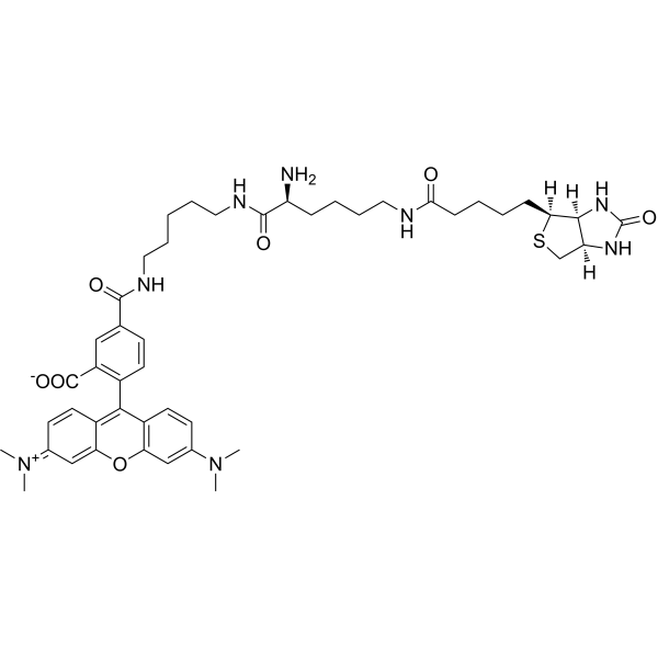
TMR Biocytin is a polar tracer used in the research of cell-cell and cell-liposome fusions, as well as membrane permeability and cellular uptake during pinocytosis. TMR Biocytin can be detected using streptavidin, and is an effective neuronal tracer in live tissue (Ex=544 nm, Em=571 nm) .
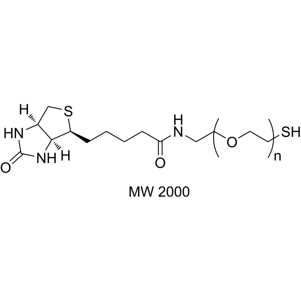
Biotin-PEG-Thiol (MW 2000) is an active compound. Biotin-PEG-Thiol (MW 2000) is pegylated by binding to streptavidin or antibiotin with high affinity and specificity. Biotin-PEG-Thiol (MW 2000) can modify biomolecules, proteins, peptides and other small molecule materials. Biotin-PEG-Thiol (MW 2000) is widely used in the research of agent release and nano new materials .
Fluorescein Biotin is used as an alternative to radioactive biotin for detecting and quantitating biotin-binding sites by either fluorescence or absorbance; the the fluorescence or absorbance of Fluorescein Biotin is quenched, upon binding to avidin or streptavidin.
TFAX 488,TFP is a green fluorescent dye and exhibits pH-insensitivity over a very broad range (pH in the 4-10). TFAX 488,TFP yields exceptionally bright, photostable conjugates with proteins or antibodies (such as goat anti-mouse IgG, streptavidin) .
TFAX 488,SE dilithium is a green fluorescent dye and exhibits pH-insensitivity over a very broad range (pH in the 4-10). TFAX 488,SE dilithium yields exceptionally bright, photostable conjugates with proteins or antibodies (such as goat anti-mouse IgG, streptavidin) .
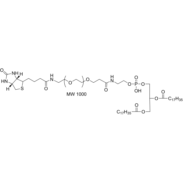
Phospholipid-PEG-Biotin (MW 1000) is a phospholipid PEG derivative that has a biotin and a phospholipid bridged by a linear PEG linker. Phospholipid-PEG-Biotin (MW 3400) can interact with avidinylated antibodies. Phospholipid-PEG-Biotin (MW 3400) can be used to modify liposome and cells surface, and pancreatic islets for cell transplantation .
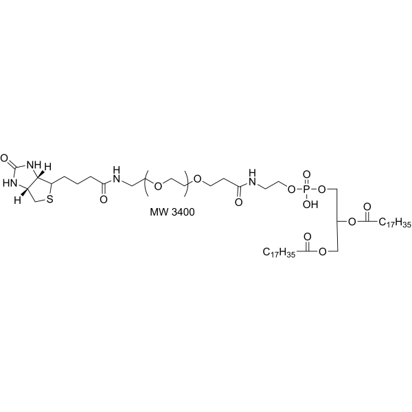
Phospholipid-PEG-Biotin (MW 3400) is a phospholipid PEG derivative that has a biotin and a phospholipid bridged by a linear PEG linker. Phospholipid-PEG-Biotin (MW 3400) can interact with avidinylated antibodies. Phospholipid-PEG-Biotin (MW 3400) can be used to modify liposome and cells surface, and pancreatic islets for cell transplantation .
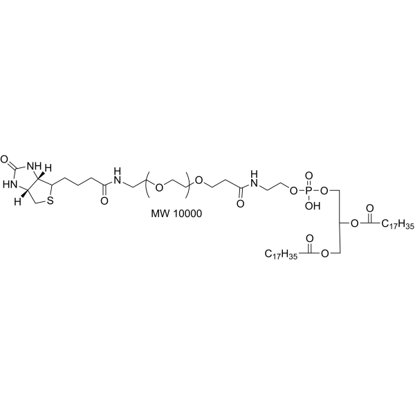
Phospholipid-PEG-Biotin (MW 10000) is a phospholipid PEG derivative that has a biotin and a phospholipid bridged by a linear PEG linker. Phospholipid-PEG-Biotin (MW 3400) can interact with avidinylated antibodies. Phospholipid-PEG-Biotin (MW 3400) can be used to modify liposome and cells surface, and pancreatic islets for cell transplantation .
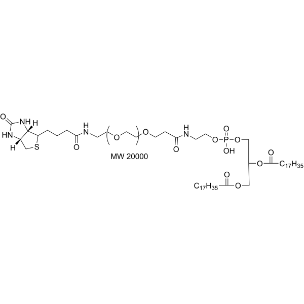
Phospholipid-PEG-Biotin (MW 20000) is a phospholipid PEG derivative that has a biotin and a phospholipid bridged by a linear PEG linker. Phospholipid-PEG-Biotin (MW 3400) can interact with avidinylated antibodies. Phospholipid-PEG-Biotin (MW 3400) can be used to modify liposome and cells surface, and pancreatic islets for cell transplantation .
SAPE (1-Stearoyl-2-arachidonoyl-sn-glycero-3-phosphorylethanolamine) is an R-Phycoerythrin (HY-D0988) labeled Streptavidin (HY-P3152) fluorescent probe. SAPE can be used for tumor detection when combined with biotin. SAPE has high sensitivity and a wide detection range .
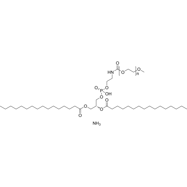
16:0 PEG350 PE is a PEG lipid functional end group used in the synthesis of liposomes (LPs) for the design of conjugated polymer nanoparticles. Through biotin modification and carboxyl terminus, lipid nanoparticles (LNPs) further coupling with other biomolecules can be achieved. Functionalized nanoparticles can be used for targeted labeling of specific cellular proteins. With streptavidin as a linker, biotinylated PEG lipid-conjugated polymer nanoparticles are able to bind to biotinylated antibodies on cell surface receptors, yielding the utility of fluorescence-based imaging and sensing.
16:0 PEG550 PE is a PEG lipid functional end group used in the synthesis of liposomes (LPs) for the design of conjugated polymer nanoparticles. Through biotin modification and carboxyl terminus, lipid nanoparticles (LNPs) further coupling with other biomolecules can be achieved. Functionalized nanoparticles can be used for targeted labeling of specific cellular proteins. With streptavidin as a linker, biotinylated PEG lipid-conjugated polymer nanoparticles are able to bind to biotinylated antibodies on cell surface receptors, yielding the utility of fluorescence-based imaging and sensing.
16:0 PEG750 PE is a PEG lipid functional end group used in the synthesis of liposomes (LPs) for the design of conjugated polymer nanoparticles. Through biotin modification and carboxyl terminus, lipid nanoparticles (LNPs) further coupling with other biomolecules can be achieved. Functionalized nanoparticles can be used for targeted labeling of specific cellular proteins. With streptavidin as a linker, biotinylated PEG lipid-conjugated polymer nanoparticles are able to bind to biotinylated antibodies on cell surface receptors, yielding the utility of fluorescence-based imaging and sensing.
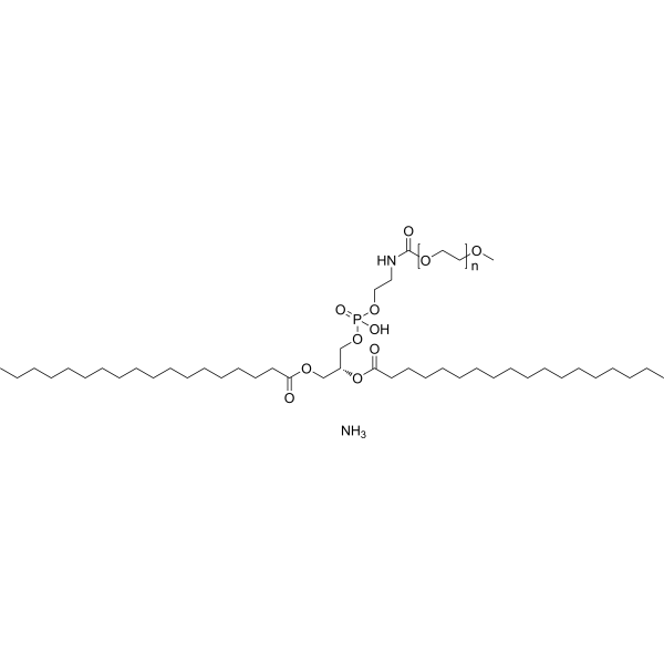
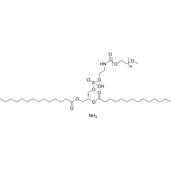
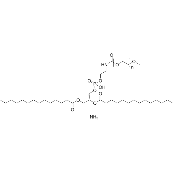
18:0 mPEG350 PE (ammonium) is a PEG lipid functional end group used in the synthesis of liposomes (LPs) for the design of conjugated polymer nanoparticles. Through biotin modification and carboxyl terminus, lipid nanoparticles (LNPs) further coupling with other biomolecules can be achieved. Functionalized nanoparticles can be used for targeted labeling of specific cellular proteins. With streptavidin as a linker, biotinylated PEG lipid-conjugated polymer nanoparticles are able to bind to biotinylated antibodies on cell surface receptors, yielding the utility of fluorescence-based imaging and sensing.
18:0 mPEG550 PE (ammonium) is a PEG lipid functional end group used in the synthesis of liposomes (LPs) for the design of conjugated polymeric nanoparticles. Through biotin modification and carboxyl terminus, lipid nanoparticles (LNPs) further coupling with other biomolecules can be achieved. Functionalized nanoparticles can be used for targeted labeling of specific cellular proteins. With streptavidin as a linker, biotinylated PEG lipid-conjugated polymer nanoparticles are able to bind to biotinylated antibodies on cell surface receptors, yielding the utility of fluorescence-based imaging and sensing.
18:0 mPEG750 PE (ammonium) is a PEG lipid functional end group used in the synthesis of liposomes (LPs) for the design of conjugated polymeric nanoparticles. Through biotin modification and carboxyl terminus, lipid nanoparticles (LNPs) further coupling with other biomolecules can be achieved. Functionalized nanoparticles can be used for targeted labeling of specific cellular proteins. With streptavidin as a linker, biotinylated PEG lipid-conjugated polymer nanoparticles are able to bind to biotinylated antibodies on cell surface receptors, yielding the utility of fluorescence-based imaging and sensing.
16:0 PEG1000 PE is a PEG lipid functional end group used in the synthesis of liposomes (LPs) for the design of conjugated polymer nanoparticles. Through biotin modification and carboxyl terminus, lipid nanoparticles (LNPs) further coupling with other biomolecules can be achieved. Functionalized nanoparticles can be used for targeted labeling of specific cellular proteins. With streptavidin as a linker, biotinylated PEG lipid-conjugated polymer nanoparticles are able to bind to biotinylated antibodies on cell surface receptors, yielding the utility of fluorescence-based imaging and sensing.
16:0 PEG3000 PE is a PEG lipid functional end group used in the synthesis of liposomes (LPs) for the design of conjugated polymer nanoparticles. Through biotin modification and carboxyl terminus, lipid nanoparticles (LNPs) further coupling with other biomolecules can be achieved. Functionalized nanoparticles can be used for targeted labeling of specific cellular proteins. With streptavidin as a linker, biotinylated PEG lipid-conjugated polymer nanoparticles are able to bind to biotinylated antibodies on cell surface receptors, yielding the utility of fluorescence-based imaging and sensing.
16:0 PEG5000 PE is a PEG lipid functional end group used in the synthesis of liposomes (LPs) for the design of conjugated polymer nanoparticles. Through biotin modification and carboxyl terminus, lipid nanoparticles (LNPs) further coupling with other biomolecules can be achieved. Functionalized nanoparticles can be used for targeted labeling of specific cellular proteins. With streptavidin as a linker, biotinylated PEG lipid-conjugated polymer nanoparticles are able to bind to biotinylated antibodies on cell surface receptors, yielding the utility of fluorescence-based imaging and sensing.
18:0 mPEG1000 PE (ammonium) is a PEG lipid functional end group used in the synthesis of liposomes (LPs) for the design of conjugated polymeric nanoparticles. Through biotin modification and carboxyl terminus, lipid nanoparticles (LNPs) further coupling with other biomolecules can be achieved. Functionalized nanoparticles can be used for targeted labeling of specific cellular proteins. With streptavidin as a linker, biotinylated PEG lipid-conjugated polymer nanoparticles are able to bind to biotinylated antibodies on cell surface receptors, yielding the utility of fluorescence-based imaging and sensing.
18:0 mPEG3000 PE (ammonium) is a PEG lipid functional end group used in the synthesis of liposomes (LPs) for the design of conjugated polymeric nanoparticles. Through biotin modification and carboxyl terminus, lipid nanoparticles (LNPs) further coupling with other biomolecules can be achieved. Functionalized nanoparticles can be used for targeted labeling of specific cellular proteins. With streptavidin as a linker, biotinylated PEG lipid-conjugated polymer nanoparticles are able to bind to biotinylated antibodies on cell surface receptors, yielding the utility of fluorescence-based imaging and sensing.
18:0 mPEG5000 PE (ammonium) is a PEG lipid functional end group used in the synthesis of liposomes (LPs) for the design of conjugated polymeric nanoparticles. Through biotin modification and carboxyl terminus, lipid nanoparticles (LNPs) further coupling with other biomolecules can be achieved. Functionalized nanoparticles can be used for targeted labeling of specific cellular proteins. With streptavidin as a linker, biotinylated PEG lipid-conjugated polymer nanoparticles are able to bind to biotinylated antibodies on cell surface receptors, yielding the utility of fluorescence-based imaging and sensing.
14:0 PEG350 PE is a PEG lipid functional end group used in the synthesis of liposomes (LPs) for the design of conjugated polymer nanoparticles. Through biotin modification and carboxyl terminus, lipid nanoparticles (LNPs) further coupling with other biomolecules can be achieved. Functionalized nanoparticles can be used for targeted labeling of specific cellular proteins. With streptavidin as a linker, biotinylated PEG lipid-conjugated polymer nanoparticles are able to bind to biotinylated antibodies on cell surface receptors, yielding the utility of fluorescence-based imaging and sensing.
14:0 PEG550 PE is a PEG lipid functional end group used in the synthesis of liposomes (LPs) for the design of conjugated polymeric nanoparticles. Through biotin modification and carboxyl terminus, lipid nanoparticles (LNPs) further coupling with other biomolecules can be achieved. Functionalized nanoparticles can be used for targeted labeling of specific cellular proteins. With streptavidin as a linker, biotinylated PEG lipid-conjugated polymer nanoparticles are able to bind to biotinylated antibodies on cell surface receptors, yielding the utility of fluorescence-based imaging and sensing.
14:0 PEG750 PE is a PEG lipid functional end group used in the synthesis of liposomes (LPs) for the design of conjugated polymeric nanoparticles. Through biotin modification and carboxyl terminus, lipid nanoparticles (LNPs) further coupling with other biomolecules can be achieved. Functionalized nanoparticles can be used for targeted labeling of specific cellular proteins. With streptavidin as a linker, biotinylated PEG lipid-conjugated polymer nanoparticles are able to bind to biotinylated antibodies on cell surface receptors, yielding the utility of fluorescence-based imaging and sensing.
14:0 PEG1000 PE is a PEG lipid functional end group used in the synthesis of liposomes (LPs) for the design of conjugated polymer nanoparticles. Through biotin modification and carboxyl terminus, lipid nanoparticles (LNPs) further coupling with other biomolecules can be achieved. Functionalized nanoparticles can be used for targeted labeling of specific cellular proteins. With streptavidin as a linker, biotinylated PEG lipid-conjugated polymer nanoparticles are able to bind to biotinylated antibodies on cell surface receptors, yielding the utility of fluorescence-based imaging and sensing.
14:0 PEG3000 PE is a PEG lipid functional end group used in the synthesis of liposomes (LPs) for the design of conjugated polymer nanoparticles. Through biotin modification and carboxyl terminus, lipid nanoparticles (LNPs) further coupling with other biomolecules can be achieved. Functionalized nanoparticles can be used for targeted labeling of specific cellular proteins. With streptavidin as a linker, biotinylated PEG lipid-conjugated polymer nanoparticles are able to bind to biotinylated antibodies on cell surface receptors, yielding the utility of fluorescence-based imaging and sensing.
14:0 PEG5000 PE is a PEG lipid functional end group used in the synthesis of liposomes (LPs) for the design of conjugated polymer nanoparticles. Through biotin modification and carboxyl terminus, lipid nanoparticles (LNPs) further coupling with other biomolecules can be achieved. Functionalized nanoparticles can be used for targeted labeling of specific cellular proteins. With streptavidin as a linker, biotinylated PEG lipid-conjugated polymer nanoparticles are able to bind to biotinylated antibodies on cell surface receptors, yielding the utility of fluorescence-based imaging and sensing.
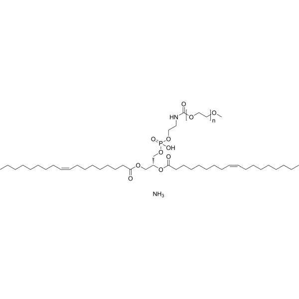
18:1 PEG350 PE is a PEG lipid functional end group used in the synthesis of liposomes (LPs) for the design of conjugated polymer nanoparticles. Through biotin modification and carboxyl terminus, lipid nanoparticles (LNPs) further coupling with other biomolecules can be achieved. Functionalized nanoparticles can be used for targeted labeling of specific cellular proteins. With streptavidin as a linker, biotinylated PEG lipid-conjugated polymer nanoparticles are able to bind to biotinylated antibodies on cell surface receptors, yielding the utility of fluorescence-based imaging and sensing.
18:1 PEG550 PE is a PEG lipid functional end group used in the synthesis of liposomes (LPs) for the design of conjugated polymer nanoparticles. Through biotin modification and carboxyl terminus, lipid nanoparticles (LNPs) further coupling with other biomolecules can be achieved. Functionalized nanoparticles can be used for targeted labeling of specific cellular proteins. With streptavidin as a linker, biotinylated PEG lipid-conjugated polymer nanoparticles are able to bind to biotinylated antibodies on cell surface receptors, yielding the utility of fluorescence-based imaging and sensing.
18:1 PEG1000 PE is a PEG lipid functional end group used in the synthesis of liposomes (LPs) for the design of conjugated polymer nanoparticles. Through biotin modification and carboxyl terminus, lipid nanoparticles (LNPs) further coupling with other biomolecules can be achieved. Functionalized nanoparticles can be used for targeted labeling of specific cellular proteins. With streptavidin as a linker, biotinylated PEG lipid-conjugated polymer nanoparticles are able to bind to biotinylated antibodies on cell surface receptors, yielding the utility of fluorescence-based imaging and sensing.
18:1 PEG3000 PE is a PEG lipid functional end group used in the synthesis of liposomes (LPs) for the design of conjugated polymer nanoparticles. Through biotin modification and carboxyl terminus, lipid nanoparticles (LNPs) further coupling with other biomolecules can be achieved. Functionalized nanoparticles can be used for targeted labeling of specific cellular proteins. With streptavidin as a linker, biotinylated PEG lipid-conjugated polymer nanoparticles are able to bind to biotinylated antibodies on cell surface receptors, yielding the utility of fluorescence-based imaging and sensing.
18:1 PEG5000 PE is a PEG lipid functional end group used in the synthesis of liposomes (LPs) for the design of conjugated polymer nanoparticles. Through biotin modification and carboxyl terminus, lipid nanoparticles (LNPs) further coupling with other biomolecules can be achieved. Functionalized nanoparticles can be used for targeted labeling of specific cellular proteins. With streptavidin as a linker, biotinylated PEG lipid-conjugated polymer nanoparticles are able to bind to biotinylated antibodies on cell surface receptors, yielding the utility of fluorescence-based imaging and sensing.
Vari Fluor 680-Streptavidin is a dye marker of Vari Fluor-streptavidin consisting of labeling streptavidin with a Vari Fluor series of fluorescent probes. Streptavidin is a high-affinity tetramer protein, each tetramer consisting of four identical streptavidin subunits. Streptavidin binds to biotin specifically via a reversible non-covalent effect. Streptavidin can achieve rapid and efficient detection of biotin markers, and is often used in immunofluorescence (IF), enzyme-linked immunosorbent assay (ELISA), immunohistochemical staining (IFH), in situ hybridization (ISH) and other experiments. Ex/Em=680 nm/701 nm.
Vari Fluor 647-Streptavidin is a dye marker of Vari Fluor-streptavidin consisting of labeling streptavidin with a Vari Fluor series of fluorescent probes. Streptavidin is a high-affinity tetramer protein, each tetramer consisting of four identical streptavidin subunits. Streptavidin binds to biotin specifically via a reversible non-covalent effect. Streptavidin can achieve rapid and efficient detection of biotin markers, and is often used in immunofluorescence (IF), enzyme-linked immunosorbent assay (ELISA), immunohistochemical staining (IFH), in situ hybridization (ISH) and other experiments. Ex/Em=650 nm/665 nm.
Vari Fluor 594-Streptavidin is a dye marker of Vari Fluor-streptavidin consisting of labeling streptavidin with a Vari Fluor series of fluorescent probes. Streptavidin is a high-affinity tetramer protein, each tetramer consisting of four identical streptavidin subunits. Streptavidin binds to biotin specifically via a reversible non-covalent effect. Streptavidin can achieve rapid and efficient detection of biotin markers, and is often used in immunofluorescence (IF), enzyme-linked immunosorbent assay (ELISA), immunohistochemical staining (IFH), in situ hybridization (ISH) and other experiments. Ex/Em=590 nm/617 nm.
Vari Fluor 555-Streptavidin is a dye marker of Vari Fluor-streptavidin consisting of labeling streptavidin with a Vari Fluor series of fluorescent probes. Streptavidin is a high-affinity tetramer protein, each tetramer consisting of four identical streptavidin subunits. Streptavidin binds to biotin specifically via a reversible non-covalent effect. Streptavidin can achieve rapid and efficient detection of biotin markers, and is often used in immunofluorescence (IF), enzyme-linked immunosorbent assay (ELISA), immunohistochemical staining (IFH), in situ hybridization (ISH) and other experiments. Ex/Em=555 nm/565 nm.
Vari Fluor 488-Streptavidin is a dye marker of Vari Fluor-streptavidin consisting of labeling streptavidin with a Vari Fluor series of fluorescent probes. Streptavidin is a high-affinity tetramer protein, each tetramer consisting of four identical streptavidin subunits. Streptavidin binds to biotin specifically via a reversible non-covalent effect. Streptavidin can achieve rapid and efficient detection of biotin markers, and is often used in immunofluorescence (IF), enzyme-linked immunosorbent assay (ELISA), immunohistochemical staining (IFH), in situ hybridization (ISH) and other experiments. Ex/Em=490 nm/515 nm.
Vari Fluor 405-Streptavidin is a dye marker of Vari Fluor-streptavidin consisting of labeling streptavidin with a Vari Fluor series of fluorescent probes. Streptavidin is a high-affinity tetramer protein, each tetramer consisting of four identical streptavidin subunits. Streptavidin binds to biotin specifically via a reversible non-covalent effect. Streptavidin can achieve rapid and efficient detection of biotin markers, and is often used in immunofluorescence (IF), enzyme-linked immunosorbent assay (ELISA), immunohistochemical staining (IFH), in situ hybridization (ISH) and other experiments. Ex/Em=405 nm/431 nm.
AF488 streptavidin is a fluorescence labeled streptavidin. AF488 streptavidin comprises a biotin-binding protein (streptavidin) covalently attached to a fluorescent label (AF488). AF488 is a bright, photostable green fluorophore .
ATTO 590 Streptavidin is a streptavidin derivative of ATTO 590, it can label protein or antibody, the maximum excitation/emission wavelength: 594/622 nm.
ATTO 700 Streptavidin is a streptavidin derivative of ATTO 700, it can label protein or antibody, the maximum excitation/emission wavelength: 700/716 nm.
ATTO 594 Streptavidin is a streptavidin derivative of ATTO 594, it can label protein or antibody, the maximum excitation/emission wavelength: 603/626 nm.
ATTO 633 Streptavidin is a streptavidin derivative of ATTO 633, it can label protein or antibody, the maximum excitation/emission wavelength: 630/651 nm.
ATTO 565 Streptavidin is a streptavidin derivative of ATTO 565, it can label protein or antibody, the maximum excitation/emission wavelength: 564/590 nm.
ATTO 610 Streptavidin is a streptavidin derivative of ATTO 610, it can label protein or antibody, the maximum excitation/emission wavelength: 616/633 nm.
ATTO 725 Streptavidin is a streptavidin derivative of ATTO 725, it can label protein or antibody, the maximum excitation/emission wavelength: 728/751 nm.
ATTO 620 Streptavidin is a streptavidin derivative of ATTO 620, it can label protein or antibody, the maximum excitation/emission wavelength: 620/642 nm.
ATTO 647 Streptavidin is a streptavidin derivative of ATTO 647, it can label protein or antibody, the maximum excitation/emission wavelength: 630/651 nm.
ATTO 740 streptavidin is a streptavidin derivative of ATTO 740, it can label protein or antibody, the maximum excitation/emission wavelength: 743/763 nm.
ATTO 488 streptavidin is a streptavidin derivative of ATTO 488, it can label protein or antibody, the maximum excitation/emission wavelength: 500/520 nm.
ATTO 532 streptavidin is a streptavidin derivative of ATTO 532, it can label protein or antibody, the maximum excitation/emission wavelength: 532/552 nm.
ATTO 550 streptavidin is a streptavidin derivative of ATTO 550, it can label protein or antibody, the maximum excitation/emission wavelength: 554/576 nm.
ATTO 514 streptavidin is a streptavidin derivative of ATTO 514, it can label protein or antibody, the maximum excitation/emission wavelength: 511/531 nm.
ATTO 665 streptavidin is a streptavidin derivative of ATTO 665, it can label protein or antibody, the maximum excitation/emission wavelength: 663/680 nm.
ATTO 680 streptavidin is a streptavidin derivative of ATTO 680, it can label protein or antibody, the maximum excitation/emission wavelength: 681/698 nm.
AF 594 streptavidin is a bioconjugating agent. It consists of AF 594 and streptomycin, a streptomycin derivative of the red fluorescent dye AF 594. AF 594 has high fluorescence quantum yield and high photostability (maximum absorption wavelength 586 nm, maximum emission wavelength 613 nm). AF 594 streptavidin can be selectively conjugated to streptavidin-modified molecules via a streptomycin-modifying group for fluorescent labeling and spectroscopic analysis.
TMR Biocytin is a polar tracer used in the research of cell-cell and cell-liposome fusions, as well as membrane permeability and cellular uptake during pinocytosis. TMR Biocytin can be detected using streptavidin, and is an effective neuronal tracer in live tissue (Ex=544 nm, Em=571 nm) .
Fluorescein Biotin is used as an alternative to radioactive biotin for detecting and quantitating biotin-binding sites by either fluorescence or absorbance; the the fluorescence or absorbance of Fluorescein Biotin is quenched, upon binding to avidin or streptavidin.
TFAX 488,TFP is a green fluorescent dye and exhibits pH-insensitivity over a very broad range (pH in the 4-10). TFAX 488,TFP yields exceptionally bright, photostable conjugates with proteins or antibodies (such as goat anti-mouse IgG, streptavidin) .
TFAX 488,SE dilithium is a green fluorescent dye and exhibits pH-insensitivity over a very broad range (pH in the 4-10). TFAX 488,SE dilithium yields exceptionally bright, photostable conjugates with proteins or antibodies (such as goat anti-mouse IgG, streptavidin) .
Streptavidin is a ∼60 kDa homotetramer. Streptavidin binds four molecules of biotin with the highest affinity. The binding affinity of biotin to streptavidin is one of the highest reported for a non-covalent interaction to date, with a KD ∼ 0.01 pM . Streptavidin has an immunosuppressive role .
Phospholipid-PEG-Biotin (MW 1000) is a phospholipid PEG derivative that has a biotin and a phospholipid bridged by a linear PEG linker. Phospholipid-PEG-Biotin (MW 3400) can interact with avidinylated antibodies. Phospholipid-PEG-Biotin (MW 3400) can be used to modify liposome and cells surface, and pancreatic islets for cell transplantation .
Phospholipid-PEG-Biotin (MW 3400) is a phospholipid PEG derivative that has a biotin and a phospholipid bridged by a linear PEG linker. Phospholipid-PEG-Biotin (MW 3400) can interact with avidinylated antibodies. Phospholipid-PEG-Biotin (MW 3400) can be used to modify liposome and cells surface, and pancreatic islets for cell transplantation .
Phospholipid-PEG-Biotin (MW 10000) is a phospholipid PEG derivative that has a biotin and a phospholipid bridged by a linear PEG linker. Phospholipid-PEG-Biotin (MW 3400) can interact with avidinylated antibodies. Phospholipid-PEG-Biotin (MW 3400) can be used to modify liposome and cells surface, and pancreatic islets for cell transplantation .
Phospholipid-PEG-Biotin (MW 20000) is a phospholipid PEG derivative that has a biotin and a phospholipid bridged by a linear PEG linker. Phospholipid-PEG-Biotin (MW 3400) can interact with avidinylated antibodies. Phospholipid-PEG-Biotin (MW 3400) can be used to modify liposome and cells surface, and pancreatic islets for cell transplantation .
16:0 PEG350 PE is a PEG lipid functional end group used in the synthesis of liposomes (LPs) for the design of conjugated polymer nanoparticles. Through biotin modification and carboxyl terminus, lipid nanoparticles (LNPs) further coupling with other biomolecules can be achieved. Functionalized nanoparticles can be used for targeted labeling of specific cellular proteins. With streptavidin as a linker, biotinylated PEG lipid-conjugated polymer nanoparticles are able to bind to biotinylated antibodies on cell surface receptors, yielding the utility of fluorescence-based imaging and sensing.
16:0 PEG550 PE is a PEG lipid functional end group used in the synthesis of liposomes (LPs) for the design of conjugated polymer nanoparticles. Through biotin modification and carboxyl terminus, lipid nanoparticles (LNPs) further coupling with other biomolecules can be achieved. Functionalized nanoparticles can be used for targeted labeling of specific cellular proteins. With streptavidin as a linker, biotinylated PEG lipid-conjugated polymer nanoparticles are able to bind to biotinylated antibodies on cell surface receptors, yielding the utility of fluorescence-based imaging and sensing.
16:0 PEG750 PE is a PEG lipid functional end group used in the synthesis of liposomes (LPs) for the design of conjugated polymer nanoparticles. Through biotin modification and carboxyl terminus, lipid nanoparticles (LNPs) further coupling with other biomolecules can be achieved. Functionalized nanoparticles can be used for targeted labeling of specific cellular proteins. With streptavidin as a linker, biotinylated PEG lipid-conjugated polymer nanoparticles are able to bind to biotinylated antibodies on cell surface receptors, yielding the utility of fluorescence-based imaging and sensing.
18:0 mPEG350 PE (ammonium) is a PEG lipid functional end group used in the synthesis of liposomes (LPs) for the design of conjugated polymer nanoparticles. Through biotin modification and carboxyl terminus, lipid nanoparticles (LNPs) further coupling with other biomolecules can be achieved. Functionalized nanoparticles can be used for targeted labeling of specific cellular proteins. With streptavidin as a linker, biotinylated PEG lipid-conjugated polymer nanoparticles are able to bind to biotinylated antibodies on cell surface receptors, yielding the utility of fluorescence-based imaging and sensing.
18:0 mPEG550 PE (ammonium) is a PEG lipid functional end group used in the synthesis of liposomes (LPs) for the design of conjugated polymeric nanoparticles. Through biotin modification and carboxyl terminus, lipid nanoparticles (LNPs) further coupling with other biomolecules can be achieved. Functionalized nanoparticles can be used for targeted labeling of specific cellular proteins. With streptavidin as a linker, biotinylated PEG lipid-conjugated polymer nanoparticles are able to bind to biotinylated antibodies on cell surface receptors, yielding the utility of fluorescence-based imaging and sensing.
18:0 mPEG750 PE (ammonium) is a PEG lipid functional end group used in the synthesis of liposomes (LPs) for the design of conjugated polymeric nanoparticles. Through biotin modification and carboxyl terminus, lipid nanoparticles (LNPs) further coupling with other biomolecules can be achieved. Functionalized nanoparticles can be used for targeted labeling of specific cellular proteins. With streptavidin as a linker, biotinylated PEG lipid-conjugated polymer nanoparticles are able to bind to biotinylated antibodies on cell surface receptors, yielding the utility of fluorescence-based imaging and sensing.
16:0 PEG1000 PE is a PEG lipid functional end group used in the synthesis of liposomes (LPs) for the design of conjugated polymer nanoparticles. Through biotin modification and carboxyl terminus, lipid nanoparticles (LNPs) further coupling with other biomolecules can be achieved. Functionalized nanoparticles can be used for targeted labeling of specific cellular proteins. With streptavidin as a linker, biotinylated PEG lipid-conjugated polymer nanoparticles are able to bind to biotinylated antibodies on cell surface receptors, yielding the utility of fluorescence-based imaging and sensing.
16:0 PEG3000 PE is a PEG lipid functional end group used in the synthesis of liposomes (LPs) for the design of conjugated polymer nanoparticles. Through biotin modification and carboxyl terminus, lipid nanoparticles (LNPs) further coupling with other biomolecules can be achieved. Functionalized nanoparticles can be used for targeted labeling of specific cellular proteins. With streptavidin as a linker, biotinylated PEG lipid-conjugated polymer nanoparticles are able to bind to biotinylated antibodies on cell surface receptors, yielding the utility of fluorescence-based imaging and sensing.
16:0 PEG5000 PE is a PEG lipid functional end group used in the synthesis of liposomes (LPs) for the design of conjugated polymer nanoparticles. Through biotin modification and carboxyl terminus, lipid nanoparticles (LNPs) further coupling with other biomolecules can be achieved. Functionalized nanoparticles can be used for targeted labeling of specific cellular proteins. With streptavidin as a linker, biotinylated PEG lipid-conjugated polymer nanoparticles are able to bind to biotinylated antibodies on cell surface receptors, yielding the utility of fluorescence-based imaging and sensing.
18:0 mPEG1000 PE (ammonium) is a PEG lipid functional end group used in the synthesis of liposomes (LPs) for the design of conjugated polymeric nanoparticles. Through biotin modification and carboxyl terminus, lipid nanoparticles (LNPs) further coupling with other biomolecules can be achieved. Functionalized nanoparticles can be used for targeted labeling of specific cellular proteins. With streptavidin as a linker, biotinylated PEG lipid-conjugated polymer nanoparticles are able to bind to biotinylated antibodies on cell surface receptors, yielding the utility of fluorescence-based imaging and sensing.
18:0 mPEG3000 PE (ammonium) is a PEG lipid functional end group used in the synthesis of liposomes (LPs) for the design of conjugated polymeric nanoparticles. Through biotin modification and carboxyl terminus, lipid nanoparticles (LNPs) further coupling with other biomolecules can be achieved. Functionalized nanoparticles can be used for targeted labeling of specific cellular proteins. With streptavidin as a linker, biotinylated PEG lipid-conjugated polymer nanoparticles are able to bind to biotinylated antibodies on cell surface receptors, yielding the utility of fluorescence-based imaging and sensing.
18:0 mPEG5000 PE (ammonium) is a PEG lipid functional end group used in the synthesis of liposomes (LPs) for the design of conjugated polymeric nanoparticles. Through biotin modification and carboxyl terminus, lipid nanoparticles (LNPs) further coupling with other biomolecules can be achieved. Functionalized nanoparticles can be used for targeted labeling of specific cellular proteins. With streptavidin as a linker, biotinylated PEG lipid-conjugated polymer nanoparticles are able to bind to biotinylated antibodies on cell surface receptors, yielding the utility of fluorescence-based imaging and sensing.
14:0 PEG350 PE is a PEG lipid functional end group used in the synthesis of liposomes (LPs) for the design of conjugated polymer nanoparticles. Through biotin modification and carboxyl terminus, lipid nanoparticles (LNPs) further coupling with other biomolecules can be achieved. Functionalized nanoparticles can be used for targeted labeling of specific cellular proteins. With streptavidin as a linker, biotinylated PEG lipid-conjugated polymer nanoparticles are able to bind to biotinylated antibodies on cell surface receptors, yielding the utility of fluorescence-based imaging and sensing.
14:0 PEG550 PE is a PEG lipid functional end group used in the synthesis of liposomes (LPs) for the design of conjugated polymeric nanoparticles. Through biotin modification and carboxyl terminus, lipid nanoparticles (LNPs) further coupling with other biomolecules can be achieved. Functionalized nanoparticles can be used for targeted labeling of specific cellular proteins. With streptavidin as a linker, biotinylated PEG lipid-conjugated polymer nanoparticles are able to bind to biotinylated antibodies on cell surface receptors, yielding the utility of fluorescence-based imaging and sensing.
14:0 PEG750 PE is a PEG lipid functional end group used in the synthesis of liposomes (LPs) for the design of conjugated polymeric nanoparticles. Through biotin modification and carboxyl terminus, lipid nanoparticles (LNPs) further coupling with other biomolecules can be achieved. Functionalized nanoparticles can be used for targeted labeling of specific cellular proteins. With streptavidin as a linker, biotinylated PEG lipid-conjugated polymer nanoparticles are able to bind to biotinylated antibodies on cell surface receptors, yielding the utility of fluorescence-based imaging and sensing.
14:0 PEG1000 PE is a PEG lipid functional end group used in the synthesis of liposomes (LPs) for the design of conjugated polymer nanoparticles. Through biotin modification and carboxyl terminus, lipid nanoparticles (LNPs) further coupling with other biomolecules can be achieved. Functionalized nanoparticles can be used for targeted labeling of specific cellular proteins. With streptavidin as a linker, biotinylated PEG lipid-conjugated polymer nanoparticles are able to bind to biotinylated antibodies on cell surface receptors, yielding the utility of fluorescence-based imaging and sensing.
14:0 PEG3000 PE is a PEG lipid functional end group used in the synthesis of liposomes (LPs) for the design of conjugated polymer nanoparticles. Through biotin modification and carboxyl terminus, lipid nanoparticles (LNPs) further coupling with other biomolecules can be achieved. Functionalized nanoparticles can be used for targeted labeling of specific cellular proteins. With streptavidin as a linker, biotinylated PEG lipid-conjugated polymer nanoparticles are able to bind to biotinylated antibodies on cell surface receptors, yielding the utility of fluorescence-based imaging and sensing.
14:0 PEG5000 PE is a PEG lipid functional end group used in the synthesis of liposomes (LPs) for the design of conjugated polymer nanoparticles. Through biotin modification and carboxyl terminus, lipid nanoparticles (LNPs) further coupling with other biomolecules can be achieved. Functionalized nanoparticles can be used for targeted labeling of specific cellular proteins. With streptavidin as a linker, biotinylated PEG lipid-conjugated polymer nanoparticles are able to bind to biotinylated antibodies on cell surface receptors, yielding the utility of fluorescence-based imaging and sensing.
18:1 PEG350 PE is a PEG lipid functional end group used in the synthesis of liposomes (LPs) for the design of conjugated polymer nanoparticles. Through biotin modification and carboxyl terminus, lipid nanoparticles (LNPs) further coupling with other biomolecules can be achieved. Functionalized nanoparticles can be used for targeted labeling of specific cellular proteins. With streptavidin as a linker, biotinylated PEG lipid-conjugated polymer nanoparticles are able to bind to biotinylated antibodies on cell surface receptors, yielding the utility of fluorescence-based imaging and sensing.
18:1 PEG550 PE is a PEG lipid functional end group used in the synthesis of liposomes (LPs) for the design of conjugated polymer nanoparticles. Through biotin modification and carboxyl terminus, lipid nanoparticles (LNPs) further coupling with other biomolecules can be achieved. Functionalized nanoparticles can be used for targeted labeling of specific cellular proteins. With streptavidin as a linker, biotinylated PEG lipid-conjugated polymer nanoparticles are able to bind to biotinylated antibodies on cell surface receptors, yielding the utility of fluorescence-based imaging and sensing.
18:1 PEG1000 PE is a PEG lipid functional end group used in the synthesis of liposomes (LPs) for the design of conjugated polymer nanoparticles. Through biotin modification and carboxyl terminus, lipid nanoparticles (LNPs) further coupling with other biomolecules can be achieved. Functionalized nanoparticles can be used for targeted labeling of specific cellular proteins. With streptavidin as a linker, biotinylated PEG lipid-conjugated polymer nanoparticles are able to bind to biotinylated antibodies on cell surface receptors, yielding the utility of fluorescence-based imaging and sensing.
18:1 PEG3000 PE is a PEG lipid functional end group used in the synthesis of liposomes (LPs) for the design of conjugated polymer nanoparticles. Through biotin modification and carboxyl terminus, lipid nanoparticles (LNPs) further coupling with other biomolecules can be achieved. Functionalized nanoparticles can be used for targeted labeling of specific cellular proteins. With streptavidin as a linker, biotinylated PEG lipid-conjugated polymer nanoparticles are able to bind to biotinylated antibodies on cell surface receptors, yielding the utility of fluorescence-based imaging and sensing.
18:1 PEG5000 PE is a PEG lipid functional end group used in the synthesis of liposomes (LPs) for the design of conjugated polymer nanoparticles. Through biotin modification and carboxyl terminus, lipid nanoparticles (LNPs) further coupling with other biomolecules can be achieved. Functionalized nanoparticles can be used for targeted labeling of specific cellular proteins. With streptavidin as a linker, biotinylated PEG lipid-conjugated polymer nanoparticles are able to bind to biotinylated antibodies on cell surface receptors, yielding the utility of fluorescence-based imaging and sensing.
MCE Streptavidin Agarose 6FF, a 6% highly cross-linked agarose reagent coupled with recombinant streptavidin, is an affinity chromatography medium for separation and purification of biotinylated peptides, antibodies, lectins, etc. The total binding capacity of Streptavidin Agarose 6FF is more than 200 nmol of D-Biotin/mL settled resin.
MCE Streptavidin Magnetic Beads provide a fast and convenient method for numerous applications, including purification of proteins and nucleic acids, protein interaction studies, immunoprecipitation, immunoassays, pull-down and cell isolation.
Streptomyces Avidinii The streptavidin protein, although its exact biological function is unknown, forms a strong non-covalent complex with biotin, binding one molecule to each subunit. Its specific affinity for biotin suggests its application in processes requiring specific binding to biotinylated molecules. Streptomyces Avidinii Streptavidin Protein is the recombinant Streptomyces Avidinii Streptavidin protein, expressed by E. coli , with tag free. The total length of Streptomyces Avidinii Streptavidin Protein is 128 a.a., with molecular weight of ~14.3 kDa.
Product Comparison
Compare
Clear All
Compare Products
Products
In-stock
-
+
Add to Cart
Cat. No.
Species
Source
Tag
Accession
Gene ID
Molecular Weight
Purity
Endotoxin Level
Biological Activity
Appearance
Formulation
Storage & Stability
Shipping
Free Sample
YesNo
Size
* This product has been "discontinued".
Optimized version of product available:
/
In-stock
-
+
Add to Cart
Get quote
Inquiry Online
Your information is safe with us. * Required Fields.