



DPPE-PEG350

Based on 1 Customer Validation
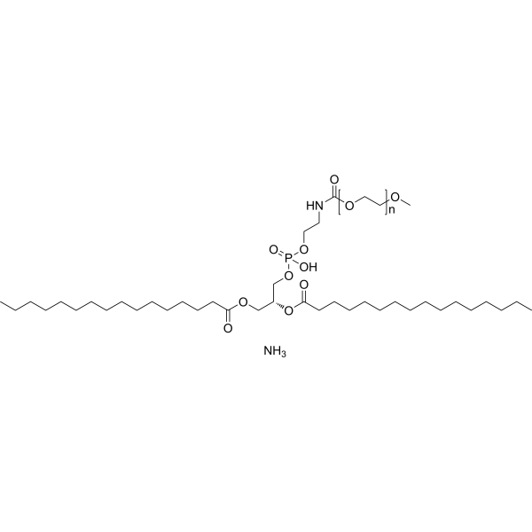
DPPE-PEG350 (16:0 PEG350 PE) is a CD1d lipid antagonist. DPPE-PEG350 blocks alpha-GalCer-dependent ERK tyrosine kinase phosphorylation in cells. DPPE-PEG350 inhibits cellular IL-4 and IFN-gamma production following alpha-GalCer stimulation. DPPE-PEG350 selectively antagonizes iNKT without affecting MHC class II-dependent OVA-specific Th2 responses. DPPE-PEG350 reduces MCP-1 and VCAM-1 protein expression and necrotic core formation in atherosclerotic lesions. DPPE-PEG350 can be used for research on asthma and atherosclerosis.
For research use only. We do not sell to patients.
- Purity : 99%
- CAS No.: 474922-84-4
- Formula: (C2H4O)nC39H76NO10P.H3N
- Molecular Weight:350(Average)
-
Storage:Powder -20°C, 3 years ; In solvent -80°C, 6 months , -20°C, 1 month

To place orders, for customer services and technical support, please contact: MedChemExpress USA
Tel: 609-228-6898 E-mail: [email protected] [email protected]


-
 Biological Activity
-
Chemical Information
- Protocols
- Purity & Documentation
- References
-
 Help & FAQs




Biological Activity
Description
IC50 & Target
[1]|
CD1d |
IL-4 |
In Vitro
DPPE-PEG350 (200 µg/mL; 4 h) inhibits α-GalCer-dependent IL-2 release from DN32.D3 cells, particularly at suboptimal α-GalCer concentrations[1].
DPPE-PEG350 (50 µg/mL) inhibits CD1d-dependent activation of iNKT by ILC2 in co-culture, while iNKT dampen IL-5 release from ILC2[4].
DPPE-PEG350 (0-100 µg/mL; 4 h) competes with α-GalCer for binding to CD1d on DN32 iNKT cell hybridomas in a dose-dependent manner[1].
DPPE-PEG350 is a chemical lipid antagonist that blocks NKT cell activation by competing with lipid antigens in CD1d binding[3].
DPPE-PEG350 (5 µg/mL) significantly inhibits the formation of CD1d-α-GalCer complexes, confirming its role as a CD1d antagonist[1].
DPPE-PEG350 (12.5-100 µg/mL; 1-60 min) completely blocks α-GalCer-induced ERK phosphorylation in iNKT cells at 100 µg/mL, thereby inhibiting TCR signaling[1].
MedChemExpress (MCE) has not independently confirmed the accuracy of these methods. They are for reference only.
-
Cell Line:DN32.D3 NKT hybridoma cells
-
Concentration:200 µg/mL
-
Incubation Time:4 h
-
Result:Inhibited IL-2 release from DN32 cells, particularly at suboptimal concentrations of α-GalCer.
In Vivo
DPPE-PEG350 (100 µg; intranasal administration; single dose) completely prevents α-GalCer-induced AHR and lung inflammation in naive mice[1].
DPPE-PEG350 (250 µg; intravenous injection; twice weekly) reduces atherosclerotic progression by 45% in mice without affecting plasma lipids or lymphocyte populations, primarily by reducing lesional T cells, B cells, MCP-1, VCAM-1, and necrotic core content[2].
DPPE-PEG350 (500 µg; intravenous injection; once weekly) completely eliminates αGalCer-aggravated atherosclerosis in mice, confirming the CD1d-dependent anti-atherosclerotic effect[2].
DPPE-PEG350 (250 µg; intravenous injection; twice weekly; 6 weeks) slows the progression of established atherosclerosis by 49%, reduces the necrotic core by 59%, and decreases lesional immune cell content in mice without affecting smooth muscle or collagen[2].
MedChemExpress (MCE) has not independently confirmed the accuracy of these methods. They are for reference only.
-
Animal Model:BALB/cByj (female, 6-8 weeks old, OVA-induced allergic airway hyperreactivity)[1]
-
Dosage:250 µg
-
Administration:i.v.; single dose
-
Result:Completely prevented the development of airway hyperreactivity upon intranasal OVA challenge.
Inhibited eosinophilic airway inflammation, significantly reducing the number of eosinophils, lymphocytes, and macrophages in the BAL fluid compared to the OVA-immunized control group.
Revealed no significant differences from saline controls in H&E- and PAS-stained lung sections, showing minimal mucus production and negligible cellular infiltration.
-
Animal Model:BALB/cByj (female, 6-8 weeks old, α-GalCer-induced airway hyperreactivity)[1]
-
Dosage:100 µg
-
Administration:i.n.; single dose
-
Result:Completely prevented the development of AHR in the group that received DPPE-PEG350 and α-GalCer when compared with α-GalCer alone.
Administration of DPPE-PEG350 alone showed no effect on the induction of AHR or pulmonary inflammation.
Revealed no significant differences between vehicle control and DPPE-PEG350-treated mice in H&E- and PAS-stained lung sections, showing minimal mucus production and minor cellular infiltration.
-
Animal Model:ApoE-/- mice on C57BL/6 background (male, 6-8 weeks old)[2]
-
Dosage:250 µg
-
Administration:i.v.; twice weekly
-
Result:Reduced total intimal atherosclerotic lesion size by 45% and ORO-stained lipid accumulation by 52% compared to PBS group.
Reduced CD4+ T cells by 54%, CD8+ T cells by 94%, and CD19+ B cell accumulation by 80% in atherosclerotic lesions.
Reduced MCP-1 and VCAM-1 protein expression by 30% and 57% respectively.
Reduced necrotic core areas by 64%.
Did not affect liver NKT cell populations (NK1.1+TCRβ+), CD4+ T cells, CD8+ T cells, B cells, or NK cells.
Did not affect plasma lipids despite ~10% reduction of body weight.
Did not affect smooth muscle and collagen content in atherosclerotic lesions.
Did not affect IFNγ mRNA expression in arterial and spleen tissues or plasma IFNγ levels.
-
Animal Model:ApoE-/- mice on C57BL/6 background (male, 6-8 weeks old)[2]
-
Dosage:250 µg
-
Administration:i.v.; twice weekly; 6 weeks
-
Result:Reduced established atherosclerosis by 49% when assessed by total intimal lesion areas.
Reduced lipid accumulation in aortic sinus lesions by 27%.
Reduced necrotic cores by 59%.
Reduced CD68+ macrophages, CD4+ T cells, CD8+ T cells, and CD19+ B cells in atherosclerotic lesions.
Decreased apoptotic cells in developed atherosclerotic lesions, with macrophages being a major target of apoptosis.
Did not affect the ratio of lipid accumulation to intima area.
Did not affect smooth muscle and collagen content in established atherosclerotic lesions.
Did not affect endothelial cell continuity.
-
Animal Model:ApoE-/- mice on C57BL/6 background (male, 6-8 weeks old)[2]
-
Dosage:500 µg (DPPE-PEG350); 2 µg (αGalCer)
-
Administration:i.v.; once weekly (DPPE-PEG350); i.p.; fortnightly (αGalCer)
-
Result:Completely abrogated αGalCer-aggravated atherosclerosis, which increased total intimal lesion by 112% without DPPE-PEG350 treatment.
Similar finding was observed with ORO-stained lipid accumulation; however lipid contents adjusted to lesion area were similar.
FACS analysis of liver lymphocytes showed that αGalCer-injection reduced CD1d-tetramer+ iNKT cells.


Chemical Information
-
CAS No. 474922-84-4
-
Appearance Solid
-
Molecular Weight 350(Average)
-
Formula (C2H4O)nC39H76NO10P.H3N
-
Color White to light yellow
-
SMILES
O=C(NCCOP(O)(OC[C@@H](COC(CCCCCCCCCCCCCCC)=O)OC(CCCCCCCCCCCCCCC)=O)=O)OCCOC.N.[n]
-
Synonyms
16:0 PEG350 PE; 1,2-Dipalmitoyl-sn-glycero-3-phosphoethanolamine-N-[methoxy(polyethylene glycol)-350] ammonium
-
Shipping
Room temperature in continental US; may vary elsewhere.
-
Storage
Powder -20°C 3 years In solvent -80°C 6 months -20°C 1 month


Purity & Documentation
-
Data Sheet (282 KB)
-
SDS (254 KB)
- English - EN (254 KB)
- Français - FR (254 KB)
- Deutsch - DE (254 KB)
- Norwegian - NO (254 KB)
- Español - ES (254 KB)
- Swedish - SV (254 KB)
- Italian - IT (254 KB)
- Korean - KR (254 KB)
- Portuguese - PT (254 KB)
-
Handling Instructions (2659 KB)
References
[1]. Lombardi V, et al. A CD1d-dependent antagonist inhibits the activation of invariant NKT cells and prevents development of allergen-induced airway hyperreactivity. Journal of immunology (Baltimore, Md. : 1950). 2010 Feb 15;184(4):2107-15. [Content Brief]
[2]. Li Y, et al. A CD1d-dependent lipid antagonist to NKT cells ameliorates atherosclerosis in ApoE-/- mice by reducing lesion necrosis and inflammation. Cardiovasc Res. 2016 Feb 1;109(2):305-17. . [Content Brief]

Calculators
Concentration (start) × Volume (start) = Concentration (final) × Volume (final)


DPPE-PEG350 Related Classifications
HY-144012A Related Classifications
 Powered by Bioz
Keywords
- DPPE-PEG350
- 474922-84-4
- 16:0 PEG350 PE
- 1,2-Dipalmitoyl-sn-glycero-3-phosphoethanolamine-N-[methoxy(polyethylene glycol)-350]
- CD1
- ERK
- Interleukin Related
- IFNAR
- asthma
- CD1d lipid antagonist
- ApoE-/- mice
- IFN-gamma
- alpha-GalCer
- iNKT cell activation
- IL-4
- atherosclerosis
- invariant TCR signaling
- ERK tyrosine kinase phosphorylation
- Inhibitor
- inhibitor
- inhibit