



5-Aminolevulinic acid
Based on 21 publication(s) in Google Scholar
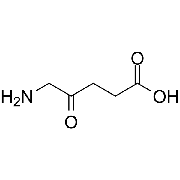
5-Aminolevulinic acid (5-ALA; δ-Aminolevulinic acid; 5-Amino-4-oxopentanoic acid) is an orally active heme precursor. 5-Aminolevulinic acid promotes aerobic energy metabolism and increases ATP levels by enhancing the activity of cytochrome c oxidase. 5-Aminolevulinic acid enhances LPS-induced proinflammatory cytokine production and gene activation, and restores the phagocytic activity and ROS generation capacity of neutrophils. 5-Aminolevulinic acid selectively accumulates protoporphyrin IX in tumor cells; as a photosensitizer and radiosensitizer, it induces ROS burst upon light or X-ray irradiation to inhibit tumor growth. 5-Aminolevulinic acid can be applied to the research of septic shock, melanoma, and cancer radiotherapy.
For research use only. We do not sell to patients.
- Purity : 99.92%
- CAS No.: 106-60-5
- Formula: C5H9NO3
- Molecular Weight:131.13
-
Storage:Powder -20°C, 3 years ; In solvent -80°C, 6 months , -20°C, 1 month

To place orders, for customer services and technical support, please contact: MedChemExpress USA
Tel: 609-228-6898 E-mail: [email protected] [email protected]


-
 Biological Activity
-
Chemical Information
-
Solvent & Solubility
- Protocols
- Purity & Documentation
- References
-
 Help & FAQs


Publications Citing Use of MedChemExpress (MCE) 5-Aminolevulinic acid
More- Nature. 2025 Jul;643(8070):192-200. [Abstract]
- Adv Sci (Weinh). 2019 Jan 20;6(5):1802057. [Abstract]
- Cell Death Dis. 2021 Oct 25;12(11):999. [Abstract]
- Pharmacol Res. 2021 Aug:170:105701. [Abstract]
- Genome Biol. 2022 Dec 15;23(1):259. [Abstract]
- EMBO Mol Med. 2023 Mar 8;15(3):e16959. [Abstract]
- Br J Pharmacol. 2026 Jun;183(11):2695-2719. [Abstract]
- PLoS Biol. 2024 Jun 27;22(6):e3002672. [Abstract]
- Mar Drugs. 2025 Mar 28;23(4):146. [Abstract]
- Int J Mol Sci. 2026 Jul 7;27(13):6091.
- Pestic Biochem Physiol. 2025 Dec 30;218:106934.
- Fish Shellfish Immunol. 2026 May:172:111209. [Abstract]
- J Inorg Biochem. 2023 Oct:247:112340. [Abstract]
- Asian J Androl. 2025 Jul 1;27(4):454-463. [Abstract]
- Photodiagnosis Photodyn Ther. 2025 Aug:54:104726. [Abstract]
- Photodiagnosis Photodyn Ther. 2025 Jun:53:104599. [Abstract]
- Photodiagnosis Photodyn Ther. 2024 Jun:47:104093. [Abstract]
- Photodiagnosis Photodyn Ther. 2023 Jun:42:103612. [Abstract]
- Photodiagnosis Photodyn Ther. 2022 Dec:40:103053. [Abstract]
- Lasers Med Sci. 2021 Dec;36(9):1873-1881. [Abstract]
- bioRxiv. 2024 Apr 3:2023.06.02.542933. [Abstract]
 Customer Validation & Images

-
Cell Imaging/Staining
-
Cell Proliferation/Viability Assay
-
Flow Cytometry
-
Cell Imaging/Staining
-
In Vivo Efficacy Study


Biological Activity
Description
IC50 & Target
|
Human Endogenous Metabolite |
Cellular Effect
|
Cell Line

|
Type | Value | Description | References |
|---|---|---|---|---|
| A-375 | IC50 |
>100 μM
Compound: ALA
|
Cytotoxicity against human A-375 cells assessed as reduction in cell viability preincubated for 4 hrs under dark condition followed by irradiation with blue light at 2.5 J.cm^-2 energy level for 5 mins and measured after 18 hrs by MTT assay
Cytotoxicity against human A-375 cells assessed as reduction in cell viability preincubated for 4 hrs under dark condition followed by irradiation with blue light at 2.5 J.cm^-2 energy level for 5 mins and measured after 18 hrs by MTT assay
|
[PMID: 38626642] |
| A-375 | IC50 |
27.24 μM
Compound: ALA
|
Cytotoxicity against human A-375 cells assessed as reduction in cell viability preincubated for 4 hrs under dark condition in presence of deferiprone followed by irradiation with blue light at 2.5 J.cm^-2 energy level for 5 mins and measured after 18 hrs
Cytotoxicity against human A-375 cells assessed as reduction in cell viability preincubated for 4 hrs under dark condition in presence of deferiprone followed by irradiation with blue light at 2.5 J.cm^-2 energy level for 5 mins and measured after 18 hrs
|
[PMID: 38626642] |
| HeLa | IC50 |
>100 μM
Compound: ALA
|
Cytotoxicity against human HeLa cells assessed as reduction in cell viability preincubated for 4 hrs under dark condition followed by irradiation with blue light at 2.5 J.cm^-2 energy level for 5 mins and measured after 18 hrs by MTT assay
Cytotoxicity against human HeLa cells assessed as reduction in cell viability preincubated for 4 hrs under dark condition followed by irradiation with blue light at 2.5 J.cm^-2 energy level for 5 mins and measured after 18 hrs by MTT assay
|
[PMID: 38626642] |
| HeLa | IC50 |
230 μM
Compound: 5-ALA
|
Photosensitization activity against human HeLa cells assessed as reduction in cell viability incubated for 4 hrs followed by LED light array irradiation followed by further incubation for 24 hrs by MTT assay
Photosensitization activity against human HeLa cells assessed as reduction in cell viability incubated for 4 hrs followed by LED light array irradiation followed by further incubation for 24 hrs by MTT assay
|
[PMID: 31398615] |
| HeLa | IC50 |
61.84 μM
Compound: ALA
|
Cytotoxicity against human HeLa cells assessed as reduction in cell viability preincubated for 4 hrs under dark condition in presence of deferiprone followed by irradiation with blue light at 2.5 J.cm^-2 energy level for 5 mins and measured after 18 hrs b
Cytotoxicity against human HeLa cells assessed as reduction in cell viability preincubated for 4 hrs under dark condition in presence of deferiprone followed by irradiation with blue light at 2.5 J.cm^-2 energy level for 5 mins and measured after 18 hrs b
|
[PMID: 38626642] |
| HL-60 | IC50 |
1570 μM
Compound: 5-ALA
|
Cytotoxicity against human HL60 cells after 72 hrs by MTT assay
Cytotoxicity against human HL60 cells after 72 hrs by MTT assay
|
[PMID: 19007111] |
| MCF7 | IC50 |
>100 μM
Compound: ALA
|
Cytotoxicity against human MCF7 cells assessed as reduction in cell viability preincubated for 4 hrs under dark condition followed by irradiation with blue light at 2.5 J.cm^-2 energy level for 5 mins and measured after 18 hrs by MTT assay
Cytotoxicity against human MCF7 cells assessed as reduction in cell viability preincubated for 4 hrs under dark condition followed by irradiation with blue light at 2.5 J.cm^-2 energy level for 5 mins and measured after 18 hrs by MTT assay
|
[PMID: 38626642] |
| MCF7 | IC50 |
1735 μM
Compound: 5-ALA
|
Cytotoxicity against human MCF7 cells after 72 hrs by Hoechst test
Cytotoxicity against human MCF7 cells after 72 hrs by Hoechst test
|
[PMID: 19007111] |
| MCF7 | IC50 |
64.95 μM
Compound: ALA
|
Cytotoxicity against human MCF7 cells assessed as reduction in cell viability preincubated for 4 hrs under dark condition in presence of deferiprone followed by irradiation with blue light at 2.5 J.cm^-2 energy level for 5 mins and measured after 18 hrs b
Cytotoxicity against human MCF7 cells assessed as reduction in cell viability preincubated for 4 hrs under dark condition in presence of deferiprone followed by irradiation with blue light at 2.5 J.cm^-2 energy level for 5 mins and measured after 18 hrs b
|
[PMID: 38626642] |
| Oocyte | IC50 |
12.9 μM
Compound: DALA
|
Antagonist activity at human GABAc Rho1 receptor expressed in Xenopus oocytes assessed as whole cell current production by two electrode voltage clamp method
Antagonist activity at human GABAc Rho1 receptor expressed in Xenopus oocytes assessed as whole cell current production by two electrode voltage clamp method
|
[PMID: 18528996] |
| U-251 | IC50 |
2665 μM
Compound: 5-ALA
|
Cytotoxicity against human U251 cells after 72 hrs by Hoechst test
Cytotoxicity against human U251 cells after 72 hrs by Hoechst test
|
[PMID: 19007111] |
| U-251 | IC50 |
2715 μM
Compound: 5-ALA
|
Antiproliferative activity against human U251 cells assessed as PpIX release pretreated 72 hrs before 360-410 nm light photosensitization measured after 48 hrs by Hoechst test
Antiproliferative activity against human U251 cells assessed as PpIX release pretreated 72 hrs before 360-410 nm light photosensitization measured after 48 hrs by Hoechst test
|
[PMID: 19007111] |
| U-251 | IC50 |
2719 μM
Compound: 5-ALA
|
Antiproliferative activity against human U251 cells assessed as PpIX release after 72 hrs by Hoechst test
Antiproliferative activity against human U251 cells assessed as PpIX release after 72 hrs by Hoechst test
|
[PMID: 19007111] |
In Vitro
5-Aminolevulinic acid (hydrochloride) (100 μM; 24 h pre-treatment) significantly enhances LPS-induced expression of pro-inflammatory cytokine (Il1b, Il6, Tnfa) and mitochondrial (mt-Nd1, Pgc1a) genes in GM-CSF-differentiated bone marrow-derived cells from C57BL/6 mice[2].
5-Aminolevulinic acid (hydrochloride) (0.1-100 μg/mL; 2-24 h) induces time- and concentration-dependent accumulation of porphyrins (predominantly PpIX) in B16-BL6 mouse melanoma cells in vitro[3].
5-Aminolevulinic acid (hydrochloride) (1-100 μg/mL; 24 h prior to X-ray irradiation) enhances X-ray-induced intracellular reactive oxygen species generation in B16-BL6 mouse melanoma cells in vitro in a concentration-dependent manner, with significant increases observed at doses as low as 1 μg/mL under high X-ray exposure[3].
MedChemExpress (MCE) has not independently confirmed the accuracy of these methods. They are for reference only. Further protocols information, click here.
In Vivo
5-Aminolevulinic acid (12 mg per mouse; p.o.; daily administration; for 2 consecutive weeks) fully restores the impaired neutrophil phagocytic activity and reactive oxygen species production in 5-ALA-deficient Alas1± mice to wild-type levels[2].
5-Aminolevulinic acid (50 mg/kg; topical administration) significantly enhances the tumor inhibitory effect in the mouse B16-BL6 melanoma model, with no systemic toxicity observed[3].
MedChemExpress (MCE) has not independently confirmed the accuracy of these methods. They are for reference only.
Clinical Trial
| NCT Number | Sponsor | Condition | Start Date |
Phase
|
|---|---|---|---|---|
| NCT01329991 | Plexxikon| | 2011-05 | PHASE1 |


Chemical Information
-
CAS No. 106-60-5
-
Appearance Solid
-
Molecular Weight 131.13
-
Formula C5H9NO3
-
Color White to light yellow
-
SMILES
O=C(O)CCC(CN)=O
-
Synonyms
5-ALA; δ-Aminolevulinic acid; 5-Amino-4-oxopentanoic acid
-
Structure Classification
-
Shipping
Room temperature in continental US; may vary elsewhere.
-
Storage
Powder -20°C 3 years In solvent -80°C 6 months -20°C 1 month


Publications (21)
-
Journal Impact Factor
-
Most Recent
-
Nature
2025 Jul;643(8070):192-200. PMID: 39695227 -
Adv Sci (Weinh)
Robust Photodynamic Therapy Using 5-ALA-Incorporated Nanocomplexes Cures Metastatic Melanoma through Priming of CD4+CD8+ Double Positive T Cells. [Abstract]2019 Jan 20;6(5):1802057. PMID: 30886812
5-Aminolevulinic acid purchased from MedChemExpress. Usage Cited in: Adv Sci (Weinh). 2019 Jan 20;6(5):1802057. [Abstract]
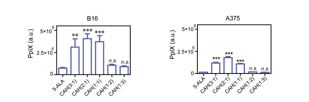
Fluorescence intensity of PpIX produced by various CAH complexes at varying molar ratios of amine to carboxyl in B16 and A375 melanoma cells, was measured in comparison with that of free 5-ALA (8 μg/mL; 12 h).
5-Aminolevulinic acid purchased from MedChemExpress. Usage Cited in: Adv Sci (Weinh). 2019 Jan 20;6(5):1802057. [Abstract]
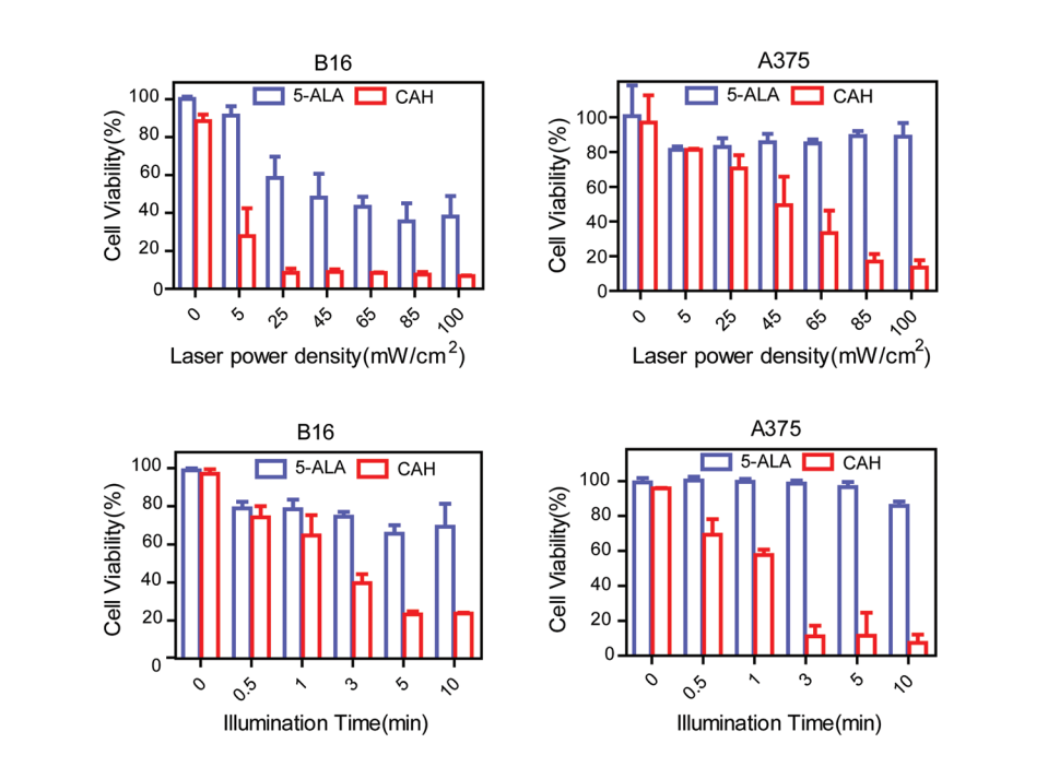
Inhibitory effects of light does and illumination time on cell viability of B16 cells and A375 cells treated with 5-ALA (8 µg/mL; 48 h) or CAH were evaluated by MTT assay.
5-Aminolevulinic acid purchased from MedChemExpress. Usage Cited in: Adv Sci (Weinh). 2019 Jan 20;6(5):1802057. [Abstract]
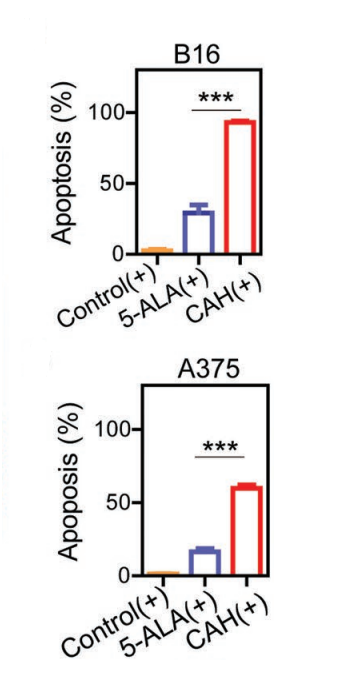
CAH specifically induced melanoma cells apoptosis in correlation with elevated intracellular ROS levels. Melanoma cell lines B16 cells and A375 cells were treated with 5-ALA (8 μg/mL; 12 h) or CAH under their optimal PDT conditions. Afterward, the cells were incubated with Annexin V-FITC (green)/PI (red), followed by FACS analysis.
5-Aminolevulinic acid purchased from MedChemExpress. Usage Cited in: Adv Sci (Weinh). 2019 Jan 20;6(5):1802057. [Abstract]
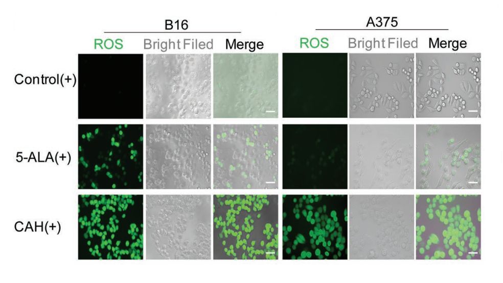
Cellular ROS levels (green) of B16 cells and A375 cells (treated with 5-ALA (8 μg/mL; 12 h) or CAH) were monitored using DCFH-DA assay with a fluorescent microscope.
5-Aminolevulinic acid purchased from MedChemExpress. Usage Cited in: Adv Sci (Weinh). 2019 Jan 20;6(5):1802057. [Abstract]
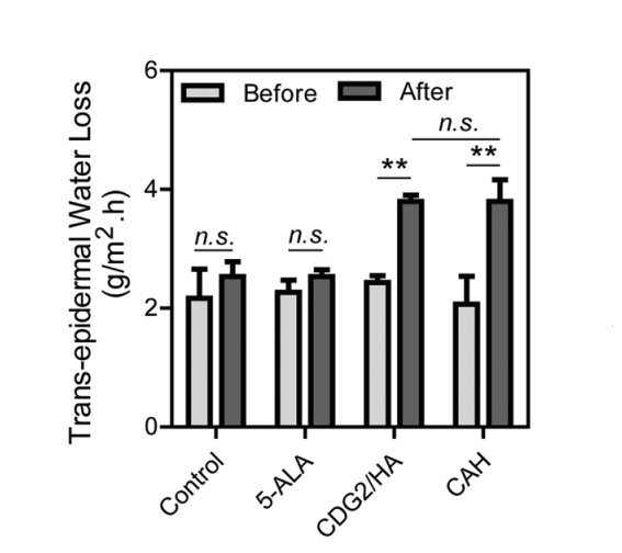
Trans-epidermal water loss (TEWL) was measured from the mice skin before and 12 h after topically applied with blank cream, 20% w/w 5-ALA cream, 20% CDG2/HA cream and 20% CAH cream with 5 min of light irradiation.
-
Cell Death Dis
2021 Oct 25;12(11):999. PMID: 34697294 -
Pharmacol Res
Dihydroartemisinin overcomes the resistance to osimertinib in EGFR-mutant non-small-cell lung cancer. [Abstract]2021 Aug:170:105701. PMID: 34087353 -
Genome Biol
G-quadruplexes sense natural porphyrin metabolites for regulation of gene transcription and chromatin landscapes. [Abstract]2022 Dec 15;23(1):259. PMID: 36522639 -
EMBO Mol Med
A whole-genome scan for Artemisinin cytotoxicity reveals a novel therapy for human brain tumors. [Abstract]2023 Mar 8;15(3):e16959. PMID: 36740985 -
Br J Pharmacol
The neuronal ALAS2/5-ala axis mitigates chemotherapy-induced neurotoxicity via the BACH1/NRF2 pathway. [Abstract]2026 Jun;183(11):2695-2719. PMID: 41652971 -
PLoS Biol
2024 Jun 27;22(6):e3002672. PMID: 38935621 -
Mar Drugs
Xanthocillin X Dimethyl Ether Exhibits Anti-Proliferative Effect on Triple-Negative Breast Cancer by Depletion of Mitochondrial Heme. [Abstract]2025 Mar 28;23(4):146. PMID: 40278267 -
-
-
Fish Shellfish Immunol
Dietary 5-aminolevulinic acid induces strain-specific effects on shell color and innate immunity in Pacific oyster (Crassostrea gigas) through divergent porphyrin metabolism. [Abstract]2026 May:172:111209. PMID: 41690531 -
J Inorg Biochem
Influence of cholesterol on kinetic parameters for human aromatase (P450 19A1) in phospholipid nanodiscs. [Abstract]2023 Oct:247:112340. PMID: 37544101 -
Asian J Androl
The addition of 5-aminolevulinic acid to HBSS protects testis grafts during hypothermic transportation: a novel preservation strategy. [Abstract]2025 Jul 1;27(4):454-463. PMID: 39589201 -
Photodiagnosis Photodyn Ther
Ferroptosis promotes 5-aminolevulinate acid-based photodynamic therapy in cervical cancer. [Abstract]2025 Aug:54:104726. PMID: 40680910 -
Photodiagnosis Photodyn Ther
The efficacy of 5-aminolevulinic acid photodynamic therapy in inducing cell death in multidrug-resistant Trichophyton mentagrophytes: An in vivo and in vitro study. [Abstract]2025 Jun:53:104599. PMID: 40258452 -
Photodiagnosis Photodyn Ther
In Vitro Modeling of Recurrent Dermatofibrosarcoma Protuberans: Assessment of 5-Aminolevulinic Acid Photodynamic Therapy Efficacy. [Abstract]2024 Jun:47:104093. PMID: 38641030 -
Photodiagnosis Photodyn Ther
5-ALA-PDT induced ferroptosis in keloid fibroblasts via ROS, accompanied by downregulation of xCT, GPX4. [Abstract]2023 Jun:42:103612. PMID: 37220842 -
Photodiagnosis Photodyn Ther
Enhancement of the cytotoxic effect of dihydroartemisinin in high-risk human papillomavirus-infected cells by aminolevulinic acid via the Bax/Bcl-2-caspase pathway. [Abstract]2022 Dec:40:103053. PMID: 35932961 -
Lasers Med Sci
Study on the mechanism of photodynamic therapy mediated by 5-aminoketovalerate in human ovarian cancer cell line. [Abstract]2021 Dec;36(9):1873-1881. PMID: 33392781 -
bioRxiv
An efficient behavioral screening platform classifies natural products and other chemical cues according to their chemosensory valence in C. elegans. [Abstract]2024 Apr 3:2023.06.02.542933. PMID: 37333363


Solvent & Solubility
In Vitro:
DMSO : 50 mg/mL (381.30 mM; Need ultrasonic; Hygroscopic DMSO has a significant impact on the solubility of product, please use newly opened DMSO)
Please refer to the solubility information to select the appropriate solvent. Once prepared, please aliquot and store the solution to prevent product inactivation from repeated freeze-thaw cycles.
Storage method and period of stock solution: -80°C, 6 months; -20°C, 1 month. When stored at -80°C, please use it within 6 months. When stored at -20°C, please use it within 1 month.
Please refer to the solubility information to select the appropriate solvent. Once prepared, please aliquot and store the solution to prevent product inactivation from repeated freeze-thaw cycles.
Storage method and period of stock solution: -80°C, 6 months; -20°C, 1 month. When stored at -80°C, please use it within 6 months. When stored at -20°C, please use it within 1 month.
Concentration (start) × Volume (start) = Concentration (final) × Volume (final)
In Vivo:
Select the appropriate dissolution method based on your experimental animal and administration route.
- For the following dissolution methods, please ensure to first prepare a clear stock solution using an In Vitro approach and then sequentially add co-solvents:
- To ensure reliable experimental results, the clarified stock solution can be appropriately stored based on storage conditions. As for the working solution for In Vivo experiments, it is recommended to prepare freshly and use it on the same day.
- The percentages shown for the solvents indicate their volumetric ratio in the final prepared solution. If precipitation or phase separation occurs during preparation, heat and/or sonication can be used to aid dissolution.
Add each solvent one by one: 10% DMSO 40% PEG300 5% Tween-80 45% Saline
Solubility: ≥ 2.5 mg/mL (19.07 mM); Clear solution
This protocol yields a clear solution of ≥ 2.5 mg/mL (saturation unknown).
Taking 1 mL working solution as an example, add 100 μL DMSO stock solution (25.0 mg/mL) to 400 μL PEG300, and mix evenly; then add 50 μL Tween-80 and mix evenly; then add 450 μL Saline to adjust the volume to 1 mL.
Preparation of Saline: Dissolve 0.9 g sodium chloride in ddH₂O and dilute to 100 mL to obtain a clear Saline solution.
Add each solvent one by one: 10% DMSO 90% (20% SBE-β-CD in Saline)
Solubility: ≥ 2.5 mg/mL (19.07 mM); Clear solution
This protocol yields a clear solution of ≥ 2.5 mg/mL (saturation unknown).
Taking 1 mL working solution as an example, add 100 μL DMSO stock solution (25.0 mg/mL) to 900 μL 20% SBE-β-CD in Saline, and mix evenly.
Preparation of 20% SBE-β-CD in Saline (4°C, storage for one week): 2 g SBE-β-CD powder is dissolved in 10 mL Saline, completely dissolve until clear.
In Vivo Dissolution Calculator
Please enter the basic information of animal experiments:
-
-
-
-
Recommended: Prepare an additional quantity of animals to account for potential losses during experiments.
Please enter your animal formula composition:
-
%DMSO +
Recommended: Keep the proportion of DMSO in working solution below 2% if your animal is weak.
-
%+
-
+%Tween-80 + +
-
%Saline +
The co-solvents required include: DMSO, . All of co-solvents are available by MedChemExpress (MCE). , Tween 80. All of co-solvents are available by MedChemExpress (MCE).
Working solution concentration: 0.22 mg/mL
Method for preparing stock solution: mg drug dissolved in μL DMSO. Stock solution concentration: mg/mL.
1. Take μL DMSO stock solution;
2. Add μL .
μL , mix evenly;
3. Then add μL Tween 80, mix evenly;
4. Then add μL
Please ensure that the stock solution in the first step is dissolved to a clear state, and add co-solvents in sequence. You can use ultrasonic heating (ultrasonic cleaner, recommended frequency 20-40 kHz), vortexing, etc. to assist dissolution.

Protocols
-
Phagocytosis Functional Assay
A phagocytosis functional assay measures the ability of phagocytic cells, such as neutrophils, macrophages, monocytes, or microglia/macrophages, to bind and internalize particulate targets including bacteria, yeast particles, beads, or myelin particles. Fluorescent flow-cytometry assays detect target uptake as fluorescence associated with gated phagocytes, while pH-sensitive dyes such as pHrodo increase signal in acidic phagosomal compartments and therefore preferentially report internalized particles rather than particles remaining outside the cell. Microscopy or high-content imaging can be used to confirm intracellular localization and, in some protocols, to follow uptake kinetics.
-
ROS/oxidative-stress fluorescent staining
ROS/oxidative-stress fluorescent staining uses cell-permeant fluorogenic probes that become fluorescent after oxidation inside cells or tissues; commonly used examples include DCFH-DA/DCFDA for broad cellular oxidant detection, DHE for superoxide-related signal detection, MitoSOX for mitochondrial superoxide-related signal detection, and CellROX probes for oxidative-stress-associated fluorescence readouts. The assay detects probe oxidation rather than a single ROS species unless the probe and analysis method have been chemically validated for that species. DCFH-DA enters cells, is deacetylated by intracellular esterases to DCFH, and produces fluorescent DCF after oxidation, so the readout is used as an operational measure of total cellular oxidative stress rather than a species-specific ROS measurement. DHE and MitoSOX can report superoxide-related oxidation, but red fluorescence alone can include non-specific ethidium-like oxidation products; HPLC or optimized spectral approaches are
-
Patient-Derived Xenograft (PDX)
Patient-derived xenograft (PDX) models are generated by engrafting primary human tumor tissue directly into immunodeficient mice, allowing in vivo propagation of patient tumor biology without initial in vitro adaptation. These models are used to preserve key histopathological and molecular characteristics of the original tumor and enable assessment of tumor growth dynamics and therapeutic response in a living organism. The biological readout is tumor engraftment and subsequent growth in the murine host, which reflects the ability of human tumor cells to survive, vascularize, and expand in an immunocompromised microenvironment.
-
Patient-Derived Orthotopic Xenograft (PDOX)
Patient-derived orthotopic xenograft (PDOX) modeling implants fresh patient tumor tissue or patient-derived tumor cells into the anatomically corresponding organ or tissue site of immunodeficient mice, usually by surgical orthotopic implantation, to preserve patient tumor histology, local microenvironmental context, invasion, metastatic behavior, and treatment-response features better than subcutaneous implantation. PDOX readouts include tumor engraftment, orthotopic tumor growth, local invasion, metastasis, recurrence after resection, histologic similarity to the donor tumor, biomarker retention, molecular concordance, survival, and response or resistance to therapy. PDOX models are used for preclinical drug testing and individualized therapy evaluation, but engraftment success varies by tumor type and specimen quality.
-
Research Protocol for Inflammation-related Diseases
The NLRP3 inflammasome is a cytosolic innate immune signaling platform that integrates priming signals and danger-signal activation to promote caspase-1 activation, maturation of IL-1β and IL-18, and gasdermin D-mediated pyroptotic cell death. The core experimental logic is to determine whether inflammatory disease phenotypes are driven by increased NLRP3 expression, ASC-containing inflammasome assembly, caspase-1 cleavage, GSDMD cleavage, and extracellular release of IL-1β/IL-18 rather than by nonspecific cell injury alone. The pathway is strongly linked to inflammation-related disease phenotypes because monosodium urate crystals activate NALP3/NLRP3 inflammasome signaling in gout-like crystal inflammation, cholesterol crystals activate NLRP3 inflammasomes in atherogenesis models, and DSS-induced intestinal inflammation has been reported to involve NLRP3 inflammasome activity. However, experimental colitis studies also show context-dependent protective effects of NLRP3 inflammasome co
-
CRISPRi/CRISPRa gene-regulation editing
CRISPRi and CRISPRa use catalytically inactive Cas9, typically SpCas9 D10A/H840A, as an RNA-guided DNA-binding platform that targets genomic loci through sgRNA complementarity and an adjacent PAM without generating Cas9 nuclease-mediated DNA cleavage. CRISPRi represses transcription by recruiting dCas9 alone or dCas9 fused to repressor domains such as KRAB to promoters or transcription start site regions, while CRISPRa activates transcription by recruiting activation domains such as VP64, VPR, or SAM components to promoter-proximal regions. The primary readout is target-gene expression change, commonly measured by RT-qPCR, RNA-seq, reporter fluorescence, or protein-level assays, and the readout reflects transcriptional repression or activation at the targeted endogenous locus.
-
Subcutaneous Cell-Line-Derived Xenograft
Subcutaneous cell-line-derived xenograft (CDX) models are established by implanting cultured human cancer cell lines into immunodeficient mice, where the injected cells form localized tumors that can be monitored in vivo as a measure of tumorigenic potential, growth kinetics, and treatment response. These models are widely used in oncology research because they allow reproducible tumor formation and enable comparative assessment of tumor growth between different cell lines or genetic manipulations in a controlled in vivo microenvironment. Subcutaneous implantation of cancer cells in immunodeficient mice is a standard approach for evaluating tumor growth behavior and therapeutic response across multiple cancer types, including prostate, esophageal, pancreatic, and colon cancer models.
-
Orthotopic Cell-Line Xenograft
Orthotopic cell-line xenograft models involve implantation of human cancer cell lines into the anatomically corresponding organ of immunodeficient mice to reproduce tumor growth within a native microenvironment, enabling more clinically relevant tumor behavior compared with subcutaneous models. These models are widely used because orthotopic placement better recapitulates tumor progression, including invasion and metastatic spread, which are often underrepresented in heterotopic implantation systems. Compared with conventional xenografts, orthotopic implantation is described as more technically complex but provides improved simulation of tumor-microenvironment interactions and metastatic behavior, making it particularly valuable for translational oncology research. Surgical orthotopic implantation approaches have been emphasized as enabling faithful reproduction of clinical cancer features, including metastasis and disease progression patterns that align with the tumor’s organ of origi


Purity & Documentation
-
Data Sheet (278 KB)
-
SDS (396 KB)
- English - EN (396 KB)
- Français - FR (396 KB)
- Deutsch - DE (396 KB)
- Norwegian - NO (396 KB)
- Español - ES (396 KB)
- Swedish - SV (396 KB)
- Italian - IT (396 KB)
- Korean - KR (396 KB)
- Portuguese - PT (396 KB)
-
Handling Instructions (2659 KB)
References
[1]. Ogura S, et al. The effect of 5-aminolevulinic acid on cytochrome c oxidase activity in mouse liver. BMC Res Notes. 2011;4:66. Published 2011 Mar 17. [Content Brief]
[2]. Saitoh S, et al. 5-Aminolevulinic Acid (5-ALA) Plays an Important Role in the Function of Innate Immune Cells. Inflammation. 2025;48(4):2588-2599. [Content Brief]
[3]. Takahashi J, et al. 5-Aminolevulinic acid enhances cancer radiotherapy in a mouse tumor model. Springerplus. 2013;2:602. Published 2013 Nov 12. [Content Brief]


Complete Stock Solution Preparation Table
Please refer to the solubility information to select the appropriate solvent. Once prepared, please aliquot and store the solution to prevent product inactivation from repeated freeze-thaw cycles.
Storage method and period of stock solution: -80°C, 6 months; -20°C, 1 month. When stored at -80°C, please use it within 6 months. When stored at -20°C, please use it within 1 month.
| Optional Solvent | Concentration Solvent Mass | 1 mg | 5 mg | 10 mg | 25 mg |
|---|---|---|---|---|---|
| DMSO | 1 mM | 7.6260 mL | 38.1301 mL | 76.2602 mL | 190.6505 mL |
| 5 mM | 1.5252 mL | 7.6260 mL | 15.2520 mL | 38.1301 mL | |
| 10 mM | 0.7626 mL | 3.8130 mL | 7.6260 mL | 19.0650 mL | |
| 15 mM | 0.5084 mL | 2.5420 mL | 5.0840 mL | 12.7100 mL | |
| 20 mM | 0.3813 mL | 1.9065 mL | 3.8130 mL | 9.5325 mL | |
| 25 mM | 0.3050 mL | 1.5252 mL | 3.0504 mL | 7.6260 mL | |
| 30 mM | 0.2542 mL | 1.2710 mL | 2.5420 mL | 6.3550 mL | |
| 40 mM | 0.1907 mL | 0.9533 mL | 1.9065 mL | 4.7663 mL | |
| 50 mM | 0.1525 mL | 0.7626 mL | 1.5252 mL | 3.8130 mL | |
| 60 mM | 0.1271 mL | 0.6355 mL | 1.2710 mL | 3.1775 mL | |
| 80 mM | 0.0953 mL | 0.4766 mL | 0.9533 mL | 2.3831 mL | |
| 100 mM | 0.0763 mL | 0.3813 mL | 0.7626 mL | 1.9065 mL |


5-Aminolevulinic acid Related Classifications
HY-W000450 Related Classifications
 Powered by Bioz