



EGFR-IN-47

Based on 1 Customer Validation
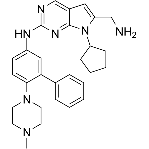
EGFR-IN-47 is a potent and orally active EGFRL858R/T790M/C797S inhibitor with an IC50 of 0.01 μM. EGFR-IN-47 induces cell cycle attest and cell apoptosis. EGFR-IN-47 has the potential for the research of NSCLC.
For research use only. We do not sell to patients.
- Purity : 96.01%
- CAS No.: 3034649-73-2
- Formula: C29H35N7
- Molecular Weight:481.64
-
Storage:Powder -20°C, 3 years , 4°C, 2 years ; In solvent -80°C, 6 months , -20°C, 1 month

To place orders, for customer services and technical support, please contact: MedChemExpress USA
Tel: 609-228-6898 E-mail: [email protected] [email protected]


-
 Biological Activity
-
Chemical Information
-
Solvent & Solubility
- Protocols
- Purity & Documentation
- References
-
 Help & FAQs


All EGFR Isoforms
More

Biological Activity
Description
IC50 & Target
[1]|
EGFRL858R/T790M/C797S 0.07 μM (IC50) |
Cellular Effect
|
Cell Line

|
Type | Value | Description | References |
|---|---|---|---|---|
| A-431 | IC50 |
2.972 μM
Compound: 14aj
|
Antiproliferative activity against human A-431 cells assessed as inhibition of cell growth incubated for 72 hrs by CCK-8 assay
Antiproliferative activity against human A-431 cells assessed as inhibition of cell growth incubated for 72 hrs by CCK-8 assay
|
[PMID: 35447433] |
| A549 | IC50 |
1.031 μM
Compound: 14aj
|
Antiproliferative activity against human A549 cells assessed as inhibition of cell growth incubated for 72 hrs by CCK-8 assay
Antiproliferative activity against human A549 cells assessed as inhibition of cell growth incubated for 72 hrs by CCK-8 assay
|
[PMID: 35447433] |
| PC-9 | IC50 |
0.013 μM
Compound: 14aj
|
Antiproliferative activity against human PC-9 cells expressing EGFR L858R/T790M/C797S mutant assessed as inhibition of cell growth incubated for 72 hrs by CCK-8 assay
Antiproliferative activity against human PC-9 cells expressing EGFR L858R/T790M/C797S mutant assessed as inhibition of cell growth incubated for 72 hrs by CCK-8 assay
|
[PMID: 35447433] |
| PC-9 | IC50 |
0.013 μM
Compound: 36
|
Antiproliferative activity against human PC-9 cells harboring EGFR L858R/T790M/C797S triple mutant assessed as cell growth inhibition
Antiproliferative activity against human PC-9 cells harboring EGFR L858R/T790M/C797S triple mutant assessed as cell growth inhibition
|
[PMID: 36417820] |
In Vitro
EGFR-IN-47 (compound 14aj) (72 h) shows anti-proliferative effects with IC50s of 0.013 μM, 2.972 μM, 1.031 μM for PC-9 (EGFRL858R/T790M/C797S), A432, A549 cells, respectively[1].
EGFR-IN-47 (0.01, 0.05, 0.25, 1 μM; 24 h) induces cell cycle attest at the G0/G1-phase[1].
EGFR-IN-47 (0.01, 0.05, 0.25 μM) induces cell deathvia apoptosis in a concentration-dependent manner[1].
EGFR-IN-47 (0.01, 0.05, 0.25, 1 μM) inhibits phosphorylation of the EGFR downstream mediators[1].
MedChemExpress (MCE) has not independently confirmed the accuracy of these methods. They are for reference only.
-
Cell Line:PC-9 (EGFRL858R/T790M/C797S), A432, A549 cells
-
Concentration:
-
Incubation Time:72 h
-
Result:Showed anti-proliferative effects with IC50s of 0.013 µM, 2.972 µM, 1.031 µM for PC-9 (EGFRL858R/T790M/C797S), A432, A549 cells, respectively.
-
Cell Line:PC-9 EGFRL858R/T790M/C797S cells
-
Concentration:0.01, 0.05, 0.25, 1 µM
-
Incubation Time:24 h
-
Result:Cells were arrest at the G0/G1-phase.
-
Cell Line:PC-9 EGFRL858R/T790M/C797S cells
-
Concentration:0.01, 0.05, 0.25 µM
-
Incubation Time:24 h
-
Result:Induced cell deathvia apoptosis in a concentration-dependent manner.
-
Cell Line:PC-9 EGFRL858R/T790M/C797S cells
-
Concentration:0.01, 0.05, 0.25, 1 µM
-
Incubation Time:
-
Result:Inhibited phosphorylation of the EGFR downstream mediators.
In Vivo
EGFR-IN-47 (20 mg/kg for i.v.; 20 mg/kg for p.o.) shows an ideal oral bioavailability of 66.05%[1].
Pharmacokinetic Parameters of JAK1/TYK2-IN-2 in Male Sprague-Dawley rats[1].
| parameter | iv (20 mg/kg) | po (20 mg/kg) |
| AUC0-∞ (mg/Lh) | 15.889 | 10.494 |
| Cmax (mg/L) | 6.845 | 0.77 |
| Tmax (h) | 0.083 | 6 |
| F (%) | 66.05 | |
| MRT0-∞ (h) | 16.439 | 16.791 |
| Vss (L/kg) | 16.015 | 28.101 |
| CL (L/h/kg) | 1.272 | 2.151 |
| t(1/2) (h) | 8.922 | 11.154 |
MedChemExpress (MCE) has not independently confirmed the accuracy of these methods. They are for reference only.
-
Animal Model:6-8 weeks, BALB/c nude mice (PC-9 EGFRL858R/T790M/C797S cells )[1]
-
Dosage:10, 20, 40 mg/kg (dissolved in 25% (v/v) PEG400 plus 5% DMSO)
-
Administration:I.g.
-
Result:Showed anti-tumor effect with low toxicity.
-
Animal Model:Male Sprague-Dawley rats[1]
-
Dosage:20 mg/kg
-
Administration:I.v.; p.o.
-
Result:Showed an ideal oral bioavailability of 66.05%.


Chemical Information
-
CAS No. 3034649-73-2
-
Appearance Solid
-
Molecular Weight 481.64
-
Formula C29H35N7
-
Color Light yellow to yellow
-
SMILES
CN(CC1)CCN1C2=CC=C(C=C2)NC3=NC=C4C=C(N(C4=N3)C5CCCC5)CNC6=CC=CC=C6
-
Shipping
Room temperature in continental US; may vary elsewhere.
-
Storage
Powder -20°C 3 years 4°C 2 years In solvent -80°C 6 months -20°C 1 month


Solvent & Solubility
In Vitro:
DMSO : 20 mg/mL (41.52 mM; ultrasonic and warming and heat to 60°C; Hygroscopic DMSO has a significant impact on the solubility of product, please use newly opened DMSO)
Please refer to the solubility information to select the appropriate solvent. Once prepared, please aliquot and store the solution to prevent product inactivation from repeated freeze-thaw cycles.
Storage method and period of stock solution: -80°C, 6 months; -20°C, 1 month. When stored at -80°C, please use it within 6 months. When stored at -20°C, please use it within 1 month.
Please refer to the solubility information to select the appropriate solvent. Once prepared, please aliquot and store the solution to prevent product inactivation from repeated freeze-thaw cycles.
Storage method and period of stock solution: -80°C, 6 months; -20°C, 1 month. When stored at -80°C, please use it within 6 months. When stored at -20°C, please use it within 1 month.
Concentration (start) × Volume (start) = Concentration (final) × Volume (final)


Purity & Documentation
-
Data Sheet (282 KB)
-
SDS (251 KB)
- English - EN (251 KB)
- Français - FR (251 KB)
- Deutsch - DE (251 KB)
- Norwegian - NO (251 KB)
- Español - ES (251 KB)
- Swedish - SV (251 KB)
- Italian - IT (251 KB)
- Korean - KR (251 KB)
- Portuguese - PT (251 KB)
-
Handling Instructions (2659 KB)
References


Complete Stock Solution Preparation Table
Please refer to the solubility information to select the appropriate solvent. Once prepared, please aliquot and store the solution to prevent product inactivation from repeated freeze-thaw cycles.
Storage method and period of stock solution: -80°C, 6 months; -20°C, 1 month. When stored at -80°C, please use it within 6 months. When stored at -20°C, please use it within 1 month.
| Optional Solvent | Concentration Solvent Mass | 1 mg | 5 mg | 10 mg | 25 mg |
|---|---|---|---|---|---|
| DMSO | 1 mM | 2.0762 mL | 10.3812 mL | 20.7624 mL | 51.9060 mL |
| 5 mM | 0.4152 mL | 2.0762 mL | 4.1525 mL | 10.3812 mL | |
| 10 mM | 0.2076 mL | 1.0381 mL | 2.0762 mL | 5.1906 mL | |
| 15 mM | 0.1384 mL | 0.6921 mL | 1.3842 mL | 3.4604 mL | |
| 20 mM | 0.1038 mL | 0.5191 mL | 1.0381 mL | 2.5953 mL | |
| 25 mM | 0.0830 mL | 0.4152 mL | 0.8305 mL | 2.0762 mL | |
| 30 mM | 0.0692 mL | 0.3460 mL | 0.6921 mL | 1.7302 mL | |
| 40 mM | 0.0519 mL | 0.2595 mL | 0.5191 mL | 1.2976 mL |


EGFR-IN-47 Related Classifications
HY-143337 Related Classifications
 Powered by Bioz