Search Result
Results for "
Mitochondrial fluorescent dye
" in MedChemExpress (MCE) Product Catalog:
1
Biochemical Assay Reagents
| Cat. No. |
Product Name |
Target |
Research Areas |
Chemical Structure |
-
- HY-D1055
-
MitoSOX Red
Maximum Cited Publications
263 Publications Verification
|
Fluorescent Dye
Reactive Oxygen Species (ROS)
Mitochondrial Metabolism
|
Cancer
|
MitoSOX Red is a live cell fluorescent probe that specifically targets mitochondria and is cell membrane permeable. MitoSOX Red enters mitochondria and is oxidized by superoxide but not by other ROS or RNS generating systems. The oxidized MitoSOX Red then binds to nucleic acids in mitochondria/nucleus, producing strong red fluorescence. MitoSOX Red can be used as a fluorescent indicator to specifically detect superoxide. In addition, superoxide dismutase (SOD) can prevent the oxidation of MitoSOX Red.
Excitation/emission wavelength: 510/580 nm.
|
-
-
- HY-D1783
-
|
MTDR FM
|
Fluorescent Dye
|
Others
|
MitoTracker Deep Red (MTDR) FM fluorescent dye that selectively accumulates in the mitochondrial matrix. MitoTracker Deep Red FM covalently binds mitochondrial proteins by reacting with free mercaptan of cysteine residues, allowing staining of mitochondrial membrane potential independent of membrane potential. Excitation/emission wavelength 644/665 nm . MitoTracker Deep Red dyes have an excitation/emission wavelength of 633/650-750 nm .
The Ex/Em of MitoTracker Deep Red (MTDR) FM is 644/665 nm.
|
-
-
- HY-D0985A
-
|
Tetramethylrhodamine ethyl ester perchlorate
|
Fluorescent Dye
|
Others
|
|
Rhodamine dyes are membrane-permeable cationic fluorescent probes that specifically recognize mitochondrial membrane potentials, thereby attaching to mitochondria and producing bright fluorescence, and at certain concentrations, rhodamine dyes have low toxicity to cells, so they are commonly used to detect mitochondria in animal cells, plant cells, and microorganisms .
|
-
-
- HY-D0984A
-
|
T668
|
Fluorescent Dye
|
Others
|
|
Rhodamine dyes are membrane-permeable cationic fluorescent probes that specifically recognize mitochondrial membrane potentials, thereby attaching to mitochondria and producing bright fluorescence, and at certain concentrations, rhodamine dyes have low toxicity to cells, so they are commonly used to detect mitochondria in animal cells, plant cells, and microorganisms .
|
-
-
- HY-135056
-
|
|
Fluorescent Dye
|
Others
|
|
Mito-Tracker Green is a green fluorescent dye that selectively accumulates in the mitochondrial matrix. MitoTracker Green FM covalently binds mitochondrial proteins by reacting with free mercaptan of cysteine residues, allowing staining of mitochondrial membrane potential independent of membrane potential. Excitation/emission wavelength 490/523 nm.
|
-
-
- HY-D0816
-
|
RH-123; R-22420
|
Fluorescent Dye
|
Cardiovascular Disease
|
|
Rhodamine dyes are membrane-permeable cationic fluorescent probes that specifically recognize mitochondrial membrane potentials, thereby attaching to mitochondria and producing bright fluorescence, and at certain concentrations, rhodamine dyes have low toxicity to cells, so they are commonly used to detect mitochondria in animal cells, plant cells, and microorganisms .
|
-
-
- HY-D0085
-
|
|
Fluorescent Dye
|
Cancer
|
|
DiSC3(5) is a fluorescent probe commonly used as a tracer dye to evaluate mitochondrial membrane potential. The excitation/emission wavelength of DiSC3(5) is up to 622/670 nm. DiSC3(5) can inhibit the respiratory system associated with mitochondrial NAD, and the IC50 value is 8 μM. DiSC3(5) in the presence of Na +/K +-ATPase inhibitor ouabain 2 can induce membrane hyperpolarization of Ehrlich ascites tumor cells .
|
-
-
- HY-D0309
-
|
Basic Red 1
|
Environmental Pollutants
Fluorescent Dye
|
Cancer
|
|
Rhodamine dyes are membrane-permeable cationic fluorescent probes that specifically recognize mitochondrial membrane potentials, thereby attaching to mitochondria and producing bright fluorescence, and at certain concentrations, rhodamine dyes have low toxicity to cells, so they are commonly used to detect mitochondria in animal cells, plant cells, and microorganisms .
|
-
-
- HY-D1696
-
|
|
Fluorescent Dye
|
Others
|
|
MitoTracker Orange CMTMRos is a fluorescent dye that labels mitochondria within live cells utilizing the mitochondrial membrane potential (Ex/Em: 551/576 nm) .
|
-
-
- HY-135009
-
|
DASPI
|
G-quadruplex
|
Others
|
|
2-Di-1-ASP (DASPI; Compound 18a) is a mono-stryryl dye, and widely used as mitochondrial stain and groove-binding fluorescent probes for double-stranded DNA. 2-Di-1-ASP is selective for G-quadruplex (G4) and double-stranded DNA .
|
-
-
- HY-D0984
-
|
|
Fluorescent Dye
|
Inflammation/Immunology
|
|
Rhodamine dyes are membrane-permeable cationic fluorescent probes that specifically recognize mitochondrial membrane potentials, thereby attaching to mitochondria and producing bright fluorescence, and at certain concentrations, rhodamine dyes have low toxicity to cells, so they are commonly used to detect mitochondria in animal cells, plant cells, and microorganisms .
|
-
-
- HY-DY1032
-
|
|
Fluorescent Dye
|
Others
|
Mito-Tracker Green (solution) is a green fluorescent dye that selectively accumulates in the mitochondrial matrix. MitoTracker Green FM covalently binds mitochondrial proteins by reacting with free mercaptan of cysteine residues, allowing staining of mitochondrial membrane potential independent of membrane potential. Excitation/emission wavelength 490/523 nm.
Solvent and concentration: DMSO: 1 mM
|
-

-
- HY-DY1042
-
|
|
Fluorescent Dye
|
Others
|
Rhodamine dyes are membrane-permeable cationic fluorescent probes that specifically recognize mitochondrial membrane potentials, thereby attaching to mitochondria and producing bright fluorescence, and at certain concentrations, rhodamine dyes have low toxicity to cells, so they are commonly used to detect mitochondria in animal cells, plant cells, and microorganisms .
Solvent and concentration: DMSO: 10 mM
|
-

-
- HY-D0060
-
|
|
Fluorescent Dye
|
Others
|
|
4-Di-2-ASP, a styryl pyridinium fluorescent dye, is a vital mitochondrial marker. 4-Di-2-ASP shows reliable and specific labelling of pulmonary NEBs (neuroepithelial bodies) .
|
-
-
- HY-137042
-
|
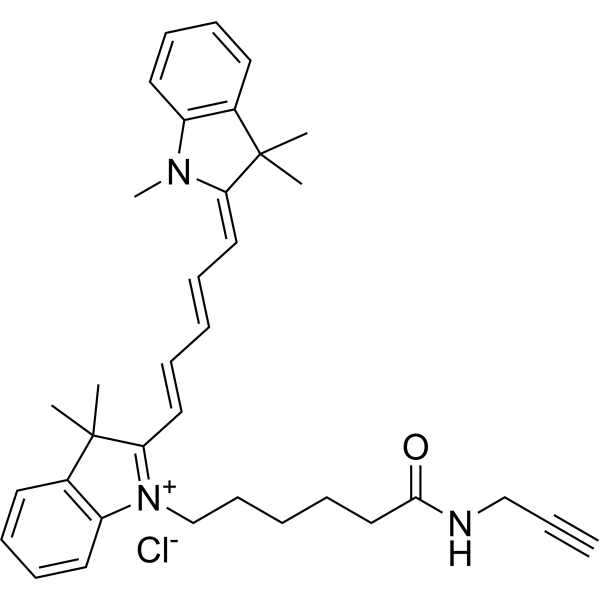
Alkyne-Cy5
|
Oxidative Phosphorylation
Mitochondrial Metabolism
|
Cancer
|
|
Cyanine5 alkyne (Alkyne-Cy5) is a fluorescent dye used to label azide proteins and can be used to analyse post-translational modifications of proteins, glycosylation etc. Cyanine5 alkyne can also be used as a mitochondrial OXPHOS inhibitor to inhibit the growth of cancer stem cells (CSC) . Cyanine5 alkyne is a click chemistry reagent, it contains an Alkyne group and can undergo copper-catalyzed azide-alkyne cycloaddition (CuAAc) with molecules containing Azide groups.
|
-
-
- HY-D1116
-
|
|
Fluorescent Dye
|
Others
|
|
MitoMark Red I is a fluorescent mitochondrial marker. MitoMark Red I is a red fluorescent dye which accumulates in mitochondria in viable cells and has an excitation wavelength of 578 nm and emission of 599 nm .
|
-
-
- HY-101876
-
|
|
Fluorescent Dye
|
Others
|
|
Rhodamine dyes are membrane-permeable cationic fluorescent probes that specifically recognize mitochondrial membrane potentials, thereby attaching to mitochondria and producing bright fluorescence, and at certain concentrations, rhodamine dyes have low toxicity to cells, so they are commonly used to detect mitochondria in animal cells, plant cells, and microorganisms .
|
-
-
- HY-DY1023
-
|
|
Fluorescent Dye
|
Others
|
Rhodamine dyes are membrane-permeable cationic fluorescent probes that specifically recognize mitochondrial membrane potentials, thereby attaching to mitochondria and producing bright fluorescence, and at certain concentrations, rhodamine dyes have low toxicity to cells, so they are commonly used to detect mitochondria in animal cells, plant cells, and microorganisms .
Solvent and concentration: DMSO: 10 mM
|
-

-
- HY-DY1021
-
|
|
Fluorescent Dye
|
Others
|
DiSC3 (5) (solution) is a fluorescent probe commonly used as a tracer dye to evaluate mitochondrial membrane potential. The excitation/emission wavelength of DiSC3 (5) is up to 622/670 nm. DiSC3 (5) can inhibit the respiratory system associated with mitochondrial NAD, and the IC50 value is 8 μM. DiSC3 (5) in the presence of Na +/K +-ATPase inhibitor ouabain 2 can induce membrane hyperpolarization of Ehrlich ascites tumor cells .
Solvent and concentration: DMSO: 1 mM
The 1 mL volume is defined as the base specification. All larger sizes correspond to incremental volumes of this base.
|
-

-
- HY-W247098
-
|
DHR 6G
|
Reactive Oxygen Species (ROS)
|
Cancer
|
|
Dihydrorhodamine 6G (DHR 6G) is the reduced form of Rhodamine 6G, which is used as fluorescent mitochondrial dye. It is nonfluorescent, but it readily enters most of the cells and is oxidized by oxidative species or by cellular redox systems to the fluorescent rhodamine 6G that accumulates in mitochondrial membranes. Dihydrorhodamine 6G is useful for detecting reactive oxygen species (ROS) including superoxide .
|
-
-
- HY-DY1054
-
|
|
Fluorescent Dye
|
Cardiovascular Disease
|
Rhodamine dyes are membrane-permeable cationic fluorescent probes that specifically recognize mitochondrial membrane potentials, thereby attaching to mitochondria and producing bright fluorescence, and at certain concentrations, rhodamine dyes have low toxicity to cells, so they are commonly used to detect mitochondria in animal cells, plant cells, and microorganisms .
Solvent and concentration: DMSO: 5 mM
|
-

-
- HY-D0309R
-
|
Basic Red 1 (Standard)
|
Fluorescent Dye
Reference Standards
|
Cancer
|
|
Rhodamine 6G (Standard) is the analytical standard of Rhodamine 6G. This product is intended for research and analytical applications. Rhodamine dyes are membrane-permeable cationic fluorescent probes that specifically recognize mitochondrial membrane potentials, thereby attaching to mitochondria and producing bright fluorescence, and at certain concentrations, rhodamine dyes have low toxicity to cells, so they are commonly used to detect mitochondria in animal cells, plant cells, and microorganisms .
|
-

-
- HY-162422
-
|
|
Fluorescent Dye
Pyroptosis
|
Cancer
|
|
Mito-DK is a small-molecule fluorescent dye with the capability of crosstalk-free response to polarity and mtDNA as well as mitochondrial morphology. Mito-DK has high photostability, low cytotoxicity, and good mitochondria-targeting properties. Mito-DK can be used for real-time tracking and multidimensional assessing of mitochondria-related pyroptosis in cancer cells .
|
-
-
- HY-W127780
-
|
Basic Red 1 perchlorate
|
Fluorescent Dye
|
Others
|
|
Rhodamine dyes are membrane-permeable cationic fluorescent probes that specifically recognize mitochondrial membrane potentials, thereby attaching to mitochondria and producing bright fluorescence, and at certain concentrations, rhodamine dyes have low toxicity to cells, so they are commonly used to detect mitochondria in animal cells, plant cells, and microorganisms .
|
-
-
- HY-167255
-
|
|
Fluorescent Dye
|
Others
|
|
JC-10 is a lipophilic mitochondrial membrane potential indicator and is a fluorescent dye. JC-10 accumulates and aggregates in healthy mitochondria to emit red fluorescence; exists as a monomer emitting green fluorescence in the cytosol or apoptotic cells with collapsed mitochondrial membrane potential, enabling measurement of mitochondrial depolarization via the green/red fluorescence ratio .
|
-

| Cat. No. |
Product Name |
Type |
-
- HY-D1055
-
MitoSOX Red
Maximum Cited Publications
263 Publications Verification
|
Fluorescent Dyes
|
MitoSOX Red is a live cell fluorescent probe that specifically targets mitochondria and is cell membrane permeable. MitoSOX Red enters mitochondria and is oxidized by superoxide but not by other ROS or RNS generating systems. The oxidized MitoSOX Red then binds to nucleic acids in mitochondria/nucleus, producing strong red fluorescence. MitoSOX Red can be used as a fluorescent indicator to specifically detect superoxide. In addition, superoxide dismutase (SOD) can prevent the oxidation of MitoSOX Red.
Excitation/emission wavelength: 510/580 nm.
|
-
- HY-D0985A
-
|
Tetramethylrhodamine ethyl ester perchlorate
|
Fluorescent Dyes
|
|
Rhodamine dyes are membrane-permeable cationic fluorescent probes that specifically recognize mitochondrial membrane potentials, thereby attaching to mitochondria and producing bright fluorescence, and at certain concentrations, rhodamine dyes have low toxicity to cells, so they are commonly used to detect mitochondria in animal cells, plant cells, and microorganisms .
|
-
- HY-D0984A
-
|
T668
|
Fluorescent Dyes
|
|
Rhodamine dyes are membrane-permeable cationic fluorescent probes that specifically recognize mitochondrial membrane potentials, thereby attaching to mitochondria and producing bright fluorescence, and at certain concentrations, rhodamine dyes have low toxicity to cells, so they are commonly used to detect mitochondria in animal cells, plant cells, and microorganisms .
|
-
- HY-135056
-
|
|
Fluorescent Dyes
|
|
Mito-Tracker Green is a green fluorescent dye that selectively accumulates in the mitochondrial matrix. MitoTracker Green FM covalently binds mitochondrial proteins by reacting with free mercaptan of cysteine residues, allowing staining of mitochondrial membrane potential independent of membrane potential. Excitation/emission wavelength 490/523 nm.
|
-
- HY-D0816
-
|
RH-123; R-22420
|
Fluorescent Dyes
|
|
Rhodamine dyes are membrane-permeable cationic fluorescent probes that specifically recognize mitochondrial membrane potentials, thereby attaching to mitochondria and producing bright fluorescence, and at certain concentrations, rhodamine dyes have low toxicity to cells, so they are commonly used to detect mitochondria in animal cells, plant cells, and microorganisms .
|
-
- HY-D0085
-
|
|
Fluorescent Dyes
|
|
DiSC3(5) is a fluorescent probe commonly used as a tracer dye to evaluate mitochondrial membrane potential. The excitation/emission wavelength of DiSC3(5) is up to 622/670 nm. DiSC3(5) can inhibit the respiratory system associated with mitochondrial NAD, and the IC50 value is 8 μM. DiSC3(5) in the presence of Na +/K +-ATPase inhibitor ouabain 2 can induce membrane hyperpolarization of Ehrlich ascites tumor cells .
|
-
- HY-D0309
-
|
Basic Red 1
|
Fluorescent Dyes
|
|
Rhodamine dyes are membrane-permeable cationic fluorescent probes that specifically recognize mitochondrial membrane potentials, thereby attaching to mitochondria and producing bright fluorescence, and at certain concentrations, rhodamine dyes have low toxicity to cells, so they are commonly used to detect mitochondria in animal cells, plant cells, and microorganisms .
|
-
- HY-D1696
-
|
|
Fluorescent Dyes
|
|
MitoTracker Orange CMTMRos is a fluorescent dye that labels mitochondria within live cells utilizing the mitochondrial membrane potential (Ex/Em: 551/576 nm) .
|
-
- HY-135009
-
|
DASPI
|
Fluorescent Dyes
|
|
2-Di-1-ASP (DASPI; Compound 18a) is a mono-stryryl dye, and widely used as mitochondrial stain and groove-binding fluorescent probes for double-stranded DNA. 2-Di-1-ASP is selective for G-quadruplex (G4) and double-stranded DNA .
|
-
- HY-D0984
-
|
|
Fluorescent Dyes
|
|
Rhodamine dyes are membrane-permeable cationic fluorescent probes that specifically recognize mitochondrial membrane potentials, thereby attaching to mitochondria and producing bright fluorescence, and at certain concentrations, rhodamine dyes have low toxicity to cells, so they are commonly used to detect mitochondria in animal cells, plant cells, and microorganisms .
|
-
- HY-DY1032
-
|
|
Fluorescent Dyes
|
Mito-Tracker Green (solution) is a green fluorescent dye that selectively accumulates in the mitochondrial matrix. MitoTracker Green FM covalently binds mitochondrial proteins by reacting with free mercaptan of cysteine residues, allowing staining of mitochondrial membrane potential independent of membrane potential. Excitation/emission wavelength 490/523 nm.
Solvent and concentration: DMSO: 1 mM
|
-
- HY-DY1042
-
|
|
Fluorescent Dyes
|
Rhodamine dyes are membrane-permeable cationic fluorescent probes that specifically recognize mitochondrial membrane potentials, thereby attaching to mitochondria and producing bright fluorescence, and at certain concentrations, rhodamine dyes have low toxicity to cells, so they are commonly used to detect mitochondria in animal cells, plant cells, and microorganisms .
Solvent and concentration: DMSO: 10 mM
|
-
- HY-D0060
-
|
|
Fluorescent Dyes
|
|
4-Di-2-ASP, a styryl pyridinium fluorescent dye, is a vital mitochondrial marker. 4-Di-2-ASP shows reliable and specific labelling of pulmonary NEBs (neuroepithelial bodies) .
|
-
- HY-137042
-
|
Alkyne-Cy5
|
Fluorescent Dyes
|
|
Cyanine5 alkyne (Alkyne-Cy5) is a fluorescent dye used to label azide proteins and can be used to analyse post-translational modifications of proteins, glycosylation etc. Cyanine5 alkyne can also be used as a mitochondrial OXPHOS inhibitor to inhibit the growth of cancer stem cells (CSC) . Cyanine5 alkyne is a click chemistry reagent, it contains an Alkyne group and can undergo copper-catalyzed azide-alkyne cycloaddition (CuAAc) with molecules containing Azide groups.
|
-
- HY-D1116
-
|
|
Fluorescent Dyes
|
|
MitoMark Red I is a fluorescent mitochondrial marker. MitoMark Red I is a red fluorescent dye which accumulates in mitochondria in viable cells and has an excitation wavelength of 578 nm and emission of 599 nm .
|
-
- HY-101876
-
|
|
Fluorescent Dyes
|
|
Rhodamine dyes are membrane-permeable cationic fluorescent probes that specifically recognize mitochondrial membrane potentials, thereby attaching to mitochondria and producing bright fluorescence, and at certain concentrations, rhodamine dyes have low toxicity to cells, so they are commonly used to detect mitochondria in animal cells, plant cells, and microorganisms .
|
-
- HY-DY1023
-
|
|
Fluorescent Dyes
|
Rhodamine dyes are membrane-permeable cationic fluorescent probes that specifically recognize mitochondrial membrane potentials, thereby attaching to mitochondria and producing bright fluorescence, and at certain concentrations, rhodamine dyes have low toxicity to cells, so they are commonly used to detect mitochondria in animal cells, plant cells, and microorganisms .
Solvent and concentration: DMSO: 10 mM
|
-
- HY-DY1021
-
|
|
Fluorescent Dyes
|
DiSC3 (5) (solution) is a fluorescent probe commonly used as a tracer dye to evaluate mitochondrial membrane potential. The excitation/emission wavelength of DiSC3 (5) is up to 622/670 nm. DiSC3 (5) can inhibit the respiratory system associated with mitochondrial NAD, and the IC50 value is 8 μM. DiSC3 (5) in the presence of Na +/K +-ATPase inhibitor ouabain 2 can induce membrane hyperpolarization of Ehrlich ascites tumor cells .
Solvent and concentration: DMSO: 1 mM
The 1 mL volume is defined as the base specification. All larger sizes correspond to incremental volumes of this base.
|
-
- HY-DY1054
-
|
|
Fluorescent Dyes
|
Rhodamine dyes are membrane-permeable cationic fluorescent probes that specifically recognize mitochondrial membrane potentials, thereby attaching to mitochondria and producing bright fluorescence, and at certain concentrations, rhodamine dyes have low toxicity to cells, so they are commonly used to detect mitochondria in animal cells, plant cells, and microorganisms .
Solvent and concentration: DMSO: 5 mM
|
-
- HY-D0309R
-
|
Basic Red 1 (Standard)
|
Fluorescent Dyes
|
|
Rhodamine 6G (Standard) is the analytical standard of Rhodamine 6G. This product is intended for research and analytical applications. Rhodamine dyes are membrane-permeable cationic fluorescent probes that specifically recognize mitochondrial membrane potentials, thereby attaching to mitochondria and producing bright fluorescence, and at certain concentrations, rhodamine dyes have low toxicity to cells, so they are commonly used to detect mitochondria in animal cells, plant cells, and microorganisms .
|
-
- HY-167255
-
|
|
Fluorescent Dyes
|
|
JC-10 is a lipophilic mitochondrial membrane potential indicator and is a fluorescent dye. JC-10 accumulates and aggregates in healthy mitochondria to emit red fluorescence; exists as a monomer emitting green fluorescence in the cytosol or apoptotic cells with collapsed mitochondrial membrane potential, enabling measurement of mitochondrial depolarization via the green/red fluorescence ratio .
|
| Cat. No. |
Product Name |
Type |
-
- HY-W127780
-
|
Basic Red 1 perchlorate
|
Biochemical Assay Reagents
|
|
Rhodamine dyes are membrane-permeable cationic fluorescent probes that specifically recognize mitochondrial membrane potentials, thereby attaching to mitochondria and producing bright fluorescence, and at certain concentrations, rhodamine dyes have low toxicity to cells, so they are commonly used to detect mitochondria in animal cells, plant cells, and microorganisms .
|
| Cat. No. |
Product Name |
|
Classification |
-
- HY-137042
-
|
Alkyne-Cy5
|
|
Labeling and Fluorescence Imaging
Alkynes
|
|
Cyanine5 alkyne (Alkyne-Cy5) is a fluorescent dye used to label azide proteins and can be used to analyse post-translational modifications of proteins, glycosylation etc. Cyanine5 alkyne can also be used as a mitochondrial OXPHOS inhibitor to inhibit the growth of cancer stem cells (CSC) . Cyanine5 alkyne is a click chemistry reagent, it contains an Alkyne group and can undergo copper-catalyzed azide-alkyne cycloaddition (CuAAc) with molecules containing Azide groups.
|
Your information is safe with us. * Required Fields.
Inquiry Information
- Product Name:
- Cat. No.:
- Quantity:
- MCE Japan Authorized Agent:


