


MitoTracker Green FM
Based on 26 publication(s) in Google Scholar
Mito-Tracker Green is a green fluorescent dye that selectively accumulates in the mitochondrial matrix. MitoTracker Green FM covalently binds mitochondrial proteins by reacting with free mercaptan of cysteine residues, allowing staining of mitochondrial membrane potential independent of membrane potential. Excitation/emission wavelength 490/523 nm.
For research use only. We do not sell to patients.
- Purity: 95.48%
- CAS No.: 201860-17-5
- Formula: C34H28Cl5N3O
- Molecular Weight:671.87
-
Storage:
-20°C, sealed storage, away from moisture and light
* In solvent : -80°C, 6 months; -20°C, 1 month (sealed storage, away from moisture and light)

To place orders, for customer services and technical support, please contact: MedChemExpress USA
Tel: 609-228-6898 E-mail: [email protected] [email protected]


-
 Biological Activity
-
Chemical Information
-
Solvent & Solubility
- Purity & Documentation
- References
-
 Help & FAQs


Publications Citing Use of MedChemExpress (MCE) MitoTracker Green FM
More- Nat Commun. 2025 Jul 24;16(1):6821. [Abstract]
- Gut Microbes. 2026 Dec 31;18(1):2657625. [Abstract]
- J Nanobiotechnology. 2025 Aug 11;23(1):559. [Abstract]
- J Nanobiotechnology. 2025 Apr 5;23(1):276. [Abstract]
- Cell Death Dis. 2023 Apr 24;14(4):287. [Abstract]
- Phytomedicine. 2025 Apr:139:156426. [Abstract]
- J Renin Angiotensin Aldosterone Syst. 2026 Feb 19.
- Acta Biomater. 2025 Apr:196:321-331. [Abstract]
- Sens Actuators B Chem. 15 December 2022, 132707.
- ACS Appl Mater Interfaces. 2025 Dec 24;17(51):69180-69195. [Abstract]
- Cell Rep. 2026 Jun 23;45(6):117504. [Abstract]
- J Agric Food Res. 2025 Mar.
- Redox Rep. 2025 Dec;30(1):2536400. [Abstract]
- J Agric Food Chem. 2025 Oct 1. [Abstract]
- ACS Biomater Sci Eng. 2024 Oct 14;10(10):6263-6285. [Abstract]
- Biomacromolecules. 2025 Aug 11;26(8):5438-5449. [Abstract]
- Glia. 2025 Aug 8. [Abstract]
- Chem Biol Interact. 2026 Jun 1:432:112059. [Abstract]
- Toxicology. 2025 Sep 17:154282. [Abstract]
- Bioorg Chem. 2025 Sep 1:165:108951. [Abstract]
- Biochim Biophys Acta Mol Basis Dis. 2025 Apr 26;1871(6):167874. [Abstract]
- Mol Med Rep. 2023 Dec;28(6):234. [Abstract]
- Cell Signal. 2025 Aug:132:111805. [Abstract]
- FASEB J. 2026 Mar 31;40(6):e71682. [Abstract]
- Biol Reprod. 2025 Aug 13;113(2):345-357. [Abstract]
- Res Sq. 2025 Jul 21.
 Customer Validation & Images

-
Flow Cytometry
-
In Vivo Imaging
-
Cell Imaging/Staining
-
Cell Imaging/Staining
-
Flow Cytometry


Biological Activity
Guide (Following is our recommended protocol. This protocol only provides a guideline, and should be modified according to your specific needs)[2].
1. Preparation of MitoTracker Green FM working solution
1.1 Preparation of the stock solution
Dissolve MitoTracker Green FM in DMSO to obtain 1 mM of stock solution.
1.2 Preparation of MitoTracker Green FM working solution
Dilute the stock solution in serum-free cell culture medium or PBS dilute to obtain 20-200 nM of working solution.
Note: Please adjust the concentration of MitoTracker Green FM working solution according to the actual situation.
2. Cell staining (Suspension cells)
2.1. Centrifuge at 1000 g at 4°C for 3-5 minutes and then discard the supernatant. Wash twice with PBS, 5 minutes each time. The cell density is 1×10⁶/mL.
2.2. Add 1 mL of working solution, and then incubate at room temperature for 15-45 minutes.
2.3. Centrifuge at 400 g at 4°C for 3-4 minutes and then discard the supernatant.
2.4. Wash twice with PBS, 5 minutes each time.
2.5. Resuspend cells with serum-free cell culture medium or PBS. Observation by fluorescence microscopy or flow cytometry.
3. Cell staining (Adherent cells)
3.1 Culture adherent cells on sterile coverslips.
3.2 Remove the coverslip from the medium and aspirate excess medium.
3.3 Add 100 μL of working solution, gently shake it to completely cover the cells, and then incubate at room temperature for 15-45 minutes.
3.4 Wash twice with medium, 5 minutes each time. Observation by fluorescence microscopy or flow cytometry.
MedChemExpress (MCE) has not independently confirmed the accuracy of these methods. They are for reference only.
523
490


Chemical Information
-
CAS No. 201860-17-5
-
Appearance Solid
-
Molecular Weight 671.87
-
Formula C34H28Cl5N3O
-
Color Yellow to orange
-
SMILES
C[N+]1=C(/C=C/C=C2N(CC3=CC=C(CCl)C=C3)C4=CC(Cl)=C(Cl)C=C4N\2CC5=CC=C(CCl)C=C5)OC6=CC=CC=C16.[Cl-]
-
Shipping
Room temperature in continental US; may vary elsewhere.
-
Storage
-20°C, sealed storage, away from moisture and light
* In solvent : -80°C, 6 months; -20°C, 1 month (sealed storage, away from moisture and light)


Publications (26)
-
Journal Impact Factor
-
Most Recent
-
Nat Commun
Impairing the interaction between Erg11 and cytochrome P450 reductase Ncp1 enhances azoles' antifungal activities. [Abstract]2025 Jul 24;16(1):6821. PMID: 40707518
MitoTracker Green FM purchased from MedChemExpress. Usage Cited in: Nat Commun. 2025 Jul 24;16(1):6821. [Abstract]
Mitochondrial misfolded proteins were determined by MitoTracker Green FM (0.1 μM; 37 °C; 30 min). Fluorescence intensity measurements were obtained using a BD FACSverse flow cytometer.
-
Gut Microbes
Dietary conjugated linoleic acid enhances resistance to Salmonella infection by promoting PPARγ-mediated metabolic reprogramming and effector function in CD8⁺ T cells. [Abstract]2026 Dec 31;18(1):2657625. PMID: 41964110 -
J Nanobiotechnology
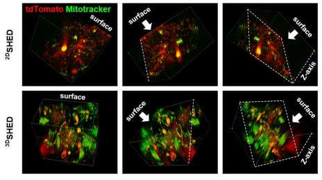
Dynamic three-dimensional culture enhances tunneling nanotubes-mediated mitochondrial transfer in mesenchymal stromal cells to accelerate wound healing. [Abstract]2025 Aug 11;23(1):559. PMID: 40790215
MitoTracker Green FM purchased from MedChemExpress. Usage Cited in: J Nanobiotechnology. 2025 Aug 11;23(1):559. [Abstract]
(A) Schematic diagram of wound modeling and live imaging for in vivo observation of mitochondrial transfer (SHED labeled with MitoTracker Green FM). (B) Representative two-photon images of the 3D-reconstructed wound area on day 2 post-treatment. Scale bar: 50 μm. Quantitative analysis of the colocalization area proportion of red and green fluorescence (n = 3).
-
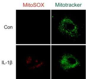
J Nanobiotechnology
Lacc1-engineered extracellular vesicles reprogram mitochondrial metabolism to alleviate inflammation and cartilage degeneration in TMJ osteoarthritis. [Abstract]2025 Apr 5;23(1):276. PMID: 40186254
MitoTracker Green FM purchased from MedChemExpress. Usage Cited in: J Nanobiotechnology. 2025 Apr 5;23(1):276. [Abstract]
Mitochondrial ROS analysis using MitoSOX Red and MitoTracker Green FM (100 nM; 37°C; 15 min) staining.
-
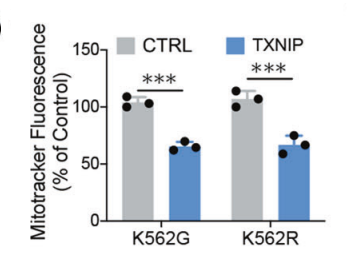
Cell Death Dis
BCR-ABL triggers a glucose-dependent survival program during leukemogenesis through the suppression of TXNIP. [Abstract]2023 Apr 24;14(4):287. PMID: 37095099
MitoTracker Green FM purchased from MedChemExpress. Usage Cited in: Cell Death Dis. 2023 Apr 24;14(4):287. [Abstract]
K562G and K562R cells were stained with the MitoTracker Green (0.1 μM; 37 °C; 30 min) and then cells were subjected to flow cytometry assays for assessing mitochondrial number.
-
Phytomedicine
Chaihuang Yishen Granule ameliorates mitochondrial homeostasis by upregulating PRDX5/TFAM axis to inhibit renal fibrosis in CKD. [Abstract]2025 Apr:139:156426. PMID: 39955823 -
-
Acta Biomater
Spatially targeted triple amplification of oxidative stress for enhanced tumor therapy via effective modulation of metal ion valence states. [Abstract]2025 Apr:196:321-331. PMID: 39983855 -
-
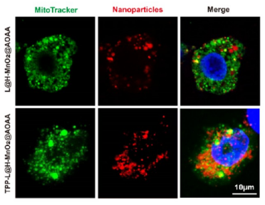
ACS Appl Mater Interfaces
Mitochondrial-Targeting Drug-Loaded Nanoparticles Reprogram Macrophage Metabolism via ROS/NO Co-elimination for Diabetic Wound Healing. [Abstract]2025 Dec 24;17(51):69180-69195. PMID: 41383171
MitoTracker Green FM purchased from MedChemExpress. Usage Cited in: ACS Appl Mater Interfaces. 2025 Dec 24;17(51):69180-69195. [Abstract]
(A) Fluorescence images of mitochondria and two nanocomposites (L@H–MnO2@AOAA and TPP-L@H–MnO2@AOAA) in LPS (1 μg/mL; 24 h)-activated RAW264.7 cells (scale bar, 10 μm). Green, mitochondria stained by MitoTracker Green FM (200 nM; 30 min); red, nanocomposites labeled by Cy3. Colocalization analysis of mitochondria with (B) L@H–MnO2@AOAA and (C)TPP-L@H–MnO2@AOAA.
-
Cell Rep
Unconventional activation of the proto-oncogene FGFR1 by extracellular phosphate via H2O2-mediated kinase oxidation. [Abstract]2026 Jun 23;45(6):117504. PMID: 42247296 -
-
Redox Rep
ROS-Drp1-mitophagy feedback loop regulates myogenic differentiation via actin cytoskeleton remodeling-mediated MRTF-A/SRF axis. [Abstract]2025 Dec;30(1):2536400. PMID: 40691128 -
J Agric Food Chem
Exploring the Mechanism of Benzyl Isothiocyanate in the Prevention and Control of Ceratocystis fimbriata and the Role of Its Response Protein PRX1. [Abstract]2025 Oct 1. PMID: 41032262 -
ACS Biomater Sci Eng
PF-PEG@ASIV-EXO Hydrogel Accelerates Diabetic Wound Healing by Ferroptosis Resistance and Promoting Angiogenesis. [Abstract]2024 Oct 14;10(10):6263-6285. PMID: 39311841 -
Biomacromolecules
Triphenylphosphine-Chitosan Functionalized MoS2 Nanosheets Delivering Elesclomol-Cu(II) Complex for Enhanced Cuproptosis-Mediated Cancer Therapy. [Abstract]2025 Aug 11;26(8):5438-5449. PMID: 40740006 -
Glia
WWP2 Overexpression Represses NLRP3 Inflammasome Activation in Cerebral Ischemia/Reperfusion Injury Through the Degradation of MAVS. [Abstract]2025 Aug 8. PMID: 40781638 -
Chem Biol Interact
Co-exposure to PS-NPs and HFPO-TA potentiates reproductive toxicity via ferroptosis and SLC1A5-Mediated glutamine deprivation in male mice. [Abstract]2026 Jun 1:432:112059. PMID: 41887313 -
Toxicology
Hexafluoropropylene oxide homologues, the novel alternatives to PFOA, induce mitochondrial dysfunction and cytotoxicity in Leydig cells through disrupting SIRT1/PGC-1α signaling pathway. [Abstract]2025 Sep 17:154282. PMID: 40972997 -
Bioorg Chem
Two in one: Acceptor engineering strategy to construct efficient type I aggregation induced emission photosensitizer for photodynamic antitumor and antibacterial therapy. [Abstract]2025 Sep 1:165:108951. PMID: 40902511 -
Biochim Biophys Acta Mol Basis Dis
PGC-1α role in rescuing ferroptosis in cerebral ischemia/reperfusion injury through promoting mitochondrial biogenesis and UCP2 expression. [Abstract]2025 Apr 26;1871(6):167874. PMID: 40294850 -
Mol Med Rep
2023 Dec;28(6):234. PMID: 37921069 -
Cell Signal
Leptin attenuates diabetic cardiomyopathy-induced cardiac remodeling via regulating cGAS/STING signaling and Opa1-mediated mitochondrial fusion. [Abstract]2025 Aug:132:111805. PMID: 40246132 -
FASEB J
Diminazene Aceturate Ameliorates Hypertension-Induced Cognitive Impairment by Disrupting the CCN1-Integrin αvβ6-TGF-β Axis and Preserving Mitochondrial Integrity. [Abstract]2026 Mar 31;40(6):e71682. PMID: 41860098 -
Biol Reprod
Astaxanthin promotes in vitro maturation of mouse oocytes by regulating mitochondrial functions and protein homeostasis. [Abstract]2025 Aug 13;113(2):345-357. PMID: 40324205 -


Solvent & Solubility
DMSO : 5 mg/mL (7.44 mM; Need ultrasonic; Hygroscopic DMSO has a significant impact on the solubility of product, please use newly opened DMSO)
Please refer to the solubility information to select the appropriate solvent. Once prepared, please aliquot and store the solution to prevent product inactivation from repeated freeze-thaw cycles.
Storage method and period of stock solution: -80°C, 6 months; -20°C, 1 month (sealed storage, away from moisture and light). When stored at -80°C, please use it within 6 months. When stored at -20°C, please use it within 1 month.
Concentration (start) × Volume (start) = Concentration (final) × Volume (final)


Purity & Documentation
-
Data Sheet (278 KB)
-
SDS (476 KB)
- English - EN (476 KB)
- Français - FR (476 KB)
- Deutsch - DE (476 KB)
- Norwegian - NO (476 KB)
- Español - ES (476 KB)
- Swedish - SV (476 KB)
- Italian - IT (476 KB)
- Korean - KR (476 KB)
- Portuguese - PT (476 KB)
-
Handling Instructions (2659 KB)
References
[1]. Gautam N, et, al. A high content imaging flow cytometry approach to study mitochondria in T cells: MitoTracker Green FM dye concentration optimization. Methods. 2018 Feb 1;134-135:11-19. [Content Brief]
[2]. Gautam N, et, al. A high content imaging flow cytometry approach to study mitochondria in T cells: MitoTracker Green FM dye concentration optimization. Methods. 2018 Feb 1;134-135:11-19. [Content Brief]


Complete Stock Solution Preparation Table
Please refer to the solubility information to select the appropriate solvent. Once prepared, please aliquot and store the solution to prevent product inactivation from repeated freeze-thaw cycles.
Storage method and period of stock solution: -80°C, 6 months; -20°C, 1 month (sealed storage, away from moisture and light). When stored at -80°C, please use it within 6 months. When stored at -20°C, please use it within 1 month.
| Optional Solvent | Concentration Solvent Mass | 1 mg | 5 mg | 10 mg | 25 mg |
|---|---|---|---|---|---|
| DMSO | 1 mM | 1.4884 mL | 7.4419 mL | 14.8838 mL | 37.2096 mL |
| 5 mM | 0.2977 mL | 1.4884 mL | 2.9768 mL | 7.4419 mL |


 Powered by Bioz