


Rhodamine 123
Based on 29 publication(s) in Google Scholar
Rhodamine dyes are membrane-permeable cationic fluorescent probes that specifically recognize mitochondrial membrane potentials, thereby attaching to mitochondria and producing bright fluorescence, and at certain concentrations, rhodamine dyes have low toxicity to cells, so they are commonly used to detect mitochondria in animal cells, plant cells, and microorganisms.
For research use only. We do not sell to patients.
- Purity: 99.47%
- CAS No.: 62669-70-9
- Formula: C21H17ClN2O3
- Molecular Weight:380.82
-
Storage:
4°C, sealed storage, away from moisture and light
* In solvent : -80°C, 2 years; -20°C, 1 year (sealed storage, away from moisture and light)

To place orders, for customer services and technical support, please contact: MedChemExpress USA
Tel: 609-228-6898 E-mail: [email protected] [email protected]


-
 Biological Activity
-
Chemical Information
-
Solvent & Solubility
- Protocol
- Purity & Documentation
- References
-
 Help & FAQs


Publications Citing Use of MedChemExpress (MCE) Rhodamine 123
More- Cancer Res. 2025 Sep 3. [Abstract]
- J Adv Res. 2025 Jul:73:743-760. [Abstract]
- Cell Rep Med. 2025 Sep 25:102371. [Abstract]
- J Control Release. 2025 Jan 8:379:45-58. [Abstract]
- Cell Death Dis. 2026 Mar 26;17(1):381. [Abstract]
- Cell Commun Signal. 2025 Nov 28;23(1):514. [Abstract]
- Phytomedicine. 2020 Nov:78:153329. [Abstract]
- J Hazard Mater. 2026 Jan 15:502:140998. [Abstract]
- J Ethnopharmacol. 2023 Oct 5:314:116566. [Abstract]
- Biochem Pharmacol. 2026 Mar 15:249:117903. [Abstract]
- Cell Mol Life Sci. 2024 May 22;81(1):226. [Abstract]
- Life Sci. 2024 Mar 15:341:122505. [Abstract]
- Foods. 2023 Dec 20;13(1):23. [Abstract]
- Chem Biol Interact. 2026 Aug 1:436:112206. [Abstract]
- Chem Biol Interact. 2025 Oct 30:111790. [Abstract]
- J Nutr Biochem. 2020 Sep:83:108404. [Abstract]
- PLoS Pathog. 2025 Sep 22;21(9):e1013509. [Abstract]
- Med Oncol. 2025 Jun 12;42(7):254. [Abstract]
- J Cell Mol Med. 2024 Oct;28(20):e70151. [Abstract]
- J Physiol. 2026 Feb 26. [Abstract]
- RSC Med Chem. 2023 Dec 7;15(2):506-518. [Abstract]
- J Biochem Mol Toxicol. 2025 Apr;39(4):e70223. [Abstract]
- Food Chem Toxicol. 2021 Sep:155:112381. [Abstract]
- Toxicol Lett. 2024 Apr:394:76-91. [Abstract]
- Fish Physiol Biochem. 2026 Apr 10;52(2):58. [Abstract]
- Asian J Androl. 2022 Sep-Oct;24(5):540-548. [Abstract]
- Heliyon. 2024 Jan 20;10(3):e24785. [Abstract]
- Oxid Med Cell Longev. 2022 Sep 5:2022:7769355. [Abstract]
- Oxid Med Cell Longev. 2020 Feb 27:2020:6431459. [Abstract]
 Customer Validation & Images

-
Flow Cytometry
-
Cell Imaging/Staining
-
Cell Imaging/Staining
-
Cell Imaging/Staining
-
Others


Biological Activity
|
Cell Line
|
Type | Value | Description | References |
|---|---|---|---|---|
| KB 3-1 | IC50 |
1.4 μM
Compound: Rhodamine 123
|
Cytotoxicity against drug-resistant human KB31 cells after 72 hrs by MTS reduction assay
Cytotoxicity against drug-resistant human KB31 cells after 72 hrs by MTS reduction assay
|
[PMID: 11087617] |
| KB 3-1 | IC50 |
1.6 μM
Compound: rhodamine 123
|
Cytotoxicity against drug-sensitive human KB-3-1 cells after 48 hrs by MTS/PMS assay
Cytotoxicity against drug-sensitive human KB-3-1 cells after 48 hrs by MTS/PMS assay
|
[PMID: 11720520] |
| KB 3-1 | IC50 |
2200 nM
Compound: rhodamine123
|
Cytotoxicity against drug-resistant human KB-3-1 cells by MTS/PMS assay
Cytotoxicity against drug-resistant human KB-3-1 cells by MTS/PMS assay
|
[PMID: 12350151] |
| KB-V1 | IC50 |
>500 μM
Compound: Rhodamine 123
|
Cytotoxicity against multidrug-resistant human KBV1 cells after 72 hrs by MTS reduction assay
Cytotoxicity against multidrug-resistant human KBV1 cells after 72 hrs by MTS reduction assay
|
[PMID: 11087617] |
| KB-V1 | IC50 |
>500 μM
Compound: rhodamine 123
|
Cytotoxicity against multidrug-resistant human KBV1 cells after 48 hrs by MTS/PMS assay
Cytotoxicity against multidrug-resistant human KBV1 cells after 48 hrs by MTS/PMS assay
|
[PMID: 11720520] |
| KB-V1 | IC50 |
>500000 nM
Compound: rhodamine123
|
Cytotoxicity against multidrug-resistant human KBV1 cells by MTS/PMS assay
Cytotoxicity against multidrug-resistant human KBV1 cells by MTS/PMS assay
|
[PMID: 12350151] |
Guide (The following is the experimental plan we recommend. This plan serves only as a reference guide. The specific operations should be adjusted according to your actual needs.)
1. Preparation of Rhodamine 123 Solution
1.1 Preparation of Reserve Solution
Dissolve Rhodamine 123 in DMSO to obtain a 5 mM reserve solution.
1.2 Preparation of Rhodamine 123 Working Solution
Dilute the reserve solution with serum-free cell culture medium or PBS to obtain a working solution of 1 - 20 μM.
Note: Please adjust the concentration of Rhodamine 123 working solution according to the actual situation.
2. Cell Staining
2.1 Suspended Cells (6-well plate)
a. Centrifuge at 1000g for 3-5 minutes at 4℃, discard the supernatant. Wash twice with PBS, each time for 5 minutes. The cell density is 1×10⁶/mL.
b. Add 1 mL of the working solution, incubate at room temperature for 5 - 30 minutes.
c. Centrifuge at 400g for 3 - 4 minutes, discard the supernatant.
d. Wash twice with PBS, each time for 5 minutes.
e. Resuspend the cells with serum-free cell culture medium or PBS. Observe with a fluorescence microscope or flow cytometer.
2.2 Attached Cells
a. Cultivate attached cells on a sterile cover glass.
b. Remove the cover glass from the culture medium and aspirate the excess medium.
c. Add 100 μL of the working solution, gently shake to completely cover the cells, then incubate at room temperature for 30 - 60 minutes.
d. Wash twice with the culture medium, each time for 5 minutes. Observe with a fluorescence microscope or flow cytometer.
Note: If using a flow cytometer for detection, the cells need to be resuspended before staining.
MedChemExpress (MCE) has not independently confirmed the accuracy of these methods. They are for reference only.
529
507


Chemical Information
-
CAS No. 62669-70-9
-
Appearance Solid
-
Molecular Weight 380.82
-
Formula C21H17ClN2O3
-
Color Brown to red
-
SMILES
O=C(C1=CC=CC=C1C2=C3C=CC(N)=CC3=[O+]C4=C2C=CC(N)=C4)OC.[Cl-]
-
Synonyms
RH-123; R-22420
-
Shipping
Room temperature in continental US; may vary elsewhere.
-
Storage
4°C, sealed storage, away from moisture and light
* In solvent : -80°C, 2 years; -20°C, 1 year (sealed storage, away from moisture and light)


Publications (29)
-
Journal Impact Factor
-
Most Recent
-
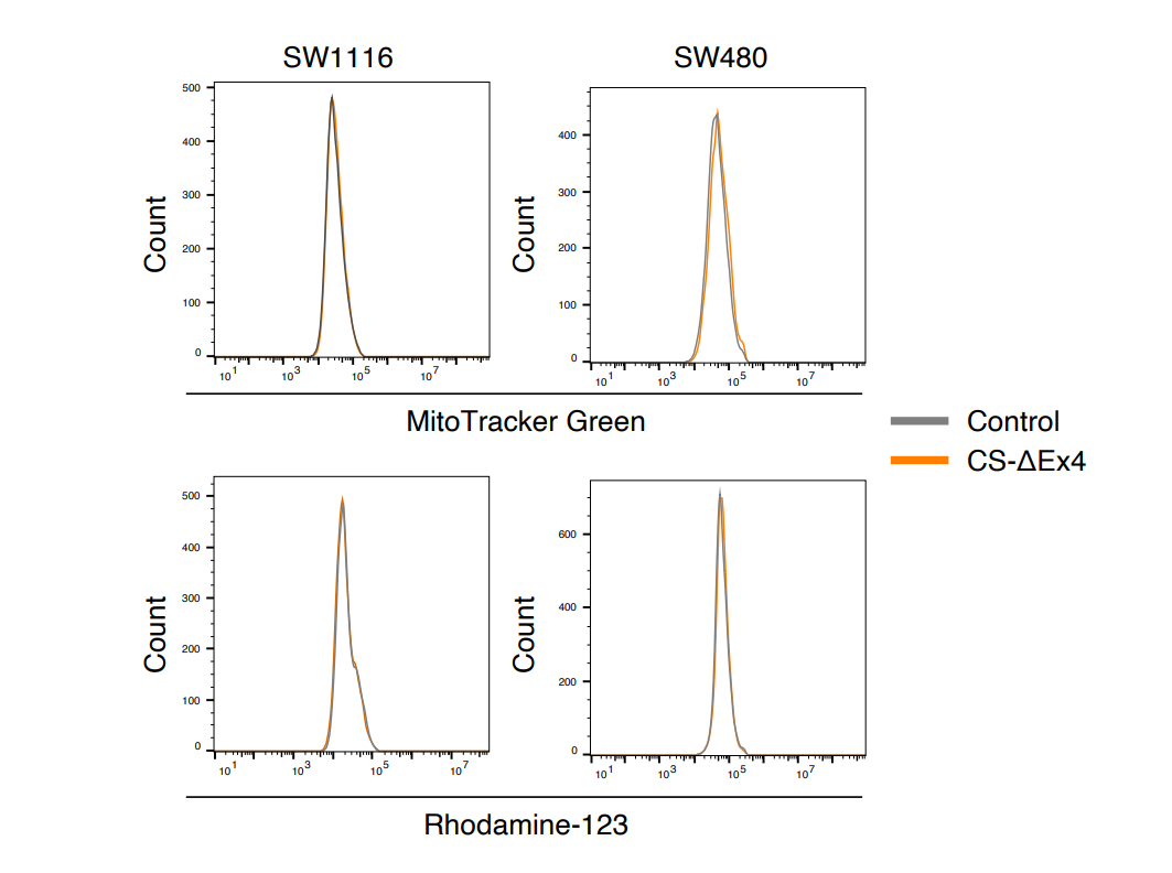
Cancer Res
A Citrate Synthase Splice Variant Rewires the TCA Cycle to Promote Colorectal Cancer Progression. [Abstract]2025 Sep 3. PMID: 40900036
Rhodamine 123 purchased from MedChemExpress. Usage Cited in: Cancer Res. 2025 Sep 3. [Abstract]
FACS detection of mitochondrial mass using MitoTracker Green and assessment of mitochondrial membrane potential using Rhodamine-123 (50 μM; 30 min).
-
J Adv Res
20(S)-Ginsenoside Rh2 overcomes gemcitabine resistance in pancreatic cancer by inhibiting LAMC2-Modulated ABC transporters. [Abstract]2025 Jul:73:743-760. PMID: 39270979 -
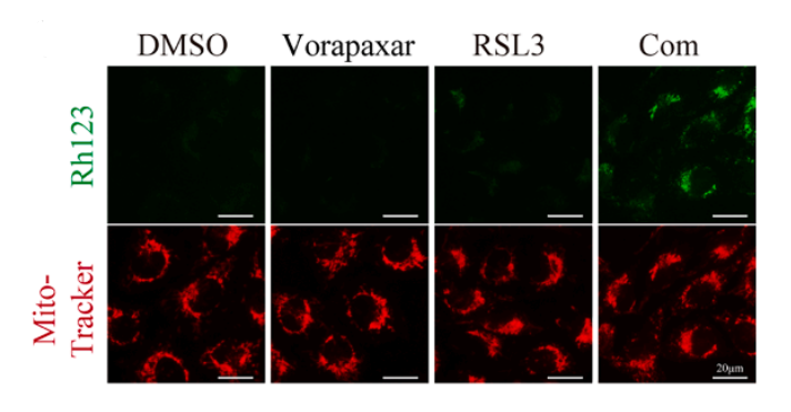
Cell Rep Med
Vorapaxar enhanced mitochondria-associated ferroptosis primes cancer immunotherapy via targeting FOXO1/HMOX1 axis. [Abstract]2025 Sep 25:102371. PMID: 41005298
Rhodamine 123 purchased from MedChemExpress. Usage Cited in: Cell Rep Med. 2025 Sep 25:102371. [Abstract]
Mitochondrial membrane potential assessment in A375 cells after 1 h treatments, using Rhodamine 123 (250 nM), with MitoTracker Red (mitochondrial marker). Scale bars, 20 μm.
-
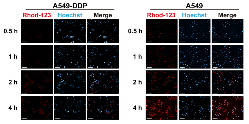
J Control Release
Combating cisplatin-resistant lung cancer using a coiled-coil lipopeptides modified membrane fused drug delivery system. [Abstract]2025 Jan 8:379:45-58. PMID: 39756686
Rhodamine 123 purchased from MedChemExpress. Usage Cited in: J Control Release. 2025 Jan 8:379:45-58. [Abstract]
Evaluation of P-gp function using Rhod-123 efflux assay. The fluorescent images for the uptake of Rhod-123 (3 μM) (red channel, Ex/Em = 507/529 nm) by A549-DDP and A549 at 0.5, 1, 2, and 4 h were obtained by the HCA System.
-
Cell Death Dis
Generation of proliferative hESC-derived grape-clustered hepatocyte organoids with multipolar architecture as regenerative counterpart via synergy of YAP and IGF2 pathways. [Abstract]2026 Mar 26;17(1):381. PMID: 41888105 -
Cell Commun Signal
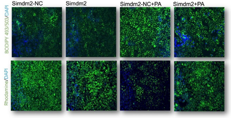
Regulatory role of sirtuin-1 by targeting MDM2 to mitochondria-associated membranes formation in the treatment of NAFLD. [Abstract]2025 Nov 28;23(1):514. PMID: 41316250
Rhodamine 123 purchased from MedChemExpress. Usage Cited in: Cell Commun Signal. 2025 Nov 28;23(1):514. [Abstract]
Rhodamine 123 (5 μM; 30 min) staining and BODIPY 493/503 staining for analyzing mitochondrial membrane potential and lipid droplets.
-
Phytomedicine
Vielanin K enhances doxorubicin-induced apoptosis via activation of IRE1α- TRAF2 - JNK pathway and increases mitochondrial Ca2 + influx in MCF-7 and MCF-7/MDR cells. [Abstract]2020 Nov:78:153329. PMID: 32896708 -
J Hazard Mater
Copper overload induces lifespan shortening through activating mitochondrial respiration in Caenorhabditis elegans. [Abstract]2026 Jan 15:502:140998. PMID: 41483520 -
J Ethnopharmacol
Reduction in gefitinib resistance mediated by Yi-Fei San-Jie pill in non-small cell lung cancer through regulation of tyrosine metabolism, cell cycle, and the MET/EGFR signaling pathway. [Abstract]2023 Oct 5:314:116566. PMID: 37169317 -
Biochem Pharmacol
Ticagrelor reverses multidrug resistance in breast cancer by inhibiting PI3K/AKT/mTOR pathway and suppressing ABCB1 expression and function. [Abstract]2026 Mar 15:249:117903. PMID: 41846011 -
Cell Mol Life Sci
Cholesterol neutralized vemurafenib treatment by promoting melanoma stem-like cells via its metabolite 27-hydroxycholesterol. [Abstract]2024 May 22;81(1):226. PMID: 38775844 -
Life Sci
Dimethyl fumarate restores Ca2+ dyshomeostasis through activation of the SIRT1 signal to treat nonalcoholic fatty liver disease. [Abstract]2024 Mar 15:341:122505. PMID: 38364937 -
Foods
Antibacterial Activity of Dihydroquercetin Separated from Fructus Polygoni orientalis against Clavibacter michiganensis subsp. sepedonicus via Damaging Cell Membrane. [Abstract]2023 Dec 20;13(1):23. PMID: 38201051 -
Chem Biol Interact
A microfluidic human blood-brain barrier model reveals neurovascular toxicity and barrier disruption induced by E-cigarette additives. [Abstract]2026 Aug 1:436:112206. PMID: 42285244 -
Chem Biol Interact
2025 Oct 30:111790. PMID: 41176046 -
J Nutr Biochem
Curcumin ameliorates CKD-induced mitochondrial dysfunction and oxidative stress through inhibiting GSK-3β activity. [Abstract]2020 Sep:83:108404. PMID: 32531667 -
PLoS Pathog
The viral BCL2 protein BHRF1 of Epstein-Barr virus promotes AIM2 inflammasome activation to facilitate lytic replication. [Abstract]2025 Sep 22;21(9):e1013509. PMID: 40982560 -
Med Oncol
Polyphyllin I inhibits ovarian cancer growth by inducing G0/G1 phase arrest and inhibiting the c-Myc signaling pathway. [Abstract]2025 Jun 12;42(7):254. PMID: 40506557 -
J Cell Mol Med
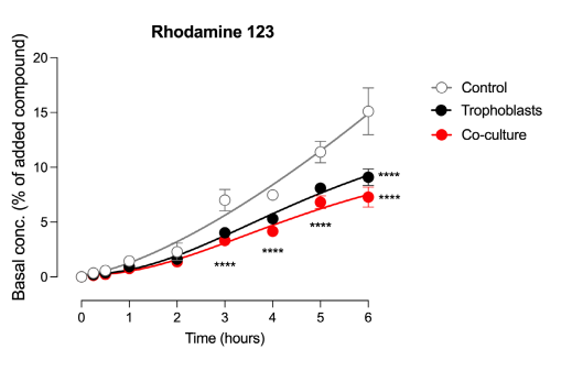
Modelling the maternal-fetal interface: An in vitro approach to investigate nutrient and drug transport across the human placenta. [Abstract]2024 Oct;28(20):e70151. PMID: 39422159
Rhodamine 123 purchased from MedChemExpress. Usage Cited in: J Cell Mol Med. 2024 Oct;28(20):e70151. [Abstract]
Rhodamine 123 (10 μM) was transported to the basal chamber within 6 hours.
-
J Physiol
A primary cell-based fluidic co-culture model to investigate drug transport across the human placenta. [Abstract]2026 Feb 26. PMID: 41746218 -
RSC Med Chem
Synthesis and evaluation of WK-X-34 derivatives as P-glycoprotein (P-gp/ABCB1) inhibitors for reversing multidrug resistance. [Abstract]2023 Dec 7;15(2):506-518. PMID: 38389882 -
J Biochem Mol Toxicol
Apelin-13 Protects Against Myocardial Hypoxia/Reoxygenation (H/R) Injury by Inhibiting Ferroptosis Via Nrf2 Activation. [Abstract]2025 Apr;39(4):e70223. PMID: 40152053 -
Food Chem Toxicol
Structure-activity relationship and mechanism of flavonoids on the inhibitory activity of P-glycoprotein (P-gp)-mediated transport of rhodamine123 and daunorubicin in P-gp overexpressed human mouth epidermal carcinoma (KB/MDR) cells. [Abstract]2021 Sep:155:112381. PMID: 34217736 -
Toxicol Lett
2024 Apr:394:76-91. PMID: 38428544 -
Fish Physiol Biochem
Ocean acidification induces neuronal hyperexcitation and anxiety-like behaviour in marine medaka via ASIC activation. [Abstract]2026 Apr 10;52(2):58. PMID: 41961137 -
Asian J Androl
2022 Sep-Oct;24(5):540-548. PMID: 35142655 -
Heliyon
A network pharmacology approach to reveal the key ingredients in Scrophulariae Radix (SR) and their effects against Alzheimer's disease. [Abstract]2024 Jan 20;10(3):e24785. PMID: 38322920 -
Oxid Med Cell Longev
Quercetin Protects against MPP+/MPTP-Induced Dopaminergic Neuron Death in Parkinson's Disease by Inhibiting Ferroptosis. [Abstract]2022 Sep 5:2022:7769355. PMID: 36105483 -
Oxid Med Cell Longev
Neuroprotective Effect of Salvianolic Acid A against Diabetic Peripheral Neuropathy through Modulation of Nrf2. [Abstract]2020 Feb 27:2020:6431459. PMID: 32184918


Solvent & Solubility
DMSO : 41.67 mg/mL (109.42 mM; Need ultrasonic; Hygroscopic DMSO has a significant impact on the solubility of product, please use newly opened DMSO)
Please refer to the solubility information to select the appropriate solvent. Once prepared, please aliquot and store the solution to prevent product inactivation from repeated freeze-thaw cycles.
Storage method and period of stock solution: -80°C, 2 years; -20°C, 1 year (sealed storage, away from moisture and light). When stored at -80°C, please use it within 2 years. When stored at -20°C, please use it within 1 year.
Concentration (start) × Volume (start) = Concentration (final) × Volume (final)

Protocol
Measurements are made at room temperature with continuous stirring of the mitochondrial suspension using spectrophotometer equipped with a magnetic stirrer with fluorescent cation R123 as probe. Excitation and emission wavelengths are 503 nm and 527 nm, respectively. The incubation medium is the respiration buffer. R123 and sodium pyruvate are added to final concentrations of 50 nM and 10 mM, respectively. Isolated mitochondria maintain a steady membrane potential (±5%) throughout the duration of the recording[1].
MedChemExpress (MCE) has not independently confirmed the accuracy of these methods. They are for reference only.


Purity & Documentation
-
Data Sheet (281 KB)
-
SDS (393 KB)
- English - EN (393 KB)
- Français - FR (393 KB)
- Deutsch - DE (393 KB)
- Norwegian - NO (393 KB)
- Español - ES (393 KB)
- Swedish - SV (393 KB)
- Italian - IT (393 KB)
- Korean - KR (393 KB)
- Portuguese - PT (393 KB)
-
Handling Instructions (2659 KB)
References


Complete Stock Solution Preparation Table
Please refer to the solubility information to select the appropriate solvent. Once prepared, please aliquot and store the solution to prevent product inactivation from repeated freeze-thaw cycles.
Storage method and period of stock solution: -80°C, 2 years; -20°C, 1 year (sealed storage, away from moisture and light). When stored at -80°C, please use it within 2 years. When stored at -20°C, please use it within 1 year.
| Optional Solvent | Concentration Solvent Mass | 1 mg | 5 mg | 10 mg | 25 mg |
|---|---|---|---|---|---|
| DMSO | 1 mM | 2.6259 mL | 13.1296 mL | 26.2591 mL | 65.6478 mL |
| 5 mM | 0.5252 mL | 2.6259 mL | 5.2518 mL | 13.1296 mL | |
| 10 mM | 0.2626 mL | 1.3130 mL | 2.6259 mL | 6.5648 mL | |
| 15 mM | 0.1751 mL | 0.8753 mL | 1.7506 mL | 4.3765 mL | |
| 20 mM | 0.1313 mL | 0.6565 mL | 1.3130 mL | 3.2824 mL | |
| 25 mM | 0.1050 mL | 0.5252 mL | 1.0504 mL | 2.6259 mL | |
| 30 mM | 0.0875 mL | 0.4377 mL | 0.8753 mL | 2.1883 mL | |
| 40 mM | 0.0656 mL | 0.3282 mL | 0.6565 mL | 1.6412 mL | |
| 50 mM | 0.0525 mL | 0.2626 mL | 0.5252 mL | 1.3130 mL | |
| 60 mM | 0.0438 mL | 0.2188 mL | 0.4377 mL | 1.0941 mL | |
| 80 mM | 0.0328 mL | 0.1641 mL | 0.3282 mL | 0.8206 mL | |
| 100 mM | 0.0263 mL | 0.1313 mL | 0.2626 mL | 0.6565 mL |


 Powered by Bioz