mRNA therapeutics use transient nucleic acid templates to direct protein production in cells. Their performance depends on the coordinated engineering of the RNA molecule and its delivery system. Optimization of modified nucleosides, the 5' cap, untranslated regions, coding sequence, and poly(A) tail can improve transcript stability, translation, and innate immune compatibility. Meanwhile, delivery systems must protect mRNA from degradation, promote cellular uptake and endosomal escape, and control tissue- or cell-specific expression.
This article reviews key advances in mRNA molecular design and delivery engineering, including nucleoside modification, cap and sequence optimization, ionizable lipid nanoparticles, organ- and cell-targeted delivery, and emerging non-LNP platforms. It also discusses major translational challenges, including extrahepatic delivery, repeat dosing, safety, manufacturing consistency, and the expansion of mRNA applications beyond vaccines.
- Advances in mRNA Therapeutic Design
- Optimization of mRNA Delivery: Toward Precise Targeting
- Challenges and Future Directions
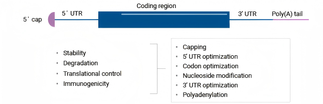
Figure 1. Composition of mRNA[1].
Messenger RNA (mRNA) functions as a carrier of genetic information, transferring the genetic protein-coding instructions encoded in DNA to the sites of protein synthesis. A synthetically engineered mRNA molecule is composed of several modular elements, including a 5' cap structure (5' Cap), a 5' untranslated region (5' UTR), a coding sequence (CDS), a 3' untranslated region (3' UTR), and a 3' polyadenylated tail (poly(A) tail). Rational design and chemical modification of these elements can enhance intracellular mRNA stability, improve translational efficiency, and optimize the expression level of the target protein[1].
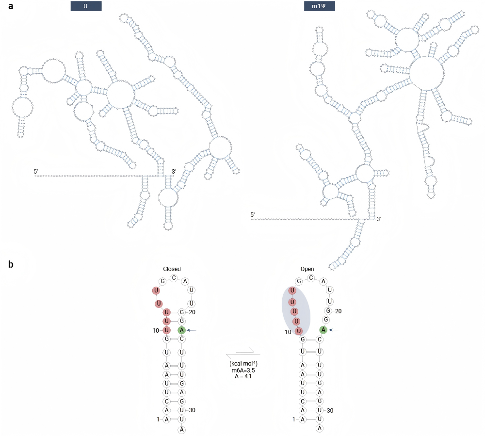
Figure 2. Modified nucleotides affect secondary structure[2].
Mammalian cells express a diverse array of pattern recognition receptors (PRRs) that recognize conserved structural features of viral RNA and initiate inflammatory responses. In the context of mRNA therapeutics, Toll-like receptors 7 and 8 (TLR7/8) are important immune-sensing receptors for exogenous RNA internalized through the endocytic pathway. Replacing natural uridine with modified nucleosides that are less readily sensed by these receptors—such as 5-methyluridine (m5U), 2-thiouridine (s2U), pseudouridine (Ψ), or 1-methylpseudouridine (m1Ψ)—can reduce innate immune recognition and improve the stability and translational performance of mRNA therapeutics[2].
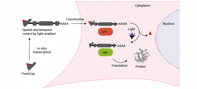
Figure 3. FlashCap modified at 5' cap was designed for optochemical control of mRNA translation. Irradiation allowed the expression of mRNA encoding eGFP with FlashCaps[7].
In eukaryotes, the mRNA cap is an N7-methylated guanosine located at the 5' terminus of the transcript. It is covalently linked to the first nucleotide through an unusual 5'-5' triphosphate bridge[3]. The 5' cap serves critical biological functions: it protects mRNA from exonuclease-mediated degradation, acts as a molecular marker for RNA processing and facilitates translation initiation. Given its pivotal role in transcript stability and translational efficiency, several in vitro capping strategies have been developed. Vaccinia virus capping enzyme and 2'-O-methyltransferase can be used for efficient post-transcriptional capping through the addition of appropriate substrates[4]. Optimized enzymatic capping processes can achieve high capping efficiencies and generate Cap1 structures, which are common in mammalian cells and are associated with efficient translation and reduced innate immune sensing. Conventional mRNA production can employ post-transcriptional enzymatic capping, whereas co-transcriptional technologies such as CleanCap® can also achieve capping efficiencies exceeding 90%. Together, these strategies can improve mRNA stability and translational expression while limiting unwanted innate immune activation[5].
Beyond their roles in mRNA stability and immune sensing, 5' cap modifications also enable precise regulation of translation initiation. Nils Klöcker and colleagues reported a class of 5' cap analogs bearing photocleavable groups, termed FlashCaps, that enable spatiotemporal control of translation through light irradiation. This design supports light-induced activation of translation while maintaining suitable stability and immune-response profiles, providing an approach for the optochemical regulation of mRNA translation[6].
Although the 5' UTR and 3' UTR do not encode proteins, they play critical roles in regulating mRNA translation and stability. The 5' UTR influences translation initiation, whereas the 3' UTR primarily modulates transcript stability and half-life. Together, these regulatory elements help determine the overall efficiency and duration of gene expression.
Current approaches to UTR optimization include selecting UTRs from highly expressed host-cell genes, such as the UTRs of human α-globin gene[8]. Another strategy is to construct UTR libraries and combine computational modelling with high-throughput screening to identify sequences with favourable expression properties[9].
In most eukaryotic mRNAs, the 3' poly(A) tail is generated through the addition of adenine residues after pre-mRNA processing. The poly(A) tail binds polyadenylate-binding protein (PABP), which interacts with the translation-initiation factor eIF4G and promotes the formation of a closed-loop mRNA structure. This interaction allows the poly(A) tail and the 5' cap to coordinate the regulation of translation initiation[10].
Poly(A)-tail engineering focuses on several strategies, including optimization of tail length, incorporation of structural elements such as poly(U) sequences, introduction of stem-loop structures upstream of the poly(A) tract, and the use of multiple or branched tails. These modifications can affect both mRNA translational efficiency and transcript stability. Chemical modification of the poly(A)-tail terminus may also stabilize its interaction with PABP and increase resistance to exonuclease-mediated degradation, thereby extending the level and duration of the target-protein expression.
Collectively, the structural elements of mRNA provide a flexible modular toolbox for mRNA engineering. Through systematic design and combinatorial optimization of these elements, researchers can improve translation efficiency and intracellular stability and achieve stronger and more sustained expression of the target protein.
Because of its high molecular weight, negative charge, and susceptibility to degradation, mRNA has limited ability to enter target cells efficiently. Naked mRNA is therefore generally unsuitable for most systemic delivery applications, highlighting the importance of delivery vectors for mRNA transfection and subsequent protein translation[11]. Delivery platforms must protect mRNA from degradation, support cellular uptake and endosomal escape, and, where possible, enable targeted delivery. A range of mRNA delivery systems has been developed, including platforms based on lipids, proteins/peptides, polymers and viruses, together with emerging delivery strategies.
Carrier for mRNA delivery | Advantages | Limitations | Clinical translation status |
|---|---|---|---|
| Lipid | High safety profile High transfection efficiency Composition Tunability | Immunogenicity risk | Marketed |
| Peptide | Favorable biocompatibility Enhanced designability Low immunogenicity of endogenous proteins | Poor stability Limited loading capacity | Early clinical trial stage |
| Polymer | High structural designability and controllability | Elevated toxicity | Preclinical trial stage |
| Viral vector | Superior transfection efficiency | Elevated toxicity Limited packaging capacity Strong immunogenicity Inherent high adjuvant property | Early clinical trial stage |
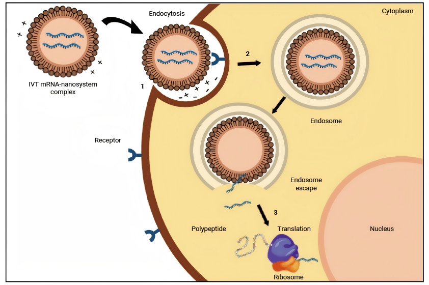
Figure 4. Intracellular barriers for in vitro transcribed (IVT) mRNA delivery[13].
Lipid nanoparticles (LNPs) are among the most widely investigated nonviral vectors for the in vivo delivery of nucleic acid vaccines[12]. Support for systemic LNP delivery was established in 2018 with the US Food and Drug Administration (FDA) approval of Onpattro® (patisiran), an siRNA-LNP therapy for polyneuropathy caused by amyloidosis. LNPs are also increasingly being investigated for gene therapy, protein replacement, and mRNA vaccines against cancer and infectious diseases.
LNPs typically consist of an ionizable lipid together with helper lipids such as phospholipids, cholesterol, and PEGylated lipids. Ionizable lipids associate with negatively charged mRNA under acidic formulation conditions, enabling efficient encapsulation within LNPs. The resulting particles are subsequently internalized by cells through endocytosis. As the endosomal lumen acidifies, ionizable lipids become protonated and interact with anionic membrane lipids, destabilizing the endosomal membrane and promoting the release of mRNA into the cytosol. Once released, the mRNA is translated by ribosomes to produce functional proteins. In vaccine applications, expression of an encoded antigen can induce an immune response and provide protection against the targeted disease[13].
In current LNP delivery systems, tissue distribution is dominated by passive targeting. Following systemic administration, LNPs often accumulate preferentially in the liver, largely because apolipoprotein E (ApoE) adsorbs onto the LNP surface and promotes low-density lipoprotein receptor (LDLR)-mediated uptake by hepatocytes. This inherent hepatic tropism can limit the delivery of mRNA therapeutics to extrahepatic tissues. Consequently, the rational design of LNPs for active targeting or specific extrahepatic tissue tropism remains a major challenge in nucleic-acid delivery.
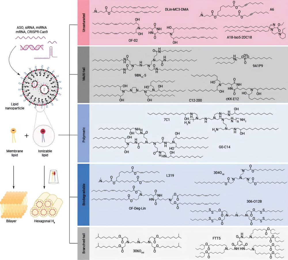
Figure 5. Five ionizable lipids have been widely applied in RNA delivery systems[14].
In LNPs, ionizable lipids are a critical component and often account for approximately 50% (molar ratio) of the formulation. They play important roles in mRNA encapsulation, protection and cytosolic delivery. At acidic pH, these lipids become positively charged, enabling efficient mRNA encapsulation; at physiological pH, they remain largely neutral, which reduces nonspecific interactions with endogenous biomolecules and can improve biocompatibility while limiting off-target toxicity.
Following cellular uptake, ionizable lipids become protonated within the acidic endosomal compartment. The protonated ionizable lipids then interact with negatively charged endosomal lipids, facilitating the formation of destabilizing cone-shaped ion pairs. This structural transition promotes membrane fusion and disruption, enabling endosomal escape and cytosolic release of the nucleic acid cargo. Rational modular design of the ionizable-lipid structures provides a route to novel lipids with improved delivery and extrahepatic targeting properties. Since 2008, a wide range of ionizable lipids with diverse chemical features has been developed. Systematic classification according to structural features can support the development of next-generation ionizable lipids. To date, five major classes have been widely applied in RNA delivery[14].
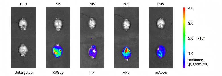
Figure 6. The transfection efficiency in the brain of LNPs conjugated with various peptides[16].
Tumor cells may overexpress specific receptors on their surfaces. By incorporating targeting moieties with affinity for these receptors onto the LNP surface, the encapsulated cargo can be preferentially delivered to target cells through receptor recognition and binding interactions. Based on the molecular type, targeting moieties can be broadly classified as peptides, antibodies, carbohydrates, ligands, and nucleic acid aptamers.
Beyond tumor-associated receptors, targeting ligands are also being investigated to overcome tissue-specific biological barriers, including the blood-brain barrier (BBB). The BBB , for instance, is an essential protective interface that shields the brain from potentially harmful circulating substances. However, it also restricts the passage of most small-molecule drugs and macromolecular therapeutics, including peptides, proteins, and gene-based agents, from the bloodstream into the brain parenchyma. This barrier substantially constrains the development and application of therapies for central nervous system (CNS) disorders[15].
Peptide-modified LNPs can engage receptors expressed on brain microvascular endothelial cells to support BBB crossing and brain delivery. For example, RVG29 peptide, which targets the nicotinic acetylcholine receptors (nAChR) , and the T7 peptide, which targets the transferrin receptor (TfR), have both been reported to improve the brain-targeting efficiency of LNPs[16].
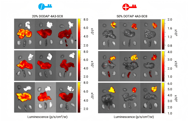
Figure 7. The tissue targeting of delivery can be altered by adjusting the ratio of DODAP[17].
Passive targeting is influenced by the physicochemical properties of nanoparticles, including particle size and surface charge. For example, smaller LNPs with negatively charged surfaces may be taken up more efficiently by lymph node dendritic cells, making them potentially useful for vaccine delivery.
Selective organ targeting (SORT) technology incorporates a fifth lipid component with defined charge or chemical properties into a conventional four-component LNP formulation. By adjusting the identity and molar ratio of this SORT molecule, researchers can alter the tissue distribution of the delivered cargo. Studies using cationic, anionic, and ionizable SORT lipids have shown that formulation composition is a key determinant of organ-selective reporter expression.
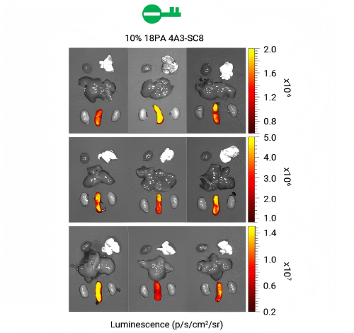
Figure 8. 10% 18PA mediates the transfer of luciferase protein expression to the spleen[17].
In addition, incorporation of 10%-40% 18PA as an anionic SORT molecule produced formulations with enhanced splenic delivery. These findings demonstrate the feasibility of achieving organ-selective targeting by modulating both the identity and proportion of SORT components within an LNP formulation[17].
For vaccines, a limited number of administrations are generally sufficient to meet prophylactic needs. However, metabolic enzyme replacement, chronic protein deficiencies, repeated immunomodulation, and long-term in vivo cell engineering may require multiple or prolonged dosing of mRNA therapeutics.
In these settings, the barriers to repeat administration are not simply a matter of accumulating immunogenicity. Instead, they involve interconnected factors, including innate immune activation, anti-PEG or anti-vector responses, complement activation, accelerated blood clearance (ABC), dose-related toxicity, and changes in pharmacokinetics and tissue distribution after repeated dosing.
Researchers have developed high-density brush-like polymer lipids (BPLs), whose comb-like structures and mushroom/brush conformations reduce antibody binding. Conventional DMG-PEG2000 LNPs can induce an anti-PEG antibody (APA) response and a marked decline in luciferase mRNA expression after a second dose in vivo, whereas BPL LNPs maintain more stable expression across repeated administrations. The immune response observed with BPL LNPs was also lower than that associated with DMG-PEG2000. These findings show that fine-tuning the polymer architecture can improve LNP performance in repeat-dosing regimens[18].
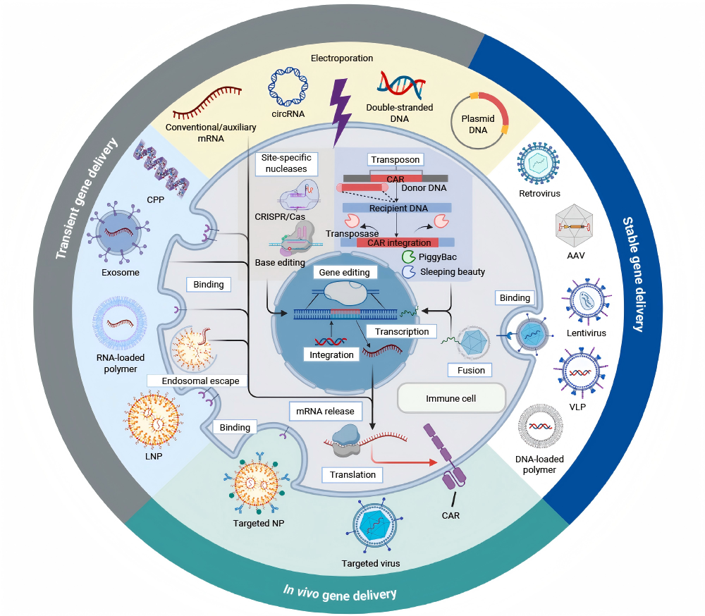
Figure 9. Various delivery routes for CAR gene therapy[19].
Chimeric antigen receptor T-cell (CAR-T) therapy involves the genetic engineering of T lymphocytes to enable them to recognize and eliminate target tumor cells. Efficient and safe delivery of the CAR transgene into immune cells is therefore a critical determinant of successful cell-based therapy.
Conventional CAR-T therapies typically rely on lentiviral vectors for gene delivery; however, genomic integration can introduce a risk of insertional mutagenesis. Lentiviral-vector production is also technically complex, costly, and may trigger host immune responses, which can limit the safety, scalability, and accessibility of this approach[19].
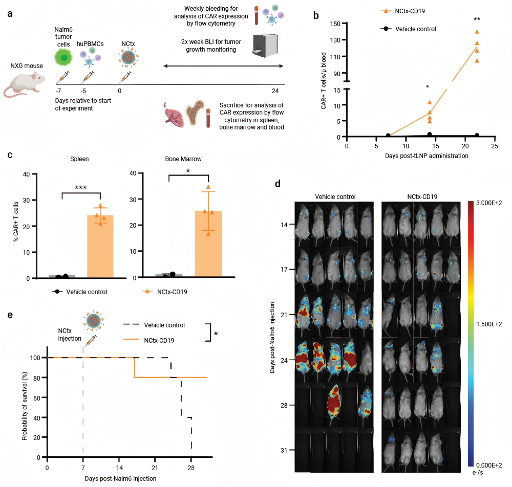
Figure 10. In vivo generation of CAR-T cells by NCtx-CD19[20].
In contrast, nonviral LNP-based approaches may offer high programmability and simplified manufacturing while avoiding some limitations associated with viral systems. For example, researchers developed an LNP formulation termed NCtx that achieves T cell-specific delivery through surface conjugation of anti-CD7 nanobodies and anti-CD3 single-chain variable fragments (scFv)[20].
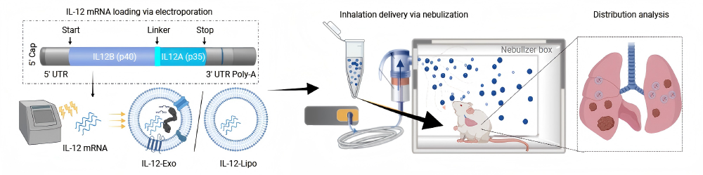
Figure 11. IL-12 mRNA loads into HEK-Exo (IL-12-Exo) or liposomes (IL-12-Lipo), followed by nebulized inhalation administration to LL/2 tumour-bearing mouse lungs[23].
Alongside established mRNA delivery carriers, several emerging platforms are being investigated. Extracellular vesicles (EVs), for example, have attracted interest as potential mRNA delivery vehicles. EVs are nanoscale vesicles secreted by cells, that mediate intercellular communication and facilitate the transfer of proteins, lipids and genetic material[21]. Cell-derived EVs can offer favourable biocompatibility and relatively low immunogenicity; however, batch-to-batch variation in composition and biological activity may affect delivery efficacy. EVs derived from different cell types can also vary substantially in their properties and functions[22].
Despite the lower maturity and narrower application breadth compared with LNP and polymers, inorganic nanoparticles have also attracted interest as potential mRNA delivery platforms. Carbon quantum dots (CQDs) have photoluminescent properties that may support imaging and tracking, whereas gold nanoparticles offer tunable size and surface chemistry. However, their mRNA delivery efficiency, safety, and translational potential require further validation[24].
The performance of mRNA therapeutics depends on coordinated optimization of both the RNA molecule and its delivery system. Improvements in nucleoside chemistry, the 5' cap, UTRs, coding sequence, poly(A) tail, and delivery platforms can enhance stability, translation, immune compatibility, cellular uptake, endosomal escape, and tissue-specific expression. Future progress will rely on more reliable extrahepatic and cell-specific delivery, improved repeat-dosing performance, and better control of formulation consistency and safety, helping expand mRNA therapeutics beyond vaccines into protein replacement, immunomodulation, and in vivo cell engineering.
Note: MCE can provide products for research use only. We do not sell to patients.
 LinkedIn
 Researchgate
 Instagram
 X
X Facebook
 WhatsApp
 Reddit
 Mail
Mail- [1]. Chien KR, et al. Nat Rev Drug Discov. 2026 Jun 3. [Content Brief]
- [2]. Metkar M, et al. Nat Rev Drug Discov. 2024 Jan;23(1):67-83. [Content Brief]
- [3]. A. Ramanathan, et al. Nucleic Acids Res. 2016 Sep 19;44(16):7511-26. [Content Brief]
- [4]. S.W. Lockless, et al. Biochemistry. 1998 Jun 9;37(23):8564-74. [Content Brief]
- [5]. S.C. Devarkar, et al. Proc Natl Acad Sci U S A. 2016 Jan 19;113(3):596-601. [Content Brief]
- [6]. N. Klöcker, et al. Nat Chem. 2022 Aug;14(8):905-913. [Content Brief]
- [7]. Zhou H, et al. Nanomicro Lett. 2026 Jan 15;18(1):118. [Content Brief]
- [8]. J.R. Babendure, et al. RNA. 2006 May;12(5):851-61. [Content Brief]
- [9]. P.J. Sample, et al. Nat Biotechnol. 2019 Jul;37(7):803-809. [Content Brief]
- [10]. Tarun SZ Jr, et al. EMBO J. 1996 Dec 16;15(24):7168-77. [Content Brief]
- [11]. Zhong Y, et al. Acta Pharm Sin B. 2023 Apr;13(4):1348-1357. [Content Brief]
- [12]. Whitehead K.A. et al. Nat Rev Drug Discov. 2009 Feb;8(2):129-38. [Content Brief]
- [13]. Aldosari BN, et al. Pharmaceutics. 2021 Feb 2;13(2):206. [Content Brief]
- [14]. Han X, et al. Nat Commun. 2021 Dec 13;12(1):7233. [Content Brief]
- [15]. Liu S, et al. J Nanobiotechnology. 2025 May 26;23(1):382. [Content Brief]
- [16]. Han EL, et al. Nano Lett. 2025 Jan 15;25(2):800-810. [Content Brief]
- [17]. Wang X, et al. Nat Protoc. 2023 Jan;18(1):265-291. [Content Brief]
- [18]. Xiao Y, et al. Nat Mater. 2025 Nov;24(11):1840-1851. [Content Brief]
- [19]. Xiao Y, et al. Trends Mol Med. 2025 Nov;31(11):1047-1064. [Content Brief]
- [20]. Bimbo JF, et al. J Immunother Cancer. 2025 Jul 13;13(7):e011759.
- [21]. Han QF, et al. Mol Cancer. 2022 Nov 1;21(1):207.
- [22]. Zhu X, et al. J Extracell Vesicles. 2017 Jun 6;6(1):1324730. [Content Brief]
- [23]. Liu M, et al. Nat Nanotechnol. 2024 Apr;19(4):565-575.
- [24]. Meixiu Li, et al. ACS Sens. 26 July 2019; 4 (7): 1732–1748.