



Firefly luciferase mRNA-LNP
Based on 1 publication(s) in Google Scholar
Firefly luciferase mRNA-LNP is a lipid nanoparticle (LNP) containing firefly luciferase mRNA. Firefly luciferase mRNA-LNP exhibits a stable nanostructure, in which LNP plays a key role in effectively protecting and transporting mRNA to cells. Luciferase is a bioluminescent reporter gene for gene regulation and functional studies. Firefly Luciferase mRNA-LNP expresses firefly luciferase protein after entering the cells, which is often used for promoter activity detection or dual fluorescent molecular complementation experiments. Firefly luciferase mRNA-LNP is studied in research for RNA delivery, cell viability, and translation efficiency.
For research use only. We do not sell to patients.
-
Storage:
-80°C


To place orders, for customer services and technical support, please contact: MedChemExpress USA
Tel: 609-228-6898 E-mail: [email protected] [email protected]


-
 Biological Activity
-
Chemical Information
- Protocols
- Purity & Documentation
- References
-
 Help & FAQs




Publications Citing Use of MedChemExpress (MCE) Firefly luciferase mRNA-LNP
More Customer Validation & Images

-
In Vivo Imaging


Biological Activity
Description
In Vitro
Firefly luciferase mRNA-LNP (5-20 ng, 24 h) significantly increases the transfection efficiency dose-dependently in HepG2 cells[3].
MedChemExpress (MCE) has not independently confirmed the accuracy of these methods. They are for reference only. Further protocols information, click here.
In Vivo
Firefly luciferase mRNA-LNP (1 μg, i.m., on day 1 and day 21) is formulated with either ALC-0315 or SM-102, and SM-102 has 60% higher expression level of luciferase protein than ALC-0315 and cKK-E12 in BALB/c mice[4].
Firefly luciferase mRNA-LNP (5-20 μg, i.m., single dose) induces activation of Aif1 in the hypothalamus but not in the prefrontal cortex of adult Balb/c male mice[5].
Firefly luciferase mRNA-LNP (5 μg, i.m./i.v./i.p./i.t., single dose) results in a large portion of the luciferase activity as well as high levels of protein production in the liver, and significant bioluminescent signal in lungs and muscles of BALB/c mouse model[6].
Firefly luciferase mRNA-LNP (5 μg, i.d./s.c., single dose) results in protein production only at the site of injection in BALB/c mouse model[6].
MedChemExpress (MCE) has not independently confirmed the accuracy of these methods. They are for reference only.
-
Animal Model:Female BALB/c mice aged 6 weeks[1]
-
Dosage:0.1, 1.0, 5.0 μg
-
Administration:Intradermal injection (i.d.)/Intraperitoneal injection (i.p.)/Subcutaneous injection (s.c.)/Intramuscular injection (i.m.)/Intravenous injection (i.v.)/Intratracheal injection (i.t.)
-
Result:Could not measure signal when mice were injected with 0.1 μg of mRNA-LNPs.
Resulted in the highest amount of peak protein production and the shortest duration of expression with intravenous delivery.
Did not result in measurable luciferase expression in the liver or other anatomical sites with subcutaneous delivery.
Resulted in the longest duration of translation with intramuscular and intradermal delivery.
Allowed saturation of the uptake or translational capacity of the intradermal space with intradermal delivery.
Observed linear dose response with intravenous injection.
Led to similar levels of peak translation at 4 h post injection with all three doses, but the increased doses extended the duration of translation from 5 (1.0 μg) to 10 (5.0 μg) days.


Chemical Information
-
Appearance Liquid
-
Color Colorless to light yellow
-
SMILES
[Firefly luciferase mRNA-LNP]
-
Shipping
Shipping with dry ice.
-
Storage
-80°C


Publications (1)
-
Journal Impact Factor
-
Most Recent
-
Sci Adv
Macrophage-targeted Mms6 mRNA-lipid nanoparticles promote locomotor functional recovery after traumatic spinal cord injury in mice. [Abstract]2025 Mar 28;11(13):eads2295. PMID: 40138430
Firefly luciferase mRNA-LNP purchased from MedChemExpress. Usage Cited in: Sci Adv. 2025 Mar 28;11(13):eads2295. [Abstract]
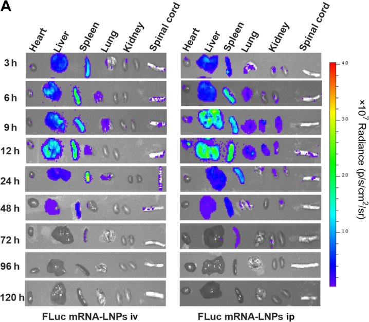
Representative in vivo imaging system images of major organs in SCI mice after being administered with firefly luciferase (FLuc) mRNA-LNPs (0.5 mg mRNA/kg). From left to right are the heart, liver, spleen, lung, two kidneys, and spinal cord. iv, intravenous; ip, intraperitoneal.

Protocols
-
RNA extraction experimental
By lysing cells, releasing RNA, and removing impurities such as proteins and DNA, high-purity RNA products are finally obtained. The commonly used traditional method is the guanidine isothiocyanate/phenol/chloroform method (Trizol), which is suitable for a variety of animal materials including animal tissues, microorganisms, cultured cells, etc., and most plant materials.
-
Dual Luciferin reporter gene assay
Luciferin reporter gene assay is a reporting system to detect the activity of Firefly Luciferase using luciferin as a substrate, which is often used in the research of miRNA target gene verification and promoter transcriptive activity regulation. Dual luciferase usually refers to Firefly luciferase and Renilla luciferase.
-
Cell Cytotoxicity Assay
Cytotoxicity assays are usually based on the assessment of cell membrane damage, which can also be indirectly detected by measuring cell viability. Detection methods include MTT assay, CKK-8 assay, LDH assay and ATP assay, etc.
-
Mammalian live/dead viability and cytotoxicity staining
Live/dead viability and cytotoxicity staining assays are based on the simultaneous detection of intracellular esterase activity in metabolically active (viable) cells and membrane integrity loss in non-viable cells. In commonly used dual-staining approaches, membrane-permeant fluorogenic substrates are converted by intracellular esterases into fluorescent products in live cells, while impermeant DNA-binding dyes selectively enter cells with compromised plasma membranes and label nucleic acids in dead or dying cells, enabling discrimination between viable and non-viable populations by fluorescence microscopy or flow cytometry.
-
CCK-8/WST-8 Cell Proliferation Assay
The CCK-8/WST-8 assay is based on the reduction of the water-soluble tetrazolium salt WST-8 to a water-soluble formazan product by cellular dehydrogenases in metabolically active cells, where the generated formazan amount is proportional to the number of living cells and is quantified by measuring absorbance in the visible range, providing a colorimetric readout for cell viability and proliferation assessment. This class of tetrazolium-based assays improves upon earlier MTT-based systems by producing a water-soluble formazan, eliminating the need for organic solubilization steps and enabling direct spectrophotometric measurement in culture medium.
-
CRISPRi/CRISPRa gene-regulation editing
CRISPRi and CRISPRa use catalytically inactive Cas9, typically SpCas9 D10A/H840A, as an RNA-guided DNA-binding platform that targets genomic loci through sgRNA complementarity and an adjacent PAM without generating Cas9 nuclease-mediated DNA cleavage. CRISPRi represses transcription by recruiting dCas9 alone or dCas9 fused to repressor domains such as KRAB to promoters or transcription start site regions, while CRISPRa activates transcription by recruiting activation domains such as VP64, VPR, or SAM components to promoter-proximal regions. The primary readout is target-gene expression change, commonly measured by RT-qPCR, RNA-seq, reporter fluorescence, or protein-level assays, and the readout reflects transcriptional repression or activation at the targeted endogenous locus.
-
MTT Cell Proliferation Assay
The MTT assay is a colorimetric endpoint assay for estimating viable cell number, cell growth, cytotoxicity, or cell activation in cultured mammalian cells. Living cells reduce the yellow tetrazolium salt MTT into purple/blue formazan, while dead cells do not generate the same signal; the resulting color can be quantified with a multiwell spectrophotometer. MTT reduction is commonly interpreted as a readout of metabolic activity that often correlates with viable cell number, but it should not be treated as a direct cell-counting method unless the assay is optimized for the cell type and experimental condition. Studies show that MTT reduction can involve mitochondrial and non-mitochondrial reducing systems, and formazan may accumulate in intracellular lipid droplets rather than simply marking mitochondria.
-
Cell Viability Determination by MTT Colorimetric Assay
The following protocol uses the MTT colorimetric assay as a classic literature-established method for assessing cell viability/metabolic activity in cultured mammalian cells. MTT[3-(4,5-dimethylthiazol-2-yl)-2,5-diphenyltetrazolium bromide] is reduced by metabolically active cells to a colored formazan product; the amount of formazan is quantified spectrophotometrically and provides an indirect measure of metabolically active viable cells. Importantly, MTT reduction reflects cellular oxidoreductase/metabolic activity rather than an absolute direct count of living cells, so changes in cellular metabolism can alter the signal independently of cell number.


Purity & Documentation
-
Data Sheet (274 KB)
-
SDS (251 KB)
- English - EN (251 KB)
- Français - FR (251 KB)
- Deutsch - DE (251 KB)
- Norwegian - NO (251 KB)
- Español - ES (251 KB)
- Swedish - SV (251 KB)
- Italian - IT (251 KB)
- Korean - KR (251 KB)
- Portuguese - PT (251 KB)
-
Handling Instructions (2659 KB)
References
[1]. Tenchov, R., et al., (2021). Lipid Nanoparticles─From Liposomes to mRNA Vaccine Delivery, a Landscape of Research Diversity and Advancement. ACS nano, 15(11), 16982-17015. [Content Brief]
[2]. Xu YZ, et al. Promoter deletion analysis using a dual-luciferase reporter system. Methods Mol Biol. 2013;977:79-93. [Content Brief]
[3]. El-Mayta, R., et al., (2023). Testing the In Vitro and In Vivo Efficiency of mRNA-Lipid Nanoparticles Formulated by Microfluidic Mixing. Journal of visualized experiments: JoVE, (191), 10.3791/64810. [Content Brief]
[4]. Zhang, L., et al., (2023). Effect of mRNA-LNP components of two globally-marketed COVID-19 vaccines on efficacy and stability. NPJ vaccines, 8(1), 156. [Content Brief]
[6]. Pardi, N., et al., (2015). Expression kinetics of nucleoside-modified mRNA delivered in lipid nanoparticles to mice by various routes. Journal of controlled release: official journal of the Controlled Release Society, 217, 345–351. [Content Brief]

Calculators
Concentration (start) × Volume (start) = Concentration (final) × Volume (final)


Firefly luciferase mRNA-LNP Related Classifications
HY-153229 Related Classifications
 Powered by Bioz