



MitoMark Red I
Based on 1 publication(s) in Google Scholar
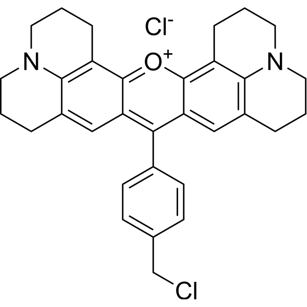
MitoMark Red I is a mitochondria-targeted fluorescent dye. MitoMark Red I accumulates in mitochondria depending on the mitochondrial membrane potential, thereby staining active mitochondria in living cells. MitoMark Red I can be used for live-cell imaging and fixed-cell studies (Ex/Em = 578/599 nm).
For research use only. We do not sell to patients.
- Purity : 97.34%
- CAS No.: 167095-09-2
- Formula: C32H32Cl2N2O
- Molecular Weight:531.52
-
Storage:
-20°C, sealed storage, away from moisture and light
* In solvent : -80°C, 6 months; -20°C, 1 month (sealed storage, away from moisture and light)

To place orders, for customer services and technical support, please contact: MedChemExpress USA
Tel: 609-228-6898 E-mail: [email protected] [email protected]


-
 Biological Activity
-
Chemical Information
- Protocols
- Purity & Documentation
- References
-
 Help & FAQs


-
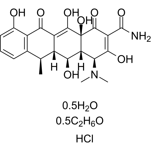
HY-N0565B
Doxycycline hyclateAntibiotic -
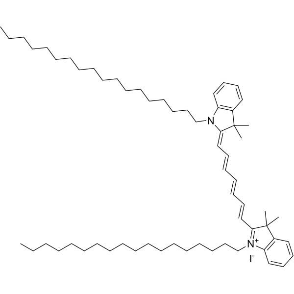
HY-D1048
DiRMembrane Dye -

HY-DY1013
DiR (solution)Membrane Dye -
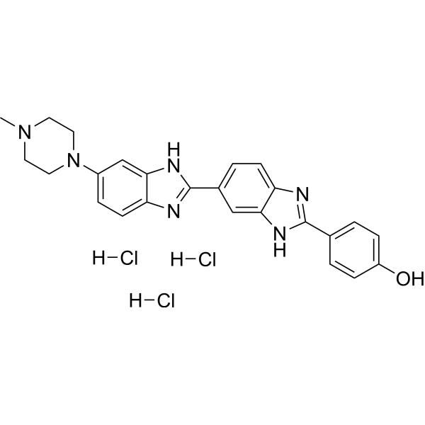
HY-15558A
Hoechst 33258 trihydrochlorideDNA Staining -
HY-135056
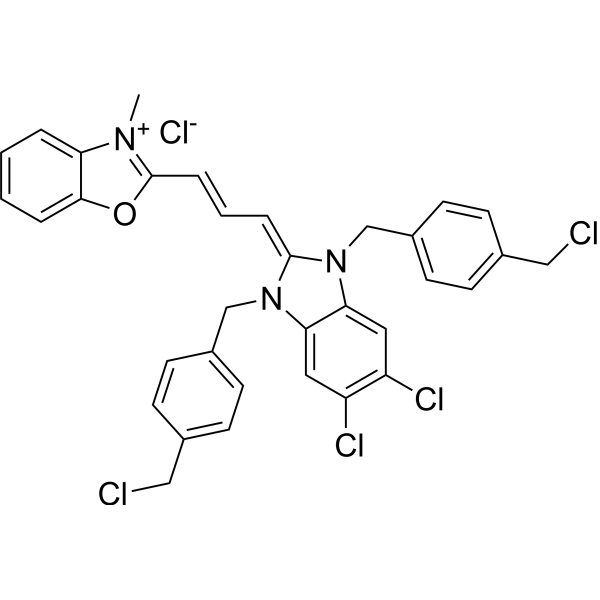
MitoTracker Green FMMitochondrial Dye -
HY-D0816
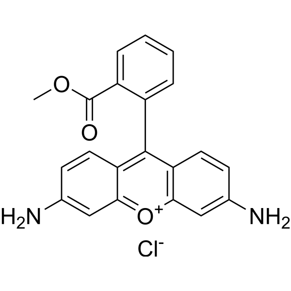
Rhodamine 123Fluorescent Dye -

HY-DY1032
MitoTracker Green FM (solution)Mitochondrial Dye -
HY-D1300
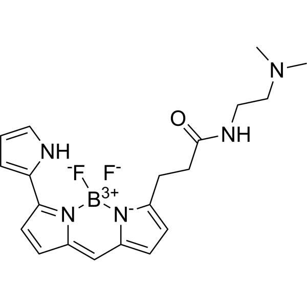
LysoTracker RedCell Death Indicator -

HY-DY1040
LysoTracker Red (solution)Cell Death Indicator -
HY-D1297
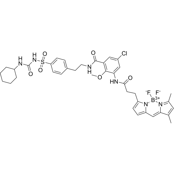
ER-Tracker GreenEndoplasmic Reticulum Fluorescent Dye -
HY-D0815
Propidium IodideFluorochrome -

HY-DY1043
ER-Tracker Green (solution)Endoplasmic Reticulum Fluorescent Dye
Publications Citing Use of MedChemExpress (MCE) MitoMark Red I
More

Biological Activity
Description
In Vitro
1. Stock Solution Preparation
1.1 Prepare a 1 mM stock solution in anhydrous DMSO.
2. Working Solution Preparation
2.1 Dilute with Hepes buffer.
2.2 Working concentration: 100-500 nM.
2.3 Notes: The working solution concentration can be adjusted according to requirements; prepare fresh before use.
3. Staining Procedure
3.1 Cell staining (suspension cells)
3.1.1 Collect cells by centrifugation, add Hepes buffer and wash twice, 5 minutes each time.
3.1.2 Gently resuspend the cells in the staining solution and incubate at 37°C for 30 minutes.
3.1.3 Centrifuge at 400 g for 3-4 minutes and discard the supernatant.
3.1.4 Add Hepes buffer to wash the cells twice, 5 minutes each time.
3.1.5 After resuspending the cells in Hepes buffer, observe using a fluorescence microscope.
3.2. Cell staining (adherent cells)
3.2.1 Culture adherent cells on sterile coverslips.
3.2.2 Remove the coverslip from the culture medium and aspirate excess medium.
3.2.3 Add the dye working solution, rock gently to ensure complete coverage of the cells, and incubate at 37°C for 30 minutes.
3.2.4 Aspirate the dye working solution and replace with 1x Hanks buffer (containing 20 mM HEPES), then observe using a fluorescence microscope.
4. Detection and Analysis
4.1 Instrument type: fluorescence microscope.
4.2 Result analysis: MitoMark Red I specifically labels mitochondria and emits red fluorescence, enabling direct visualization of the relative localization of mitochondria and free radicals within cells.
MedChemExpress (MCE) has not independently confirmed the accuracy of these methods. They are for reference only.


Chemical Information
-
CAS No. 167095-09-2
-
Appearance Solid
-
Molecular Weight 531.52
-
Formula C32H32Cl2N2O
-
Color Brown to black
-
SMILES
ClCC1=CC=C(C(C2=CC3=C4N(CCC3)CCCC4=C2[O+]=C56)=C5C=C7CCCN8CCCC6=C87)C=C1.[Cl-]
-
Shipping
Room temperature in continental US; may vary elsewhere.
-
Storage
-20°C, sealed storage, away from moisture and light
* In solvent : -80°C, 6 months; -20°C, 1 month (sealed storage, away from moisture and light)


Publications (1)
-
Journal Impact Factor
-
Most Recent
-
Redox Biol
TRPV4 drives macrophage pyroptosis via mitochondrial dysfunction and mtROS-dependent NLRP3 inflammasome activation in acute lung injury. [Abstract]2026 Jun 11:95:104255. PMID: 42314332


Purity & Documentation
-
Data Sheet (278 KB)
-
SDS (252 KB)
- English - EN (252 KB)
- Français - FR (252 KB)
- Deutsch - DE (252 KB)
- Norwegian - NO (252 KB)
- Español - ES (252 KB)
- Swedish - SV (252 KB)
- Italian - IT (252 KB)
- Korean - KR (252 KB)
- Portuguese - PT (252 KB)
-
Handling Instructions (2659 KB)
References

Calculators
Concentration (start) × Volume (start) = Concentration (final) × Volume (final)


MitoMark Red I Related Classifications
HY-D1116 Related Classifications
 Powered by Bioz