



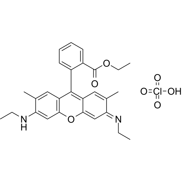
Rhodamine 6G perchlorate
Based on 4 publication(s) in Google Scholar
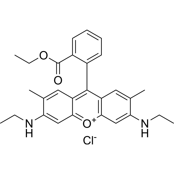
Rhodamine dyes are membrane-permeable cationic fluorescent probes that specifically recognize mitochondrial membrane potentials, thereby attaching to mitochondria and producing bright fluorescence, and at certain concentrations, rhodamine dyes have low toxicity to cells, so they are commonly used to detect mitochondria in animal cells, plant cells, and microorganisms.
For research use only. We do not sell to patients.
- CAS No.: 13161-28-9
- Formula: C28H31ClN2O7
- Molecular Weight:543.01
-
Storage:
Please store the product under the recommended conditions in the Certificate of Analysis.

To place orders, for customer services and technical support, please contact: MedChemExpress USA
Tel: 609-228-6898 E-mail: [email protected] [email protected]


-
 Biological Activity
-
Chemical Information
- Purity & Documentation
- References
-
 Help & FAQs


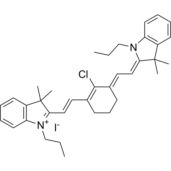
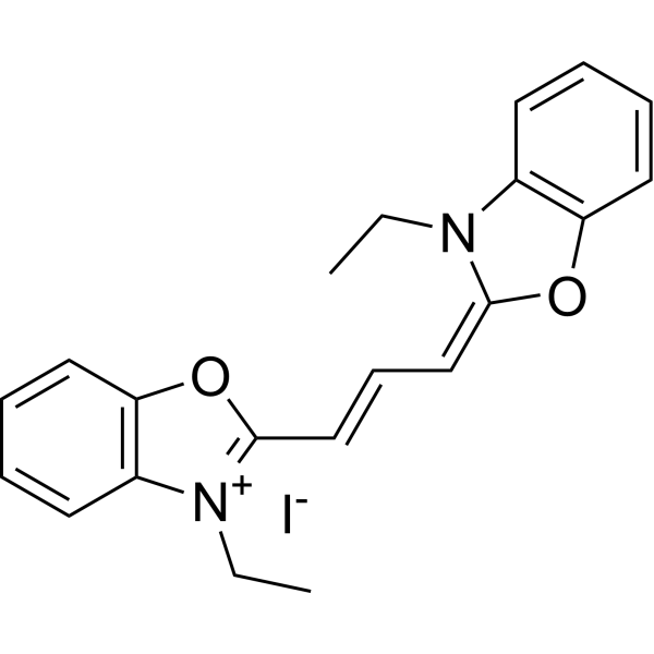
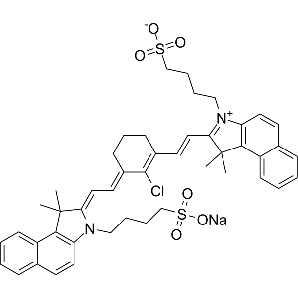

Publications Citing Use of MedChemExpress (MCE) Rhodamine 6G perchlorate
More Customer Validation & Images

-
Cell Imaging/Staining


Biological Activity
Description
In Vitro
Guide (The following is our recommended experimental protocol. This protocol serves merely as a reference guide; specific procedures should be adjusted according to your actual requirements.)
1. Preparation of Rhodamine 6G perchlorate Working Solution
1.1 Preparation of Stock Solution
Dilute 1 mg of Rhodamine 6G perchlorate with 525 μL of anhydrous DMSO to prepare a 5 mM stock solution.
1.2 Preparation of Working Solution
Dilute the stock solution with pre-warmed serum-free cell culture medium or PBS to prepare a Rhodamine 6G perchlorate working solution with a concentration of 1-20 μM.
Note: Please adjust the concentration of the Rhodamine 6G working solution according to your specific needs, and prepare it immediately before use.
2. Cell Staining (Suspension Cells)
2.1 Centrifuge to harvest the cells, then wash twice with PBS (5 minutes per wash). Adjust the cell density to 1 × 10⁶ cells/mL.
2.2 Add 1 mL of the Rhodamine 6G working solution and incubate at room temperature for 30–60 minutes.
2.3 Centrifuge at 400 × g for 3-4 minutes, and discard the supernatant.
2.4 Wash the cells twice with PBS (5 minutes per wash).
2.5 Resuspend the cells in 1 mL of serum-free medium or PBS, then observe using a fluorescence microscope or flow cytometer.
3. Cell Staining (Adherent Cells)
3.1 Culture the adherent cells on sterile coverslips.
3.2 Remove the coverslips from the culture medium and aspirate any excess medium.
3.3 Add 100 μL of the dye working solution, gently agitate to ensure the solution completely covers the cells, and incubate for 5-30 minutes.
3.4 Aspirate the working dye solution, wash with culture medium 2-3 times (5 minutes each), and observe using a fluorescence microscope or flow cytometer.
MedChemExpress (MCE) has not independently confirmed the accuracy of these methods. They are for reference only.


Chemical Information
-
CAS No. 13161-28-9
-
Molecular Weight 543.01
-
Formula C28H31ClN2O7
-
SMILES
CCNC1=CC2=C(C=C1C)C(=C3C=C(C)/C(=N/CC)/C=C3O2)C4=CC=CC=C4C(=O)OCC.Cl(=O)(=O)(=O)O
-
Synonyms
Basic Red 1 perchlorate
-
Shipping
Room temperature in continental US; may vary elsewhere.
-
Storage
Please store the product under the recommended conditions in the Certificate of Analysis.


Publications (4)
-
Journal Impact Factor
-
Most Recent
-
Int J Biol Macromol
Low molecular weight protein tyrosine phosphatase: A driver of lipid metabolic remodeling in Caenorhabditis elegans. [Abstract]2025 May;306(Pt 1):141332. PMID: 39988157
Rhodamine 6G perchlorate purchased from MedChemExpress. Usage Cited in: Int J Biol Macromol. 2025 May;306(Pt 1):141332. [Abstract]
Representative images and relative fluorescence intensity of membrane potential in control and lmwptp RNAi-treated C. elegans, stained with Rhodamine 6G.
-
Thromb Haemost
Inhibition of IP3 (Inositol 1,4,5-Trisphosphate) Receptors Retards SARS-CoV-2-Induced Endothelial von Willebrand Factor Secretion and Thrombosis. [Abstract]2025 Mar 6. PMID: 40049586 -
J Mol Cell Cardiol
Homocysteine activates endothelial TP receptor to promote von Willebrand factor secretion and thrombosis. [Abstract]2025 Nov 1:210:59-71. PMID: 41183610 -


Purity & Documentation
-
Data Sheet (273 KB)
-
SDS (252 KB)
- Français - FR (252 KB)
- Deutsch - DE (252 KB)
- Norwegian - NO (252 KB)
- Español - ES (252 KB)
- Swedish - SV (252 KB)
- Italian - IT (252 KB)
- Korean - KR (252 KB)
- Portuguese - PT (252 KB)
-
Handling Instructions (2659 KB)
References
[1]. Zehentbauer FM, et al. Fluorescence spectroscopy of Rhodamine 6G: concentration and solvent effects. Spectrochim Acta A Mol Biomol Spectrosc. 2014;121:147-51. [Content Brief]
[2]. Kutushov M, et al. Low concentrations of Rhodamine 6G selectively destroy tumor cells and improve survival of melanoma transplanted mice. Neoplasma. 2013;60(3):262-73. [Content Brief]
[3]. Kutushov M, et al. Low concentrations of Rhodamine 6G selectively destroy tumor cells and improve survival of melanoma transplanted mice. Neoplasma. 2013;60(3):262-73. [Content Brief]
[4]. Jain RK, et al. Measuring leukocyte-endothelial interactions in mice. Cold Spring Harb Protoc. 2013 Jun 1;2013(6):561-3. [Content Brief]

Calculators
Concentration (start) × Volume (start) = Concentration (final) × Volume (final)


Rhodamine 6G perchlorate Related Classifications
HY-W127780 Related Classifications
 Powered by Bioz