



PACAP (1-38), human, ovine, rat
Based on 5 publication(s) in Google Scholar
PACAP (1-38), human, ovine, rat is a PAC1 receptor agonist. PACAP (1-38), human, ovine, rat binds to PACAP type I receptor, PACAP type II receptor VIP1, and PACAP type II receptor VIP2 with IC50s of 4 nM, 2 nM, and 1 nM, respectively. PACAP (1-38), human, ovine, rat increases the α-secretase activity. PACAP (1-38), human, ovine, rat elevates cytosolic Ca2+, increases proliferation and increases phosphorylation of extracellular regulates kinase (ERK) and the epidermal growth factor receptor (EGFR). PACAP (1-38), human, ovine, rat demonstrates potent, efficacious, and sustained stimulatory effects on sympathetic neuronal NPY and catecholamine production. PACAP (1-38), human, ovine, rat can be used for neurotrophic and neuroprotective research.
For research use only. We do not sell to patients.
- Purity : 99.93%
- CAS No.: 137061-48-4
- Formula: C203H331N63O53S
- Molecular Weight:4534.26
-
Storage:
Sealed storage, away from moisture.
Powder -80°C, 2 years , -20°C, 1 year* In solvent : -80°C, 6 months; -20°C, 1 month (sealed storage, away from moisture)


To place orders, for customer services and technical support, please contact: MedChemExpress USA
Tel: 609-228-6898 E-mail: [email protected] [email protected]


-
 Biological Activity
-
Chemical Information
-
Solvent & Solubility
- Protocols
- Purity & Documentation
- References
-
 Help & FAQs


-

HY-P0176
PACAP (1-27), human, ovine, ratPACAP Receptor Agonist -

HY-P0220
PACAP (6-38), human, ovine, ratPACAP Receptor Antagonist -

HY-P1071
α-CGRP (human)Vasodilator -
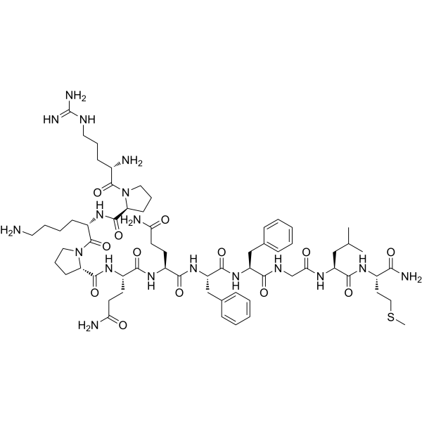
HY-P0201
Substance PNeurotransmitter -

HY-P0012
AviptadilVasodilator Agent -
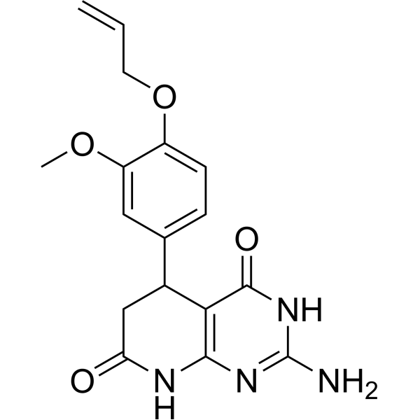
HY-133529
PA-8PAC1 Receptor Antagonist -

HY-P0220A
PACAP (6-38), human, ovine, rat TFAPACAP Receptor Antagonist -
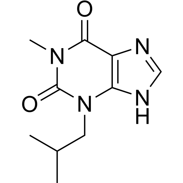
HY-12318
IBMXPDE Inhibitor -

HY-114118
SemaglutideGLP-1R Agonist -
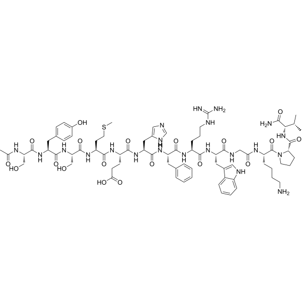
HY-P0252
α-MSHMC4R Agonist -

HY-Y0320C
Dimethyl sulfoxideAprotic Solvent -
HY-P0234
NeurotensinNeurotensin Receptor Agonist
Publications Citing Use of MedChemExpress (MCE) PACAP (1-38), human, ovine, rat
More Customer Validation & Images

-
In Vivo Efficacy Study
-
In Vivo Efficacy Study
-
IF
-
WB
-
Bio/Physico-chemical Assay
All PACAP Receptor Isoforms
MoreAll EGFR Isoforms
MoreAll Calcium Channel Isoforms
More

Biological Activity
Description
IC50 & Target
IC50: 4 nM (PACAP type I receptor), 2 nM (PACAP type II receptor VIP1), and 1 nM (PACAP type II receptor VIP2)[1]
In Vitro
PACAP (1-38), human, ovine, rat is a fragment of pituitary adenylate cyclase activating polypeptide[1].
PACAP (1-38), human, ovine, rat shows high affinity for PACAP specific (PAC1) receptor in membranes from various tissues including the endocrine pancreas[2].
In vitro, PACAP (1-38), human, ovine, rat relaxes guinea-pig and rabbit tracheal smooth muscle precontracted by histamine and by acetylcholine. PACAP (1-38) also increases adenosine 3':5'-cyclic monophosphate (cyclic AMP) in tracheal smooth muscle, providing a possible mechanism for the relaxant effect of PACAP (1-38)[3].
PACAP (1-38), human, ovine, rat (100 nM; 2 min; NCI-H838 cells) induces EGFR, HER2 and ERK tyrosine phosphorylation[5].
PACAP (1-38), human, ovine, rat (100 nM; 30 min; NCI-H838 cells) induces EGFR tyrosine phosphorylation with generates ROS and increases ROS levels by 51%[5].
PACAP (1-38), human, ovine, rat (10 nM; 48 h) stimulates the growth of NCI-H838 cells[5].
PACAP (1-38), human, ovine, rat (300 nM; 4 h) stimulates generation of APPsα in neural cells[6].
PACAP (1-38), human, ovine, rat (0.01-10 nM; HEK 293 cells) stimulates neural cells express endogenous PAC1 receptors by cAMP accumulation and by an increase in cytosolic free calcium and induces elevation of the intracellular Ca2+ concentration in a dose-dependent manner with an EC50 value of 0.81 nM[6].
PACAP (1-38), human, ovine, rat (0.01 nM; 48 h) elicits potent and efficacious stimulation of NPY secretion from SCG neuronal cultures[7].
PACAP (1-38), human, ovine, rat (100 nM; 14 d) produces sustained stimulated NPY and catecholamine secretionmpathetic neurons[7].
MedChemExpress (MCE) has not independently confirmed the accuracy of these methods. They are for reference only. Further protocols information, click here.
-
Cell Line:NCI-H838 cells
-
Concentration:10 nM
-
Incubation Time:48 hours
-
Result:Increased the number of NCI-H838 cells by 72%.
-
Cell Line:NCI-H838 cells
-
Concentration:100 nM
-
Incubation Time:2 minutes
-
Result:Increased tyrosine phosphorylation of the EGFR, HER2, and ERK by 377, 299 and 216%, respectively.
In Vivo
MedChemExpress (MCE) has not independently confirmed the accuracy of these methods. They are for reference only.


Chemical Information
-
CAS No. 137061-48-4
-
Appearance Solid
-
Molecular Weight 4534.26
-
Formula C203H331N63O53S
-
Color White to off-white
-
Synonyms
Pituitary Adenylate Cyclase Activating Polypeptide 38
-
Sequence
His-Ser-Asp-Gly-Ile-Phe-Thr-Asp-Ser-Tyr-Ser-Arg-Tyr-Arg-Lys-Gln-Met-Ala-Val-Lys-Lys-Tyr-Leu-Ala-Ala-Val-Leu-Gly-Lys-Arg-Tyr-Lys-Gln-Arg-Val-Lys-Asn-Lys-NH2
-
Sequence Shortening
HSDGIFTDSYSRYRKQMAVKKYLAAVLGKRYKQRVKNK-NH2
-
Shipping
Room temperature in continental US; may vary elsewhere.
-
Storage
Sealed storage, away from moisture
Powder -80°C 2 years -20°C 1 year * In solvent : -80°C, 6 months; -20°C, 1 month (sealed storage, away from moisture)


Publications (5)
-
Journal Impact Factor
-
Most Recent
-
Mil Med Res
2024 Jul 23;11(1):49. PMID: 39044298
PACAP (1-38), human, ovine, rat purchased from MedChemExpress. Usage Cited in: Mil Med Res. 2024 Jul 23;11(1):49. [Abstract]
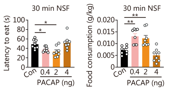
Latency to eat and food consumption in NSF at 30 min post-PACAP (1-38) (0.4, 2.0, 4.0 ng/site; administrated intrahippocampally in a volume of 0.2 μL) [F (3, 28) = 5.940, P = 0.0029; F (3, 28) = 14.16, P < 0.0001]. The results showed that 30 minutes after the administration of low and medium doses of PACAP (1-38), a shortened feeding latency and increased food intake were observed in the Novelty Suppressed Feeding (NSF) paradigm.
PACAP (1-38), human, ovine, rat purchased from MedChemExpress. Usage Cited in: Mil Med Res. 2024 Jul 23;11(1):49. [Abstract]
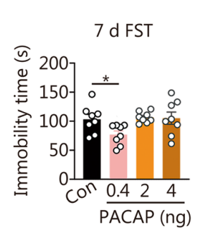
Immobility time in FST at 7 d post-PACAP (1-38) [F (3, 28) = 3.415, P = 0.0309] (n = 8). One-way ANOVA, *P < 0.05, **P < 0.01, ***P < 0.001. The immediate and lasting antidepressant activity with the infusion of 0.4 ng/site of hippocampal PACAP (1-38) (administrated intrahippocampally in a volume of 0.2 μL) in mice subjected to chronic mild stress (CMS).
-
J Nanobiotechnology
Exosome-sheathed ROS-responsive nanogel to improve targeted therapy in perimenopausal depression. [Abstract]2023 Aug 8;21(1):261. PMID: 37553718
PACAP (1-38), human, ovine, rat purchased from MedChemExpress. Usage Cited in: J Nanobiotechnology. 2023 Aug 8;21(1):261. [Abstract]
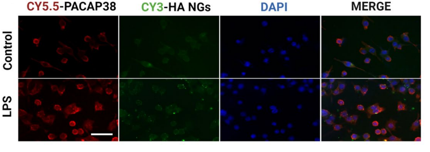
Colocalization of CY5.5-PACAP (4 h), CY3-HA NGs in HA NGs@exosomes from primary neuronal cells by fluorescent microscopy. Scale bar: 50 μm. Immunofluorescence staining showed the co-localization of PACAP (1-38) (200 µg/mL; overnight) and HA nanogels, indicating the successful encapsulation of PACAP (1-38) into HA NGs@exosomes in primary neuronal cells.
-
J Cell Mol Med
Dedifferentiated Schwann cells promote perineural invasion mediated by the PACAP paracrine signalling in cervical cancer. [Abstract]2023 Dec;27(23):3692-3705. PMID: 37830980
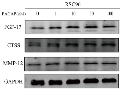
PACAP (1-38), human, ovine, rat purchased from MedChemExpress. Usage Cited in: J Cell Mol Med. 2023 Dec;27(23):3692-3705. [Abstract]
SCs were treated with various concentrations of PACAP (1-38) (PACAP, 1-100 nM) for 48 h and then FGF17, CTSS and MMP‐12 expression was measured by western blotting. Western blotting showed that PACAP (1-38) (rPACAP) induced expression of FGF17, CTSS and MMP‐12 in SCs.
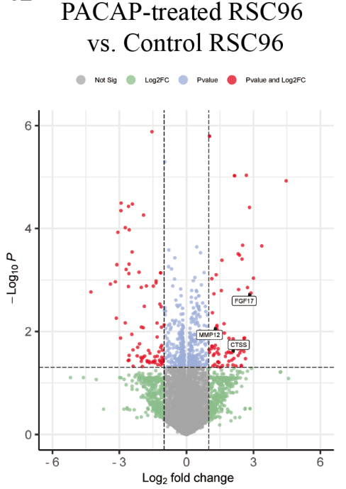
PACAP (1-38), human, ovine, rat purchased from MedChemExpress. Usage Cited in: J Cell Mol Med. 2023 Dec;27(23):3692-3705. [Abstract]
Volcano plot of DEGs in SCs after PACAP (1-38) (100 nM; 48 h) treatment. Analysis of upregulated DEGs induced by PACAP (1-38) showed that fibroblast growth factor 17 (FGF17) expression was significantly upregulated in the treatment group (log2 fold‐change = 2.89, p = 0.0019).
-
-


Solvent & Solubility
In Vitro:
H2O : 50 mg/mL (11.03 mM; Need ultrasonic)
Please refer to the solubility information to select the appropriate solvent. Once prepared, please aliquot and store the solution to prevent product inactivation from repeated freeze-thaw cycles.
Storage method and period of stock solution: -80°C, 6 months; -20°C, 1 month (sealed storage, away from moisture). When stored at -80°C, please use it within 6 months. When stored at -20°C, please use it within 1 month.
* Note: If you choose water as the stock solution, please dilute it to the working solution, then filter and sterilize it with a 0.22 μm filter before use.
Please refer to the solubility information to select the appropriate solvent. Once prepared, please aliquot and store the solution to prevent product inactivation from repeated freeze-thaw cycles.
Storage method and period of stock solution: -80°C, 6 months; -20°C, 1 month (sealed storage, away from moisture). When stored at -80°C, please use it within 6 months. When stored at -20°C, please use it within 1 month.
* Note: If you choose water as the stock solution, please dilute it to the working solution, then filter and sterilize it with a 0.22 μm filter before use.
Concentration (start) × Volume (start) = Concentration (final) × Volume (final)

Protocols
-
Kinase activity and phosphorylation assays
Kinase activity assays measure the ability of kinases to transfer phosphate groups from ATP to specific substrates, while phosphorylation assays detect the presence and levels of phosphorylated proteins. Common methods include radiolabeled ATP incorporation (e. g. ,), ADP release detection via bioluminescence (e. g. ,[3]), enzyme-linked immunosorbent assays (ELISA) for phospho-specific epitopes (e. g. ,[6]), and microtiter-based formats for high-throughput screening (e. g. ,[8]). The ADP-Glo assay quantifies kinase activity by measuring ADP produced during phosphorylation using a luciferase-based system. Radiometric assays involve autoradiography or scintillation counting after incorporation of 32P-labeled ATP into substrate proteins. ELISA-based approaches rely on phospho-specific antibodies to detect activated kinases in cell lysates or purified samples.
-
Western Blot
Western blotting (WB) is a commonly used experimental method in molecular biology, biochemistry, and immunogenetics for identifying and quantifying target proteins. It combines gel electrophoresis with immunoassay, enabling researchers to analyze protein expression, post-translational modifications, and molecular weight.
-
Ca2+ Staining Technique
Ca2+ staining is an experimental technique that utilizes specific fluorescent probes (such as Fluo-4 AM, Fura-2, etc.) to qualitatively or quantitatively detect dynamic changes in intracellular Ca2+ concentrations; this is achieved by monitoring the changes in fluorescent signals generated when these probes bind to free intracellular calcium ions. The underlying principle relies primarily on the presence of chelating groups within the probe's molecular structure that possess high affinity for calcium ions.
-
Protocol for Kinase activity and phosphorylation assays
Kinase activity assays measure transfer of phosphate from ATP to a protein or peptide substrate, generating phosphorylated substrate, ADP, or incorporated radiolabeled phosphate as the readout; phosphorylation assays measure site-specific phosphorylation in cells or tissues as a proxy for kinase-pathway activation, inhibition, or substrate regulation. Phosphorylation can be detected by phospho-specific Western blot, immunoprecipitation kinase assay, phospho-immunofluorescence, phospho-flow cytometry, luminescent ADP detection, radiolabeled ATP incorporation, or reporter-based pathway assays, and these readouts can be applied to cancer cells, primary neurons, mouse tumors, organoids, inflammatory macrophages, ferroptosis studies, and mitophagy studies when the kinase target is biologically relevant.
-
Cell Viability Determination by MTT Colorimetric Assay
The following protocol uses the MTT colorimetric assay as a classic literature-established method for assessing cell viability/metabolic activity in cultured mammalian cells. MTT[3-(4,5-dimethylthiazol-2-yl)-2,5-diphenyltetrazolium bromide] is reduced by metabolically active cells to a colored formazan product; the amount of formazan is quantified spectrophotometrically and provides an indirect measure of metabolically active viable cells. Importantly, MTT reduction reflects cellular oxidoreductase/metabolic activity rather than an absolute direct count of living cells, so changes in cellular metabolism can alter the signal independently of cell number.


Purity & Documentation
-
Data Sheet (295 KB)
-
SDS (252 KB)
- English - EN (252 KB)
- Français - FR (252 KB)
- Deutsch - DE (252 KB)
- Norwegian - NO (252 KB)
- Español - ES (252 KB)
- Swedish - SV (252 KB)
- Italian - IT (252 KB)
- Korean - KR (252 KB)
- Portuguese - PT (252 KB)
-
Handling Instructions (2659 KB)
References
[1]. Gourlet P, et al. Fragments of pituitary adenylate cyclase activating polypeptide discriminate between type I and II recombinant receptors. Eur J Pharmacol. 1995 Dec 4;287(1):7-11. [Content Brief]
[2]. Yamaguchi N. Pituitary adenylate cyclase activating polypeptide enhances glucose-evoked insulin secretion in the canine pancreas in vivo. JOP. 2001 Sep;2(5):306-16. [Content Brief]
[3]. Lindén A, et al. Inhibition of bronchoconstriction by pituitary adenylate cyclase activating polypeptide (PACAP 1-27) in guinea-pigs in vivo. Br J Pharmacol. 1995 Jul;115(6):913-6. [Content Brief]
[4]. Werling D, et al. Passage through the Ocular Barriers and Beneficial Effects in Retinal Ischemia of Topical Application of PACAP (1-38) in Rodents. Int J Mol Sci. 2017 Mar 21;18(3). pii: E675. [Content Brief]
[5]. Moody TW, et, al. PAC1 regulates receptor tyrosine kinase transactivation in a reactive oxygen species-dependent manner. Peptides. 2019 Oct;120:170017. [Content Brief]
[6]. Kojro E, et, al. The neuropeptide PACAP promotes the alpha-secretase pathway for processing the Alzheimer amyloid precursor protein. FASEB J. 2006 Mar;20(3):512-4. [Content Brief]
[7]. Braas KM, et, al. Pituitary adenylate cyclase-activating polypeptides, PACAP-38 and PACAP-27, regulation of sympathetic neuron catecholamine, and neuropeptide Y expression through activation of type I PACAP/VIP receptor isoforms. Ann N Y Acad Sci. 1996 Dec 26;805:204-16; discussion 217-8. [Content Brief]


Complete Stock Solution Preparation Table
Please refer to the solubility information to select the appropriate solvent. Once prepared, please aliquot and store the solution to prevent product inactivation from repeated freeze-thaw cycles.
Storage method and period of stock solution: -80°C, 6 months; -20°C, 1 month (sealed storage, away from moisture). When stored at -80°C, please use it within 6 months. When stored at -20°C, please use it within 1 month.
| Optional Solvent | Concentration Solvent Mass | 1 mg | 5 mg | 10 mg | 25 mg |
|---|---|---|---|---|---|
| H2O | 1 mM | 0.2205 mL | 1.1027 mL | 2.2054 mL | 5.5136 mL |
| 5 mM | 0.0441 mL | 0.2205 mL | 0.4411 mL | 1.1027 mL | |
| 10 mM | 0.0221 mL | 0.1103 mL | 0.2205 mL | 0.5514 mL |
* Note: If you choose water as the stock solution, please dilute it to the working solution, then filter and sterilize it with a 0.22 μm filter before use.


PACAP (1-38), human, ovine, rat Related Classifications
HY-P0221 Related Classifications
 Powered by Bioz
Keywords
- PACAP (1-38), human, ovine, rat
- 137061-48-4
- Pituitary Adenylate Cyclase Activating Polypeptide 38
- Pituitary Adenylate Cyclase Activating Polypeptide38
- Pituitary Adenylate Cyclase Activating Polypeptide-38
- PACAP Receptor
- ERK
- EGFR
- Reactive Oxygen Species (ROS)
- Calcium Channel
- PAC1
- cAMP
- Ca2+
- HER2
- ERK
- ROS
- NPY
- catecholamine
- Inhibitor
- inhibitor
- inhibit