



GAPDH Antibody (HRP) (YA874)
(Synonyms: GAPDH; GAPD; CDABP0047; OK/SW-cl.12; Glyceraldehyde-3-phosphate dehydrogenase; Peptidyl-cysteine S-nitrosylase GAPDH)Based on 3 publication(s) in Google Scholar
GAPDH Antibody (HRP) (YA874) is a HRP-conjugated, mouse-derived, anti-GAPDH (HRP) (YA874) IgG1 monoclonal antibody.
-
Host:
Mouse
-
Isotype:
IgG1
-
Application:
WB, ELISA
-
Reactivity :
Human, Mouse, Rat
-
Formulation:
Supplied in PBS containing 50% glycerol, 0.5% BSA and 0.02% sodium azide, pH 7.3.
-
Conjugation:
HRP


To place orders, for customer services and technical support, please contact: MedChemExpress USA
Tel: 609-228-6898 E-mail: [email protected] [email protected]



Publications Citing Use of MedChemExpress (MCE) GAPDH Antibody (HRP) (YA874)
More

Applications
| Application |
WB

WB: Western Blot
|
ELISA
ELISA: Enzyme Linked Immunosorbent Assay
|
|---|---|---|
| Dilution Ratio | 1:1000-1:5000 | 1:10000 |


Product Details
GAPDH Antibody (HRP) (YA874) is a HRP-conjugated, mouse-derived, anti-GAPDH (HRP) (YA874) IgG1 monoclonal antibody.
-
Host Mouse

-
Clonality Monoclonal

-
Species ReactivityHuman, Mouse, Rat
-
Observed Molecular WeightObserved band size: 36 kDa
Note: Due to possible protein modifications or aggregation, the molecular weight should be confirmed by actual measurement, and the predicted value is for reference only.

-
Calculated Molecular Weight Predicted band size: 36 kDa

Entrez Gene: 374193 Chicken ; 2597 Human ; 100042025 Mouse ; 14433 Mouse ; 24383 Rat685186
SwissProt: P00356 Chicken ; P04406 Human ; P16858 Mouse ; P04797 Rat
OMIM: 138400 Human
A synthetic peptide corresponding to residues near the amino terminus of human GAPDH protein.
Endogenous
Affinity Purified
HRP
Unmodified
IgG1


Product Properties
-
Appearance
Solution
-
Formulation
Supplied in PBS containing 50% glycerol, 0.5% BSA and 0.02% sodium azide, pH 7.3.
-
Concentration
Batch-dependent, Please check the COA for the concentration of each lot. Check Lot Concentration
-
Shipping
Shipping with blue ice.


Publications (3)
-
Journal Impact Factor
-
Most Recent
-
Acta Pharm Sin B
THBru ameliorates atherosclerosis by inhibiting endothelial ferroptosis via regulation of the super-enhancer-associated ABCC1. [Abstract]2025 Dec;15(12):6461-6477. PMID: 41477343 -
Carbohydr Polym
Comparative study on the mechanisms of citrus pectin and polygalacturonic acid in alleviating food allergies. [Abstract]2026 Feb 1:373:124651. PMID: 41320410 -
Eur J Cell Biol
TGF-β drives pulmonary fibrosis through dual dysregulation of TNF pathway transcription and splicing. [Abstract]2026 Feb 27;105(2):151534. PMID: 41764855


Verification Images
-
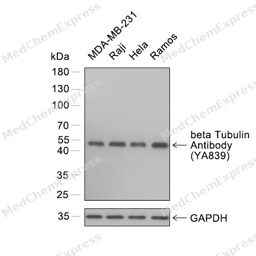 Western blot analysis of extracts from MDA-MB-231 (lane 1) and Raji (lane 2) and Hela (lane 3) and Ramos (lane 4) using beta Tubulin antibody. Proteins were transferred to a PVDF membrane and blocked with 5% nonfat powdered milk in PBST for 2 hour at room temperature. The primary antibody (1/3000) and Loading control antibody (GAPDH, HY-P80954A, 1/3000) was diluted with 5% nonfat powdered milk in PBST at 4°C overnight. Goat Anti-Rabbit IgG-HRP Secondary Antibody (1/8,000) was incubated for 45min at room temperature.
 -
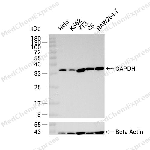 Western blot analysis was performed on extracts from Hela (lane 1, 15 μg), K562 (lane 2, 15 μg), 3T3 (lane 3, 15 μg), C6 (lane 4, 15 μg), and RAW264.7 (lane 5, 15 μg), using GAPDH Mouse mAb (HRP).Proteins were transferred to a PVDF membrane and blocked with 5% non-fat milk in TBST at 4°C overnight.The primary antibody (1:10000 dilution) and the loading control antibody (beta-Actin, HY-P83730, 1:20000 dilution) were incubated in 5% non-fat milk in TBST for 1 hour at 37°C.Goat Anti-Rabbit IgG-HRP Secondary Antibody (1:20000 dilution) was then applied for 40 minutes at 37°C.
 -
 HeLa cells were cultured in 6 cm dishes to approximately 70% confluence and then treated with human IFN-α (HY-P70241) at concentration of 20 ng/mL for 30 mins (Serum free). Phospho-IκB-α (Ser32) protein expression was subsequently analyzed by Western blot. The results demonstrated that IFN-α induced phosphorylation of IκB-α (Ser32). Primary antibodies used: Phospho-IκB-α (Ser32) Antibody (HY-P87716) and GAPDH Antibody (HY-P80954A).
 -
 HeLa cells were cultured in 6 cm dishes to approximately 70% confluence and then treated with human IFN-α (HY-P70241) at concentration of 20 and 50 ng/mL for 30 mins (Serum free). Phospho-STAT3 (Tyr705) protein expression was subsequently analyzed by Western blot. The results demonstrated that IFN-α induced phosphorylation of STAT3 (Tyr705). Primary antibodies used: Phospho-STAT3 (Tyr705) Antibody (HY-P87666) and GAPDH Antibody (HY-P80954A).
 -
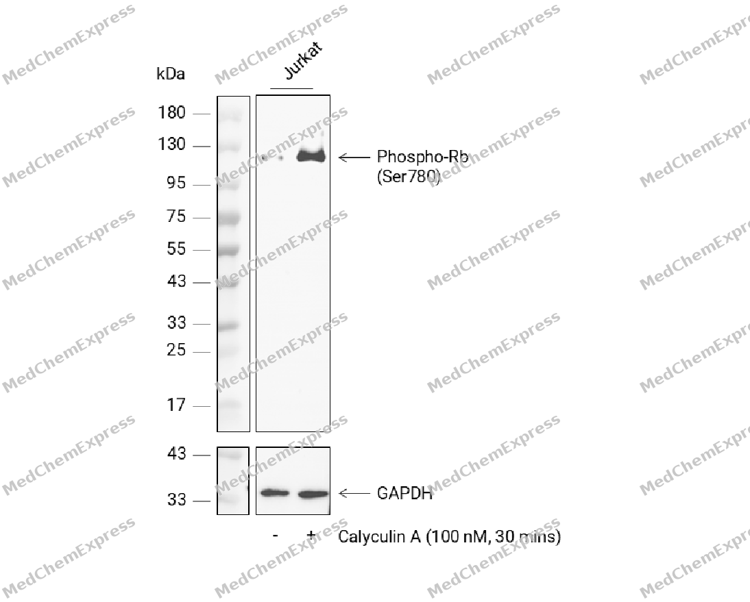 HeLa cells were cultured in 6 cm dishes to approximately 70% confluence and then treated with Calyculin A (HY-18983) at concentration of 100 nM for 30 mins. Phospho-Rb (Thr356) protein expression was subsequently analyzed by Western blot. The results demonstrated that Calyculin A induced phosphorylation of Rb (Thr356). Primary antibodies used: Phospho-Rb (Thr356) Antibody (HY-P81301) and GAPDH Antibody (HY-P80954A).
 -
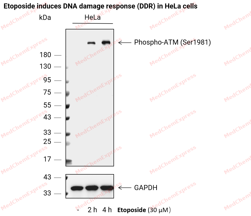 HeLa cells were cultured in 6 cm dishes to approximately 70% confluence and then treated with Etoposide (HY-13629) at concentration of 30 μM for 2 h and 4 h, respectively. Phospho-ATM (Ser1981) protein expression was subsequently analyzed by Western blot. The results demonstrated that Etoposide induced DNA damage in HeLa cells, indicated by phosphorylation of ATM (Ser1981). Primary antibodies used: Phospho-ATM (Ser1981) Antibody (HY-P87805) and GAPDH Antibody (HY-P80954A).
 -
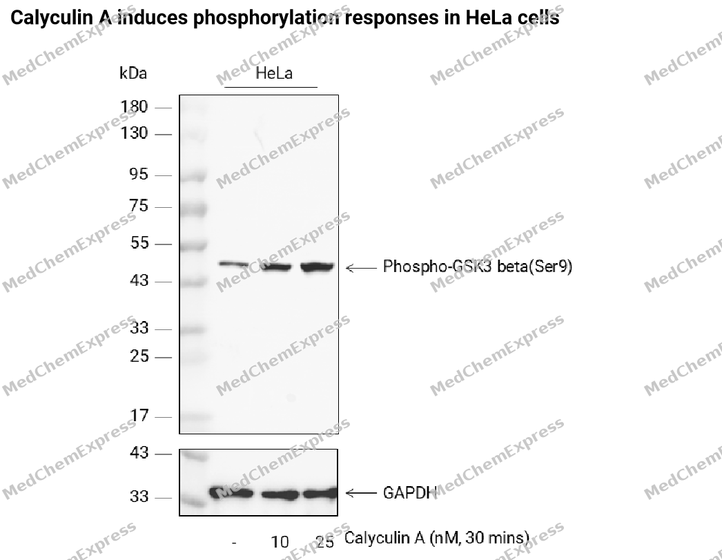 HeLa cells were cultured in 6 cm dishes to approximately 70% confluence and then treated with Calyculin A (HY-18983) at concentration of 10 and 25 nM for 30 mins. Phospho-GSK3 beta (Ser9) protein expression was subsequently analyzed by Western blot. The results demonstrated that Calyculin A induced phosphorylation of GSK3 beta (Ser9). Primary antibodies used: Phospho-GSK3 beta(Ser9) Antibody (HY-P86595) and GAPDH Antibody (HY-P80954A).
 -
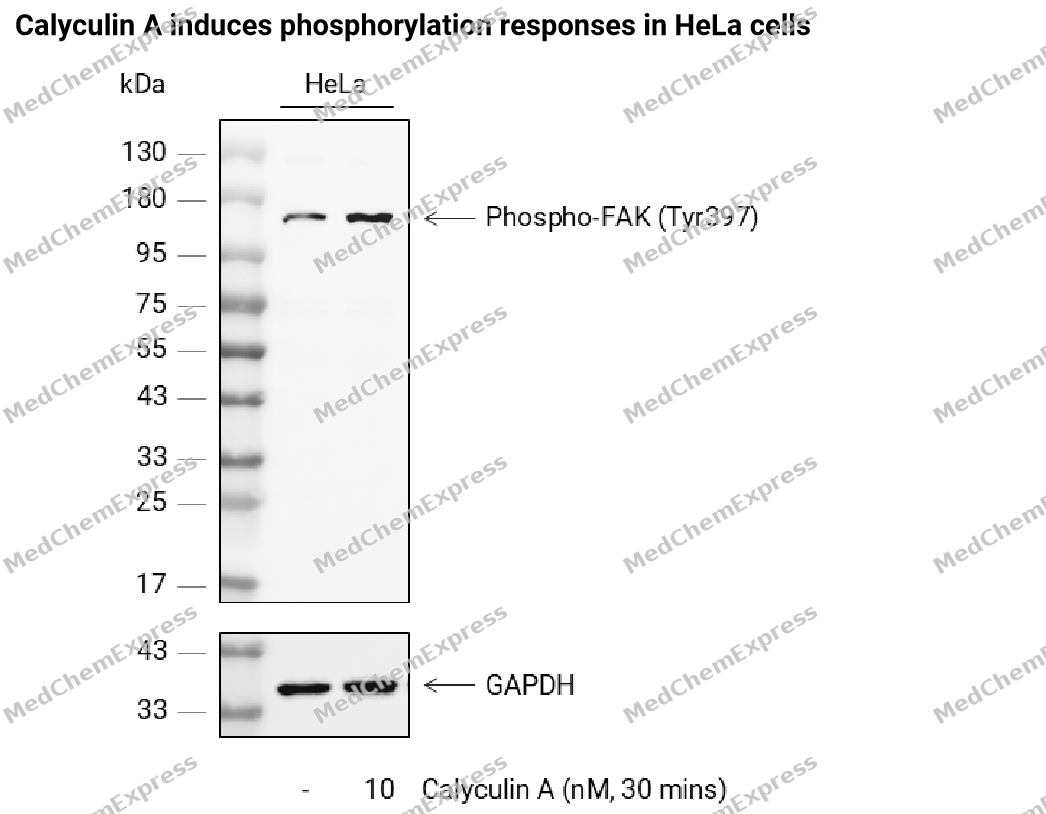 HeLa cells were cultured in 6 cm dishes to approximately 70% confluence and then treated with Calyculin A (HY-18983) at concentration of 10 nM for 30 mins. Phospho-FAK (Tyr397) protein expression was subsequently analyzed by Western blot. The results demonstrated that Calyculin A induced phosphorylation of FAK (Tyr397). Primary antibodies used: Phospho-FAK (Tyr397) Antibody (HY-P87691) and GAPDH Antibody (HY-P80954A).
 -
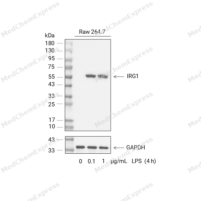 RAW264.7 cells were cultured in 6 cm dishes to approximately 70% confluence and then treated with LPS (HY-D1056) at concentrations ranging from 0 to 1 μg/mL for 4 h. IRG1 protein expression was subsequently analyzed by Western blot. The results demonstrated that LPS induced IRG1 expression in a dose-dependent manner, confirming the responsiveness of the model. Primary antibodies used: IRG1 Antibody (HY-P84946) and GAPDH Antibody (HY-P80954A).


Background
-
Function
GAPDH is a Has both glyceraldehyde-3-phosphate dehydrogenase and nitrosylase activities, thereby playing a role in glycolysis and nuclear functions, respectively. Glyceraldehyde-3-phosphate dehydrogenase is a key enzyme in glycolysis that catalyzes the first step of the pathway by converting D-glyceraldehyde 3-phosphate (G3P) into 3-phospho-D-glyceroyl phosphate. Modulates the organization and assembly of the cytoskeleton. Facilitates the CHP1-dependent microtubule and membrane associations through its ability to stimulate the binding of CHP1 to microtubules. Component of the GAIT (gamma interferon-activated inhibitor of translation) complex which mediates interferon-gamma-induced transcript-selective translation inhibition in inflammation processes. Upon interferon-gamma treatment assembles into the GAIT complex which binds to stem loop-containing GAIT elements in the 3'-UTR of diverse inflammatory mRNAs (such as ceruplasmin) and suppresses their translation. Also plays a role in innate immunity by promoting TNF-induced NF-kappa-B activation and type I interferon production, via interaction with TRAF2 and TRAF3, respectively. Participates in nuclear events including transcription, RNA transport, DNA replication and apoptosis. Nuclear functions are probably due to the nitrosylase activity that mediates cysteine S-nitrosylation of nuclear target proteins such as SIRT1, HDAC2 and PRKDC[1][2][3][4][5].
-
Subcellular Localization
Cytoplasm, cytosol; Nucleus; Cytoplasm, perinuclear region; Membrane; Cytoplasm, cytoskeleton
-
Isoforms & Post-Translational Modification
P04406 has 2 isomers: P04406-1: 36053 Da (predicted); P04406-2: 31548 Da (predicted).
S-nitrosylation of Cys-152 leads to interaction with SIAH1, followed by translocation to the nucleus (By similarity). S-nitrosylation of Cys-247 is induced by interferon-gamma and LDL(ox) implicating the iNOS-S100A8/9 transnitrosylase complex and seems to prevent interaction with phosphorylated RPL13A and to interfere with GAIT complex activity (PubMed:22771119, PubMed:25417112);ISGylated;Sulfhydration at Cys-152 increases catalytic activity;Oxidative stress can promote the formation of high molecular weight disulfide-linked GAPDH aggregates, through a process called nucleocytoplasmic coagulation. Such aggregates can be observed in vivo in the affected tissues of patients with Alzheimer disease or alcoholic liver cirrhosis, or in cell cultures during necrosis. Oxidation at Met-46 may play a pivotal role in the formation of these insoluble structures. This modification has been detected in vitro following treatment with free radical donor (+/-)-(E)-4-ethyl-2-[(E)-hydroxyimino]-5-nitro-3-hexenamide. It has been proposed to destabilize nearby residues, increasing the likelihood of secondary oxidative damages, including oxidation of Tyr-45 and Met-105. This cascade of oxidations may augment GAPDH misfolding, leading to intermolecular disulfide cross-linking and aggregation;Succination of Cys-152 and Cys-247 by the Krebs cycle intermediate fumarate, which leads to S-(2-succinyl)cysteine residues, inhibits glyceraldehyde-3-phosphate dehydrogenase activity. Fumarate concentration as well as succination of cysteine residues in GAPDH is significantly increased in muscle of diabetic mammals. It was proposed that the S-(2-succinyl)cysteine chemical modification may be a useful biomarker of mitochondrial and oxidative stress in diabetes and that succination of GAPDH and other thiol proteins by fumarate may contribute to the metabolic changes underlying the development of diabetes complications;(Microbial infection) Glycosylated by C.rodentium protein NleB, enteropathogenic E.coli protein NleB1 and S.typhimurium protein Ssek1: arginine GlcNAcylation prevents the interaction with TRAF2 and TRAF3 (PubMed:23332158, PubMed:27387501, PubMed:28522607). This leads to reduced ubiquitination of TRAF2 and TRAF3, and subsequent inhibition of NF-kappa-B signaling and type I interferon production, respectively (PubMed:23332158, PubMed:27387501) -
Subunit
Homotetramer (PubMed:16239728, PubMed:16510976).
-
SwissProt ID
-
Synonyms
GAPDH; GAPD; CDABP0047; OK/SW-cl.12; Glyceraldehyde-3-phosphate dehydrogenase; Peptidyl-cysteine S-nitrosylase GAPDH
-
Research Field
Neuroscience
Documentation
-
Data Sheet (261 KB)
-
SDS (252 KB)
- English - EN (252 KB)
- Français - FR (252 KB)
- Deutsch - DE (252 KB)
- Norwegian - NO (252 KB)
- Español - ES (252 KB)
- Swedish - SV (252 KB)
- Italian - IT (252 KB)
- Korean - KR (252 KB)
- Portuguese - PT (252 KB)
-
User Guide for Antibodies (1077 KB)
References
[1]. Tisdale EJ, et al. Glyceraldehyde-3-phosphate dehydrogenase is phosphorylated by protein kinase Ciota /lambda and plays a role in microtubule dynamics in the early secretory pathway. J Biol Chem. 2002 Feb 1;277(5):3334-41. [Content Brief]
[2]. Ercolani L, et al. Isolation and complete sequence of a functional human glyceraldehyde-3-phosphate dehydrogenase gene. J Biol Chem. 1988 Oct 25;263(30):15335-41. [Content Brief]
[3]. Arif A, et al. Heterotrimeric GAIT complex drives transcript-selective translation inhibition in murine macrophages. Mol Cell Biol. 2012 Dec;32(24):5046-55. [Content Brief]
[4]. Gao X, et al. NleB, a bacterial effector with glycosyltransferase activity, targets GAPDH function to inhibit NF-κB activation. Cell Host Microbe. 2013 Jan 16;13(1):87-99. [Content Brief]
[5]. Gao X, et al. Citrobacter rodentium NleB Protein Inhibits Tumor Necrosis Factor (TNF) Receptor-associated Factor 3 (TRAF3) Ubiquitination to Reduce Host Type I Interferon Production. J Biol Chem. 2016 Aug 26;291(35):18232-8. [Content Brief]

 Powered by Bioz
