



Top/HDAC-IN-2
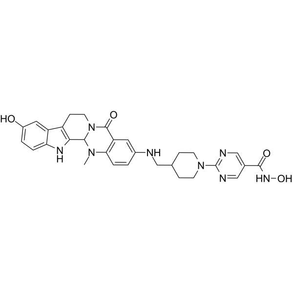
Top/HDAC-IN-2 (45b), a Top and HDAC dual inhibitor, exhibits potent antitumor activities and induces apoptosis.
For research use only. We do not sell to patients.
- CAS No.: 2775446-64-3
- Formula: C30H32N8O4
- Molecular Weight:568.63
-
Storage:
Please store the product under the recommended conditions in the Certificate of Analysis.

To place orders, for customer services and technical support, please contact: MedChemExpress USA
Tel: 609-228-6898 E-mail: [email protected] [email protected]


-
 Biological Activity
-
Chemical Information
- Protocols
- Purity & Documentation
- References
-
 Help & FAQs


All Topoisomerase Isoforms
More

Biological Activity
Description
IC50 & Target
|
HDAC1 0.004 μM (IC50) |
HDAC2 0.005 μM (IC50) |
HDAC6 0.15 μM (IC50) |
HDAC3 1.10 μM (IC50) |
HDAC8 4.5 μM (IC50) |
Top1 |
Topoisomerase II |
Cellular Effect
|
Cell Line

|
Type | Value | Description | References |
|---|---|---|---|---|
| A549 | IC50 |
0.23 μM
Compound: 16b
|
Antiproliferative activity against human A549 cells
Antiproliferative activity against human A549 cells
|
[PMID: 37875056] |
| HCT-116 | IC50 |
0.23 μM
Compound: 16b
|
Antiproliferative activity against human HCT-116 cells
Antiproliferative activity against human HCT-116 cells
|
[PMID: 37875056] |
| HCT-116 | IC50 |
0.23 μM
Compound: 71
|
Antiproliferative activity against human HCT-116 cells
Antiproliferative activity against human HCT-116 cells
|
[PMID: 36549115] |
| MCF7 | IC50 |
0.23 μM
Compound: 16b
|
Antiproliferative activity against human MCF7 cells
Antiproliferative activity against human MCF7 cells
|
[PMID: 37875056] |
In Vitro
Top/HDAC-IN-2 (45b) shows antitumor activities against the HCT116 (IC50 = 0.23 µM), MCF-7 and A549 cells[1].
Top/HDAC-IN-2 (45b) (0-5 µM, 24h) arrests the HCT116 cell cycle at the G2 phase in a concentration-dependent manner[1].
Top/HDAC-IN-2 (45b) (0-2.5 µM, 24h) induces the apoptosis in HCT116 cell line in a concentration-dependent manner[1].
Top/HDAC-IN-2 (45b) (0-5 μM, 24h) inhibits HDAC in living cells[1].
MedChemExpress (MCE) has not independently confirmed the accuracy of these methods. They are for reference only.
-
Cell Line:HCT116
-
Concentration:0.1, 2.5 and 5 μM
-
Incubation Time:24 h
-
Result:Arrested the HCT116 cell cycle at the G2 phase in a concentration-dependent manner.
-
Cell Line:HCT116
-
Concentration:0.1, 0.5 and 2.5 μM
-
Incubation Time:24 h
-
Result:Effectively induced the apoptosis in HCT116 cell line in a concentration-dependent manner.
-
Cell Line:HCT116
-
Concentration:1 μM and 5 μM
-
Incubation Time:24 h
-
Result:Concentration-dependently increased the level of acetyl-H3 and acetyl-H4 in HCT116 cells.


Chemical Information
-
CAS No. 2775446-64-3
-
Molecular Weight 568.63
-
Formula C30H32N8O4
-
SMILES
CN1C2=C(C(N3CCC4=C(C13)NC5=CC=C(C=C54)O)=O)C=C(C=C2)NCC6CCN(CC6)C7=NC=C(C=N7)C(NO)=O
-
Shipping
Room temperature in continental US; may vary elsewhere.
-
Storage
Please store the product under the recommended conditions in the Certificate of Analysis.


Purity & Documentation
References

Calculators
Concentration (start) × Volume (start) = Concentration (final) × Volume (final)


Top/HDAC-IN-2 Related Classifications
HY-145852 Related Classifications
 Powered by Bioz