


Afatinib
Based on 107 publication(s) in Google Scholar
Afatinib (BIBW 2992) is an orally active, potent and irreversible dual specificity inhibitor of ErbB family (EGFR and HER2), with IC50 values of 0.5 nM, 0.4 nM, 10 nM and 14 nM for EGFRwt, EGFRL858R, EGFRL858R/T790M and HER2, respectively. Afatinib can be used for the research of esophageal squamous cell carcinoma (ESCC), non-small cell lung cancer (NSCLC) and gastric cancer.
For research use only. We do not sell to patients.
- Purity: 99.92%
- CAS No.: 850140-72-6
- Formula: C24H25ClFN5O3
- Molecular Weight:485.94
-
Storage:Powder -20°C, 3 years , 4°C, 2 years ; In solvent -80°C, 2 years , -20°C, 1 year


-
 Biological Activity
-
Chemical Information
-
Solvent & Solubility
- Purity & Documentation
- References
-
 Help & FAQs
-


Apoptosis Compound Library
HY-L003
-
Immunology/Inflammation Compound Library
HY-L007
-
JAK/STAT Compound Library
HY-L008
-
Kinase Inhibitor Library
HY-L009
-
MAPK Compound Library
HY-L010
-
PI3K/Akt/mTOR Compound Library
HY-L015
-
Protein Tyrosine Kinase Compound Library
HY-L016
-
Stem Cell Signaling Compound Library
HY-L017
-
FDA-Approved Drug Library
HY-L022
-
Anti-Cancer Compound Library
HY-L025
-
Autophagy Compound Library
HY-L029
-
Anti-Aging Compound Library
HY-L034
-
Drug Repurposing Compound Library
HY-L035
-
Covalent Screening Library
HY-L036
-
Differentiation Inducing Compound Library
HY-L038
-
Reprogramming Compound Library
HY-L039
-
Oxygen Sensing Compound Library
HY-L045
-
Glycolysis Compound Library
HY-L058
-
Pyroptosis Compound Library
HY-L059
-
Cytoskeleton Compound Library
HY-L060
-
Orally Active Compound Library
HY-L061
-
Chemical Probe Library
HY-L063
-
Glutamine Metabolism Compound Library
HY-L064
-
FDA Approved & Pharmacopeial Drug Library
HY-L066
-
Anti-Hepatitis C Virus Compound Library
HY-L073
-
Anti-Breast Cancer Compound Library
HY-L074
-
Anti-Lung Cancer Compound Library
HY-L075
-
Anti-Pancreatic Cancer Compound Library
HY-L077
-
Anti-Blood Cancer Compound Library
HY-L079
-
Targeted Therapy Drug Library
HY-L080
-
Anti-Cancer Metabolism Compound Library
HY-L083
-
Neurodegenerative Disease-related Compound Library
HY-L086
-
Anti-Obesity Compound Library
HY-L087
-
Angiogenesis-Related Compound Library
HY-L088
-
Glucose Metabolism Compound Library
HY-L092
-
Anti-Liver Cancer Compound Library
HY-L101
-
Rare Diseases Drug Library
HY-L102
-
Anti-Colorectal Cancer Compound Library
HY-L103
-
Antidepressant Compound Library
HY-L108
-
Chemotherapy Drug Library
HY-L112
-
EMA-Approved Drug Library
HY-L116
-
FDA-Approved Anticancer Drug Library
HY-L122
-
Anti-Prostate Cancer Compound Library
HY-L124
-
Anti-Pulmonary Fibrosis Compound Library
HY-L125
-
Cancer Stem Cells Compound Library
HY-L135
-
Heterocyclic Compound Library
HY-L138
-
Pain-Related Compound Library
HY-L139
-
Off-patent Drug Library
HY-L141
-
Membrane Protein-targeted Compound Library
HY-L149
-
Membrane Receptor-targeted Compound Library
HY-L150
-
Cysteine Targeted Covalent Library
HY-L153
-
Highly Selective Inhibitors Library
HY-L158
-
Highly Selective Activators Library
HY-L159
-
Cytokine Inhibitors Library
HY-L161
-
Cell Death Library
HY-L162
-
Serine/Threonine Kinase Inhibitor Library
HY-L164
-
Anti-Hematopathy Compound Library
HY-L171
-
Anti-Ovarian Cancer Compound Library
HY-L173
-
Multi-Target Compound Library
HY-L176
-
Bioactive Compound Library Max
HY-L181
-
Anti-Aging Compound Library Mini
HY-L034M
-
MCE Bioactive Compound Library
HY-L001V
-
Covalent Screening Library Plus
HY-L036P
-
Drug Repurposing Compound Library Plus
HY-L035P
-
FDA-Approved Drug Library Plus
HY-L022P
-
FDA-Approved Drug Library Mini
HY-L022M
-
Bioactive Compound Library
HY-L001
-
Anti-Gastric Cancer Compound Library
HY-L184
-
Anti-Fibrosis Compound Library
HY-L185
-
Anti-Brain Cancer Compound Library
HY-L188
-
Protein Kinase Compound Library
HY-L196
-
Non-Alcoholic Fatty Liver Disease (NAFLD) Compound Library
HY-L199
-
Cell Proliferation Compound Library
HY-L201
-
High-Throughput Bioactive Compound Library
HY-L205
-
Anti-Rheumatic Arthritis Compound Library
HY-L210
-
Anti-Cancer Approved Drug Library
HY-L213
-
Posttranslational Modification Library
HY-L226
-
Nephrotoxicity Compound Library
HY-L229
-
FDA Kinase Inhibitor Library
HY-L230
-
RNA Binding Bioactive Compound Library
HY-L248
-
Lactylation Compound Library
HY-L249


Publications Citing Use of MedChemExpress (MCE) Afatinib
More- Nature. 2026 Jan;649(8098):1032-1041. [Abstract]
- Cancer Cell. 2026 May 21:S1535-6108(26)00220-5.
- Cancer Cell. 2024 Jul 8;42(7):1286-1300.e8. [Abstract]
- Cancer Cell. 2023 Jan 9;41(1):88-105.e8. [Abstract]
- J Hematol Oncol. 2024 Jul 30;17(1):58. [Abstract]
- Cancer Discov. 2025 Feb 7;15(2):346-362. [Abstract]
- Nat Genet. 2025 Jan;57(1):165-179. [Abstract]
- Nat Cell Biol. 2025 Mar;27(3):449-463. [Abstract]
- Cancer Res. 2025 Dec 29. [Abstract]
- Cancer Res. 2025 Jun 10:10.1158/0008-5472.CAN-24-3904. [Abstract]
- Mol Cell. 2025 Mar 21:S1097-2765(25)00194-7. [Abstract]
- Cancer Res. 2021 Sep 15;81(18):4822-4834. [Abstract]
- Nat Commun. 2026 Feb 12;17(1):1214. [Abstract]
- Nat Commun. 2026 Jan 21;17(1):1902. [Abstract]
- Nat Commun. 2025 Nov 23;16(1):11115. [Abstract]
- Nat Commun. 2025 Feb 1;16(1):1237. [Abstract]
- Nat Commun. 2019 Apr 18;10(1):1812 [Abstract]
- Sci Transl Med. 2018 Jul 18;10(450):eaaq1093. [Abstract]
- Chem Eng J. 2024 May 15, 488, 150822.
- Biomaterials. 2022 Oct:289:121800. [Abstract]
- J Exp Clin Cancer Res. 2025 Aug 19;44(1):245. [Abstract]
- J Exp Clin Cancer Res. 2024 Nov 20;43(1):308. [Abstract]
- J Exp Clin Cancer Res. 2023 Oct 27;42(1):284. [Abstract]
- Redox Biol. 2026 Jun:93:104172. [Abstract]
- Cell Rep Med. 2025 Apr 2:102053. [Abstract]
- Cell Rep Med. 2023 Feb 21;4(2):100911. [Abstract]
- Pharmacol Res. 2020 Sep;159:104934. [Abstract]
- Pharmacol Res. 2019 Jun:144:79-89. [Abstract]
- Cancer Lett. 2025 Apr 10:217715. [Abstract]
- Cell Death Dis. 2021 Jan 7;12(1):42. [Abstract]
- Osteoarthritis Cartilage. 2022 Jun;30(6):862-874. [Abstract]
- J Pharm Anal. 2019 Feb;9(1):49-54. [Abstract]
- Acta Pharmacol Sin. 2023 Jul;44(7):1475-1486. [Abstract]
- EMBO Mol Med. 2021 Apr 9;13(4):e13144. [Abstract]
- NPJ Parkinsons Dis. 2025 Jun 7;11(1):157. [Abstract]
- ACS Appl Mater Interfaces. 2022 May 25;14(20):23152-23163. [Abstract]
- NPJ Precis Oncol. 2026 Apr 8;10(1):211. [Abstract]
- J Transl Med. 2022 Jun 25;20(1):286. [Abstract]
- Oncogene. 2022 Aug;41(32):3953-3968. [Abstract]
- Oncogene. 2018 Aug;37(31):4300-4312. [Abstract]
- Clin Transl Med. 2026 Mar;16(3):e70638. [Abstract]
- Sci Signal. 2021 Jun 22;14(688):eabe6156. [Abstract]
- Acta Neuropathol Commun. 2025 Jun 28;13(1):143. [Abstract]
- J Mater Chem B. 2016 Nov 7;4(41):6652-6661. [Abstract]
- Mol Cancer Ther. 2025 Oct 23. [Abstract]
- Pharmaceutics. 2022 Jan 12;14(1):176. [Abstract]
- Mol Cancer Ther. 2018 Mar;17(3):603-613. [Abstract]
- Cancer Drug Resist. 2025 Nov 5:8:57. [Abstract]
- Biol Direct. 2025 Jul 2;20(1):77. [Abstract]
- Cancer Nanotechnol. 12, 13 (2021).
- Int Immunopharmacol. 2026 Mar 1:172:116131. [Abstract]
- Eur J Pharmacol. 2023 Dec 5:960:176114. [Abstract]
- Mol Cancer Res. 2019 Nov;17(11):2233-2243. [Abstract]
- Toxicology. 2024 Nov 26:154018. [Abstract]
- Cancers (Basel). 2024 Aug 7;16(16):2785. [Abstract]
- Cancer Sci. 2018 Apr;109(4):1166-1176. [Abstract]
- Adv Ther. 2024 Jul 18.
- Sci Rep. 2026 Feb 8;16(1):7792. [Abstract]
- PLoS Comput Biol. 2020 Feb 26;16(2):e1007701. [Abstract]
- Viruses. 2021 Jun 28;13(7):1255. [Abstract]
- Exp Cell Res. 2020 Aug 1;393(1):112054. [Abstract]
- Cell Biol Int. 2020 Feb;44(2):621-629. [Abstract]
- J Pharm Biomed Anal. 2018 May 30:154:321-331. [Abstract]
- Genes (Basel). 2024 Dec 19;15(12):1624. [Abstract]
- PLoS One. 2024 Nov 1;19(11):e0308647. [Abstract]
- PLoS One. 2018 Jun 4;13(6):e0198364. [Abstract]
- Fundam Clin Pharmacol. 2021 Oct;35(5):919-929. [Abstract]
- Eur J Drug Metab Pharmacokinet. 2021 Sep;46(5):625-635. [Abstract]
- Int J Radiat Biol. 2021;97(2):170-178. [Abstract]
- Acta Histochem. 2019 Nov;121(8):151439. [Abstract]
- GEN Biotechnol. 2025 Apr 17.
- J Oral Biosci. 2024 Sep 6:S1349-0079(24)00198-1. [Abstract]
- Biochem Biophys Res Commun. 2026 Feb 12:800:153165.
- Oncol Lett. 2021 Nov;22(5):754. [Abstract]
- Cryobiology. 2019 Feb:86:71-76. [Abstract]
- Cell Physiol Biochem. 2018;47(3):1259-1273. [Abstract]
- Bioanalysis. 2018 Sep 1;10(18):1511-1523. [Abstract]
- Biomed Chromatogr. 2016 Jul;30(7):1150-4. [Abstract]
- Biol Methods Protoc. 2025 Feb 13;10(1):bpaf012. [Abstract]
- Xenobiotica. 2018 Oct;48(10):1059-1071. [Abstract]
- bioRxiv. 2026 Jun 19.
- bioRxiv. 2026 Mar 12.
- bioRxiv. 2026 Jan 9.
- bioRxiv. 2026 Jan 12.
- Technical University of Dresden. 2025.
- bioRxiv. 2025 Dec 5.
- bioRxiv. 2025 Nov 24.
- SSRN. 2025 Jul 4.
- bioRxiv. 2025 Jun 7:2025.06.04.657911. [Abstract]
- World J Exp Med. 2025 Jun 20;15(2):100443. [Abstract]
- State University of New York at Buffalo. 2025.
- bioRxiv. 2025 May 9:2025.05.04.652129. [Abstract]
- Patent. US20240344020A1
- Patent. US20240344020A1.
- bioRxiv. 2024 October 04.
- Research Square Preprint. 2024 Nov 26.
- bioRxiv. 2024 Feb 12.
- bioRxiv. 2024 Jan 4:2023.10.06.561161. [Abstract]
- University of Cádiz. 2023 Oct.
- bioRxiv. 2023 Sep 13.
- Research Square Print. 2023 Jan 6.
- bioRxiv. October 28, 2021.
- Research Square Preprint. 2021 Jul.
- Chem Rep. 2019, 1(1): 3-12.
- Patent. US20190010159A1.
- Oncotarget. 2015 Oct 13;6(31):31313-22. [Abstract]
- Oncotarget. 2014 Dec 15;5(23):11971-85. [Abstract]
 Customer Validation & Images

-
Flow Cytometry

-
Bio/Physico-chemical Assay

-
WB
-
Others
-
IHC

All EGFR Isoforms
More

Biological Activity
|
EGFRL858R 0.4 nM (IC50) |
EGFR 0.5 nM (IC50) |
EGFRL858R/T790M 10 nM (IC50) |
HER2 14 nM (IC50) |
HER3 |
|
Cell Line
|
Type | Value | Description | References |
|---|---|---|---|---|
| A-431 | IC50 |
0.023 nM
Compound: 6, BIBW2992
|
Antiproliferative activity against human A431 cells after 72 hrs by SRB assay
Antiproliferative activity against human A431 cells after 72 hrs by SRB assay
|
[PMID: 24183742] |
| A-431 | IC50 |
0.02 μM
Compound: 5, BIBW2992
|
Antiproliferative activity against human A431 cells assessed as growth inhibition after 72 hrs by SRB assay
Antiproliferative activity against human A431 cells assessed as growth inhibition after 72 hrs by SRB assay
|
[PMID: 24565969] |
| A-431 | IC50 |
0.9 μM
Compound: Afatinib
|
Antiproliferative activity against human A431 cells after 48 hrs by Celltiter-Glo assay
Antiproliferative activity against human A431 cells after 48 hrs by Celltiter-Glo assay
|
[PMID: 25409491] |
| A-431 | EC50 |
0.6 μM
Compound: Afatinib
|
Antiproliferative activity against human A431 cells harboring wild type EGFR assessed as growth inhibition after 96 hrs by CellTiter-Glo assay
Antiproliferative activity against human A431 cells harboring wild type EGFR assessed as growth inhibition after 96 hrs by CellTiter-Glo assay
|
[PMID: 28603991] |
| A-431 | EC50 |
0.2 μM
Compound: Afatinib
|
Antiproliferative activity against human A431 cells expressing wild type EGFR incubated for 96 hrs measured on day 5 by CellTiterGlo assay
Antiproliferative activity against human A431 cells expressing wild type EGFR incubated for 96 hrs measured on day 5 by CellTiterGlo assay
|
[PMID: 28853575] |
| A-431 | IC50 |
10.21 nM
Compound: Afatinib
|
Antiproliferative activity against human A431 cells harboring wild-type EGFR after 72 hrs by CCK8 assay
Antiproliferative activity against human A431 cells harboring wild-type EGFR after 72 hrs by CCK8 assay
|
[PMID: 30600209] |
| A-431 | IC50 |
1.4 μM
Compound: Afatinib
|
Antiproliferation activity against human A431 cells assessed as reduction in cell viability incubated incubated for 48 hrs by MTT assay
Antiproliferation activity against human A431 cells assessed as reduction in cell viability incubated incubated for 48 hrs by MTT assay
|
[PMID: 32912431] |
| A-431 | EC50 |
0.535 μM
Compound: Afatinib
|
Antiproliferative activity against human A-431 cells harboring wild type EGFR measured after 72 hrs by CellTiter-Glo assay
Antiproliferative activity against human A-431 cells harboring wild type EGFR measured after 72 hrs by CellTiter-Glo assay
|
[PMID: 35486541] |
| A-431 | GI50 |
0.594 μM
Compound: Afatinib
|
Antiproliferative activity against human A-431 cells harboring wild type EGFR measured after 72 hrs by CellTiter-Glo assay
Antiproliferative activity against human A-431 cells harboring wild type EGFR measured after 72 hrs by CellTiter-Glo assay
|
[PMID: 35486541] |
| A549 | IC50 |
1.811 μM
Compound: Afatinib
|
Cytotoxicity against human A549 cells after 48 hrs by MTT assay
Cytotoxicity against human A549 cells after 48 hrs by MTT assay
|
[PMID: 24900830] |
| A549 | IC50 |
0.05 μM
Compound: BIBW-2992
|
Cytotoxicity against human A549 cells after 72 hrs by MTT assay
Cytotoxicity against human A549 cells after 72 hrs by MTT assay
|
[PMID: 26906472] |
| A549 | IC50 |
1.4 μM
Compound: BIBW-2992
|
Cytotoxicity against human A549 cells assessed as decrease in cell viability after 72 hrs by MTT assay
Cytotoxicity against human A549 cells assessed as decrease in cell viability after 72 hrs by MTT assay
|
[PMID: 28428040] |
| A549 | EC50 |
1.49 μM
Compound: Afatinib
|
Antiproliferative activity against human A549 cells harboring KRAS-G12S mutant incubated for 96 hrs measured on day 5 by CellTiterGlo assay
Antiproliferative activity against human A549 cells harboring KRAS-G12S mutant incubated for 96 hrs measured on day 5 by CellTiterGlo assay
|
[PMID: 28853575] |
| A549 | IC50 |
3766 nM
Compound: Afatinib
|
Antiproliferative activity against human A549 cells harboring wild-type EGFR after 72 hrs by EZ-Cytox assay
Antiproliferative activity against human A549 cells harboring wild-type EGFR after 72 hrs by EZ-Cytox assay
|
[PMID: 30554954] |
| A549 | IC50 |
5 μM
Compound: Afatinib
|
Antiproliferative activity against wild type human A549 cells assessed as reduction in cell viability incubated for 72 hrs by MTT assay
Antiproliferative activity against wild type human A549 cells assessed as reduction in cell viability incubated for 72 hrs by MTT assay
|
[PMID: 31446247] |
| A549 | IC50 |
6.32 μM
Compound: Afatinib
|
Antiproliferative activity against human A549 cells assessed as reduction in cell viability after 24 hrs by MTT assay
Antiproliferative activity against human A549 cells assessed as reduction in cell viability after 24 hrs by MTT assay
|
[PMID: 31648877] |
| A549 | IC50 |
1.2 μM
Compound: Afatinib
|
Antiproliferative activity against human A549 cells expressing wild type EGFR assessed as inhibition of cell growth incubated for 72 hrs by MTT assay
Antiproliferative activity against human A549 cells expressing wild type EGFR assessed as inhibition of cell growth incubated for 72 hrs by MTT assay
|
[PMID: 35728507] |
| A549 | IC50 |
5.4 μM
Compound: Afatinib
|
Antiproliferative activity against wild type human A549 cells assessed as reduction in cell viability incubated for 72 hrs by CCK8 assay
Antiproliferative activity against wild type human A549 cells assessed as reduction in cell viability incubated for 72 hrs by CCK8 assay
|
[PMID: 37141440] |
| A549 | IC50 |
1.24 μM
Compound: Afatinib
|
Antiproliferative activity against human A549 cells overexpressing c-Met and EGFR assessed as reduction in cell viability incubated for 72 hrs by MTT assay
Antiproliferative activity against human A549 cells overexpressing c-Met and EGFR assessed as reduction in cell viability incubated for 72 hrs by MTT assay
|
[PMID: 37984296] |
| BaF3 | IC50 |
>1 μM
Compound: 6
|
Antiproliferative activity against mouse BA/F3 cells expressing TEL-JAK3 after 72 hrs by cell titer glo assay
Antiproliferative activity against mouse BA/F3 cells expressing TEL-JAK3 after 72 hrs by cell titer glo assay
|
[PMID: 26258521] |
| BaF3 | IC50 |
381.3 nM
Compound: Afatinib
|
Cytotoxicity against mouse BaF3 cells expressing IL-3 assessed as inhibition of cell viability incubated for 72 hrs by CellTiter-Glo assay
Cytotoxicity against mouse BaF3 cells expressing IL-3 assessed as inhibition of cell viability incubated for 72 hrs by CellTiter-Glo assay
|
[PMID: 28287083] |
| BaF3 | EC50 |
0.097 μM
Compound: Afatinib
|
Antiproliferative activity against mouse Ba/F3 cells harbouring Her2-A775_G776insYVMA mutant measured after 72 hrs by CellTiter-Glo assay
Antiproliferative activity against mouse Ba/F3 cells harbouring Her2-A775_G776insYVMA mutant measured after 72 hrs by CellTiter-Glo assay
|
[PMID: 35486541] |
| BaF3 | GI50 |
0.015 μM
Compound: Afatinib
|
Antiproliferative activity against mouse Ba/F3 cells harbouring Her2-A775_G776insYVMA mutant measured after 72 hrs by CellTiter-Glo assay
Antiproliferative activity against mouse Ba/F3 cells harbouring Her2-A775_G776insYVMA mutant measured after 72 hrs by CellTiter-Glo assay
|
[PMID: 35486541] |
| BaF3 | IC50 |
1.58 nM
Compound: Afatinib
|
Antiproliferative activity against mouse BAF3 cells harboring FL-EGFR A763-Y764 ins FQEA mutant after 72 hrs by celltiter-Glo luminescence assay
Antiproliferative activity against mouse BAF3 cells harboring FL-EGFR A763-Y764 ins FQEA mutant after 72 hrs by celltiter-Glo luminescence assay
|
[PMID: 36804726] |
| BaF3 | IC50 |
1.61 nM
Compound: Afatinib
|
Antiproliferative activity against mouse BAF3 cells harboring HER2-P780-Y781 ins GSP mutant after 72 hrs by celltiter-Glo luminescence assay
Antiproliferative activity against mouse BAF3 cells harboring HER2-P780-Y781 ins GSP mutant after 72 hrs by celltiter-Glo luminescence assay
|
[PMID: 36804726] |
| BaF3 | IC50 |
37.8 nM
Compound: Afatinib
|
Antiproliferative activity against mouse BAF3 cells harboring FL-EGFR D770-N771 ins NPG mutant after 72 hrs by celltiter-Glo luminescence assay
Antiproliferative activity against mouse BAF3 cells harboring FL-EGFR D770-N771 ins NPG mutant after 72 hrs by celltiter-Glo luminescence assay
|
[PMID: 36804726] |
| BaF3 | IC50 |
9.9 nM
Compound: Afatinib
|
Antiproliferative activity against mouse BAF3 cells harboring HER2-A775-G776 ins YVMA mutant after 72 hrs by celltiter-Glo luminescence assay
Antiproliferative activity against mouse BAF3 cells harboring HER2-A775-G776 ins YVMA mutant after 72 hrs by celltiter-Glo luminescence assay
|
[PMID: 36804726] |
| BaF3 | IC50 |
3.8 nM
Compound: Afatinib
|
Antiproliferative activity against mouse BaF3 cells harboring wild type EGFR incubated for 3 days by Cell Titer-Glo assay
Antiproliferative activity against mouse BaF3 cells harboring wild type EGFR incubated for 3 days by Cell Titer-Glo assay
|
[PMID: 37669428] |
| BaF3 | IC50 |
40 nM
Compound: Afatinib
|
Antiproliferative activity against mouse BaF3 cells harboring EGFR-D770-N771insNPG mutant incubated for 3 days by Cell Titer-Glo assay
Antiproliferative activity against mouse BaF3 cells harboring EGFR-D770-N771insNPG mutant incubated for 3 days by Cell Titer-Glo assay
|
[PMID: 37669428] |
| BaF3 | IC50 |
19.96 nM
Compound: Afatinib
|
Antiproliferative activity against mouse BaF3 cells harboring EGFR D770_N771insSVD by CellTiter-Glo assay
Antiproliferative activity against mouse BaF3 cells harboring EGFR D770_N771insSVD by CellTiter-Glo assay
|
[PMID: 38518121] |
| BXPC-3 | EC50 |
>1000 nM
Compound: Afatinib
|
Antiproliferative activity against human BxPC3 cells after 72 hrs by MTT assay
Antiproliferative activity against human BxPC3 cells after 72 hrs by MTT assay
|
[PMID: 27010810] |
| Caco-2 | IC50 |
7.7 μM
Compound: Afatinib
|
Antiproliferative activity against human Caco2 cells by MTT assay
Antiproliferative activity against human Caco2 cells by MTT assay
|
[PMID: 30216848] |
| Calu-3 | GI50 |
2.77 nM
Compound: cpd A
|
Antiproliferative activity against human Calu-3 cells assessed as reduction in cell viability after 72 hrs by CCK-8 assay
Antiproliferative activity against human Calu-3 cells assessed as reduction in cell viability after 72 hrs by CCK-8 assay
|
[PMID: 35364239] |
| CL97 | IC50 |
0.1 μM
Compound: Afatinib
|
Antiproliferative activity against human CL97 cells harbouring EGFR G719A/T790M mutant assessed as reduction in cell viability incubated for 72 hrs by MTT assay
Antiproliferative activity against human CL97 cells harbouring EGFR G719A/T790M mutant assessed as reduction in cell viability incubated for 72 hrs by MTT assay
|
[PMID: 31446247] |
| HCC827 | IC50 |
8 nM
Compound: 9, BIBW-2992
|
Antiproliferative activity against human HCC827 cells after 96 hrs by MTS assay
Antiproliferative activity against human HCC827 cells after 96 hrs by MTS assay
|
[PMID: 20550212] |
| HCC827 | EC50 |
<0.014 μM
Compound: Afatinib
|
Antiproliferative activity against human HCC827 cells harboring EGFR-delE746_A750 mutant incubated for 96 hrs measured on day 5 by CellTiterGlo assay
Antiproliferative activity against human HCC827 cells harboring EGFR-delE746_A750 mutant incubated for 96 hrs measured on day 5 by CellTiterGlo assay
|
[PMID: 28853575] |
| HCC827 | IC50 |
0.4376 nM
Compound: Afatinib
|
Antiproliferative activity against human HCC827 cells harboring EGFR L858R mutant after 72 hrs by CCK8 assay
Antiproliferative activity against human HCC827 cells harboring EGFR L858R mutant after 72 hrs by CCK8 assay
|
[PMID: 30600209] |
| HCC827 | CC50 |
14 nM
Compound: Afatinib
|
Cytotoxicity in human HCC827 cells assessed as reduction in cell viability incubated for 96 hrs by MTS method
Cytotoxicity in human HCC827 cells assessed as reduction in cell viability incubated for 96 hrs by MTS method
|
[PMID: 31560541] |
| HCC827 | IC50 |
0.23 nM
Compound: Afatinib
|
Antiproliferative activity against human HCC827 cells by CellTiter-Glo assay
Antiproliferative activity against human HCC827 cells by CellTiter-Glo assay
|
[PMID: 38518121] |
| HCT-116 | IC50 |
11.4 μM
Compound: Afatinib
|
Antiproliferative activity against human HCT116 cells by MTT assay
Antiproliferative activity against human HCT116 cells by MTT assay
|
[PMID: 30216848] |
| HCT-116 | IC50 |
5.3 μg/mL
Compound: Afatinib
|
Cytotoxicity against human HCT-116 cells
Cytotoxicity against human HCT-116 cells
|
[PMID: 34655985] |
| HeLa | IC50 |
6.81 μM
Compound: BIBW-2992
|
Cytotoxicity against human HeLa cells after 72 hrs by MTT assay
Cytotoxicity against human HeLa cells after 72 hrs by MTT assay
|
[PMID: 26906472] |
| HeLa | IC50 |
6.2 μM
Compound: Afatinib
|
Antiproliferative activity against human HeLa cells by MTT assay
Antiproliferative activity against human HeLa cells by MTT assay
|
[PMID: 30216848] |
| HeLa | IC50 |
6.39 μM
Compound: Afatinib
|
Antiproliferative activity against human HeLa cells assessed as reduction in cell viability after 24 hrs by MTT assay
Antiproliferative activity against human HeLa cells assessed as reduction in cell viability after 24 hrs by MTT assay
|
[PMID: 31648877] |
| HepG2 | IC50 |
1.33 μM
Compound: BIBW-2992
|
Cytotoxicity against human HepG2 cells assessed as decrease in cell viability after 72 hrs by MTT assay
Cytotoxicity against human HepG2 cells assessed as decrease in cell viability after 72 hrs by MTT assay
|
[PMID: 28428040] |
| HepG2 | IC50 |
5.3 μg/mL
Compound: Afatinib
|
Cytotoxicity against human HepG2 cells
Cytotoxicity against human HepG2 cells
|
[PMID: 34655985] |
| L02 | CC50 |
25.3 μM
Compound: Afatinib
|
Cytotoxicity against human LO2 cells assessed as reduction in cell viability after 24 hrs by MTT assay
Cytotoxicity against human LO2 cells assessed as reduction in cell viability after 24 hrs by MTT assay
|
[PMID: 31648877] |
| LoVo | IC50 |
0.012 μM
Compound: 3
|
Inhibition of wild type EGFR phosphorylation in human LoVo cells after 2 hrs by fluorescence assay
Inhibition of wild type EGFR phosphorylation in human LoVo cells after 2 hrs by fluorescence assay
|
[PMID: 23930994] |
| LoVo | IC50 |
15 nM
Compound: 11; BIBW-2992
|
Growth inhibition of human LoVo cells harboring wild type EGFR
Growth inhibition of human LoVo cells harboring wild type EGFR
|
[PMID: 32432477] |
| MCF7 | IC50 |
5.83 μM
Compound: BIBW-2992
|
Cytotoxicity against human MCF7 cells after 72 hrs by MTT assay
Cytotoxicity against human MCF7 cells after 72 hrs by MTT assay
|
[PMID: 26906472] |
| MCF7 | IC50 |
2.63 μM
Compound: BIBW-2992
|
Cytotoxicity against human MCF7 cells assessed as decrease in cell viability after 72 hrs by MTT assay
Cytotoxicity against human MCF7 cells assessed as decrease in cell viability after 72 hrs by MTT assay
|
[PMID: 28428040] |
| MCF7 | IC50 |
7.1 μM
Compound: Afatinib
|
Antiproliferative activity against human MCF7 cells by MTT assay
Antiproliferative activity against human MCF7 cells by MTT assay
|
[PMID: 30216848] |
| MCF7 | IC50 |
7.62 μM
Compound: Afatinib
|
Antiproliferative activity against human MCF7 cells assessed as reduction in cell viability after 24 hrs by MTT assay
Antiproliferative activity against human MCF7 cells assessed as reduction in cell viability after 24 hrs by MTT assay
|
[PMID: 31648877] |
| MDA-MB-231 | EC50 |
>1000 nM
Compound: Afatinib
|
Antiproliferative activity against human MDA-MB-231 cells after 72 hrs by MTT assay
Antiproliferative activity against human MDA-MB-231 cells after 72 hrs by MTT assay
|
[PMID: 27010810] |
| MDA-MB-231 | IC50 |
7.5 μM
Compound: Afatinib
|
Antiproliferative activity against human MDA231 cells by MTT assay
Antiproliferative activity against human MDA231 cells by MTT assay
|
[PMID: 30216848] |
| NCI-H1975 | IC50 |
0.023 μM
Compound: 3
|
Inhibition of EGFR L858R/T790M double mutant phosphorylation in human NCI-H1975 cells after 2 hrs by fluorescence assay
Inhibition of EGFR L858R/T790M double mutant phosphorylation in human NCI-H1975 cells after 2 hrs by fluorescence assay
|
[PMID: 23930994] |
| NCI-H1975 | IC50 |
0.148 μM
Compound: 6, BIBW2992
|
Antiproliferative activity against human NCI-H1975 cells expressing EGFR T790M mutant after 72 hrs by SRB assay
Antiproliferative activity against human NCI-H1975 cells expressing EGFR T790M mutant after 72 hrs by SRB assay
|
[PMID: 24183742] |
| NCI-H1975 | IC50 |
0.15 μM
Compound: 5, BIBW2992
|
Antiproliferative activity against gefitinib-resistant human NCI-H1975 cells assessed as growth inhibition after 72 hrs by SRB assay
Antiproliferative activity against gefitinib-resistant human NCI-H1975 cells assessed as growth inhibition after 72 hrs by SRB assay
|
[PMID: 24565969] |
| NCI-H1975 | IC50 |
0.392 μM
Compound: Afatinib
|
Cytotoxicity against human NCI-H1975 cells after 48 hrs by MTT assay
Cytotoxicity against human NCI-H1975 cells after 48 hrs by MTT assay
|
[PMID: 24900830] |
| NCI-H1975 | IC50 |
0.9 μM
Compound: Afatinib
|
Antiproliferative activity against human NCI-H1975 cells after 48 hrs by Celltiter-Glo assay
Antiproliferative activity against human NCI-H1975 cells after 48 hrs by Celltiter-Glo assay
|
[PMID: 25409491] |
| NCI-H1975 | GI50 |
107.1 nM
Compound: Afatinib
|
Antiproliferative activity against human NCI-H1975 cells assessed as growth inhibition
Antiproliferative activity against human NCI-H1975 cells assessed as growth inhibition
|
[PMID: 26310890] |
| NCI-H1975 | IC50 |
0.9 μM
Compound: 39
|
Antiproliferative activity against human NCI-H1975 cells expressing EGFR L858R/T790M double mutant assessed as decrease in cell viability after 48 hrs by WST assay
Antiproliferative activity against human NCI-H1975 cells expressing EGFR L858R/T790M double mutant assessed as decrease in cell viability after 48 hrs by WST assay
|
[PMID: 26882288] |
| NCI-H1975 | EC50 |
116 nM
Compound: Afatinib
|
Antiproliferative activity against human NCI-H1975 cells harboring EGFR L858R/T790M double mutant after 72 hrs by MTT assay
Antiproliferative activity against human NCI-H1975 cells harboring EGFR L858R/T790M double mutant after 72 hrs by MTT assay
|
[PMID: 27010810] |
| NCI-H1975 | IC50 |
1.97 μM
Compound: Afatinib
|
Cytotoxicity against human NCI-H1975 cells assessed as decrease in cell viability after 72 hrs by CellTiter-Glo assay
Cytotoxicity against human NCI-H1975 cells assessed as decrease in cell viability after 72 hrs by CellTiter-Glo assay
|
[PMID: 28274675] |
| NCI-H1975 | EC50 |
0.7 μM
Compound: Afatinib
|
Antiproliferative activity against human NCI-H1975 cells harboring EGFR L858R/T790M double mutant assessed as growth inhibition after 96 hrs by CellTiter-Glo assay
Antiproliferative activity against human NCI-H1975 cells harboring EGFR L858R/T790M double mutant assessed as growth inhibition after 96 hrs by CellTiter-Glo assay
|
[PMID: 28603991] |
| NCI-H1975 | EC50 |
0.6 μM
Compound: Afatinib
|
Antiproliferative activity against human NCI-H1975 cells harboring EGFR-L858R/T790M double mutant incubated for 96 hrs measured on day 5 by CellTiterGlo assay
Antiproliferative activity against human NCI-H1975 cells harboring EGFR-L858R/T790M double mutant incubated for 96 hrs measured on day 5 by CellTiterGlo assay
|
[PMID: 28853575] |
| NCI-H1975 | IC50 |
131 nM
Compound: Afatinib
|
Antiproliferative activity against human NCI-H1975 cells harboring EGFR T790M/L858R double mutant after 72 hrs by EZ-Cytox assay
Antiproliferative activity against human NCI-H1975 cells harboring EGFR T790M/L858R double mutant after 72 hrs by EZ-Cytox assay
|
[PMID: 30554954] |
| NCI-H1975 | IC50 |
96.07 nM
Compound: Afatinib
|
Antiproliferative activity against human NCI-H1975 cells harboring EGFR T790M mutant after 72 hrs by CCK8 assay
Antiproliferative activity against human NCI-H1975 cells harboring EGFR T790M mutant after 72 hrs by CCK8 assay
|
[PMID: 30600209] |
| NCI-H1975 | IC50 |
1 μM
Compound: Afatinib
|
Antiproliferative activity against human NCI-H1975 cells harbouring EGFR L858R/T790M double mutant assessed as reduction in cell viability incubated for 72 hrs by MTT assay
Antiproliferative activity against human NCI-H1975 cells harbouring EGFR L858R/T790M double mutant assessed as reduction in cell viability incubated for 72 hrs by MTT assay
|
[PMID: 31446247] |
| NCI-H1975 | CC50 |
224 nM
Compound: Afatinib
|
Cytotoxicity in human NCI-H1975 cells assessed as reduction in cell viability incubated for 96 hrs by MTS method
Cytotoxicity in human NCI-H1975 cells assessed as reduction in cell viability incubated for 96 hrs by MTS method
|
[PMID: 31560541] |
| NCI-H1975 | IC50 |
5.55 μM
Compound: Afatinib
|
Antiproliferative activity against human NCI-H1975 cells assessed as reduction in cell viability after 24 hrs by MTT assay
Antiproliferative activity against human NCI-H1975 cells assessed as reduction in cell viability after 24 hrs by MTT assay
|
[PMID: 31648877] |
| NCI-H1975 | IC50 |
22 nM
Compound: 11; BIBW-2992
|
Growth inhibition of human NCI-H1975 cells harboring EGFR L858R/T790M double mutant
Growth inhibition of human NCI-H1975 cells harboring EGFR L858R/T790M double mutant
|
[PMID: 32432477] |
| NCI-H1975 | IC50 |
12.68 μM
Compound: Afatinib
|
Antiproliferative activity against human NCI-H1975 cells assessed as cell growth inhibition by MTT assay
Antiproliferative activity against human NCI-H1975 cells assessed as cell growth inhibition by MTT assay
|
[PMID: 32739648] |
| NCI-H1975 | IC50 |
0.92 μM
Compound: Afatinib
|
Antiproliferation activity against human NCI-H1975 cells expressing EGFR L858R/T790M mutant assessed as reduction in cell viability incubated incubated for 48 hrs by MTT assay
Antiproliferation activity against human NCI-H1975 cells expressing EGFR L858R/T790M mutant assessed as reduction in cell viability incubated incubated for 48 hrs by MTT assay
|
[PMID: 32912431] |
| NCI-H1975 | IC50 |
0.37 μM
Compound: Afatinib
|
Antiproliferative activity against human NCI-H1975 cells harboring EGFR T790M mutant assessed as reduction in cell viability measured after 72 hrs by CCK8 assay
Antiproliferative activity against human NCI-H1975 cells harboring EGFR T790M mutant assessed as reduction in cell viability measured after 72 hrs by CCK8 assay
|
[PMID: 35178185] |
| NCI-H1975 | IC50 |
93 nM
Compound: Afatinib
|
Antiproliferative activity against human NCI-H1975 cells expressing EGFR T790M mutant assessed as inhibition of EGFR phosphorylation
Antiproliferative activity against human NCI-H1975 cells expressing EGFR T790M mutant assessed as inhibition of EGFR phosphorylation
|
[PMID: 35728507] |
| NCI-H1975 | IC50 |
0.9 μM
Compound: Afatinib
|
Antiproliferative activity against human NCI-H1975 cells overexpressing c-Met and EGFR assessed as reduction in cell viability incubated for 72 hrs by MTT assay
Antiproliferative activity against human NCI-H1975 cells overexpressing c-Met and EGFR assessed as reduction in cell viability incubated for 72 hrs by MTT assay
|
[PMID: 37984296] |
| NCI-H1993 | IC50 |
>12.5 μM
Compound: Afatinib
|
Cytotoxicity against human NCI-H1993 cells after 48 hrs by MTT assay
Cytotoxicity against human NCI-H1993 cells after 48 hrs by MTT assay
|
[PMID: 24900830] |
| NCI-H358 | GI50 |
1 nM
Compound: cpd A
|
Antiproliferative activity against human NCI-H358 cells assessed as reduction in cell viability after 72 hrs by CCK-8 assay
Antiproliferative activity against human NCI-H358 cells assessed as reduction in cell viability after 72 hrs by CCK-8 assay
|
[PMID: 35364239] |
| NCI-H460 | IC50 |
6.14 μM
Compound: Afatinib
|
Antiproliferative activity against human H460 cells assessed as reduction in cell viability after 24 hrs by MTT assay
Antiproliferative activity against human H460 cells assessed as reduction in cell viability after 24 hrs by MTT assay
|
[PMID: 31648877] |
| NIH3T3 | IC50 |
3.605 μM
Compound: Afatinib
|
Cytotoxicity against mouse NIH/3T3 cells after 48 hrs by MTT assay
Cytotoxicity against mouse NIH/3T3 cells after 48 hrs by MTT assay
|
[PMID: 24900830] |
| NSCLC | IC50 |
0.5 nM
Compound: BIBW-2992
|
Cytotoxicity against human NSCLC cells
Cytotoxicity against human NSCLC cells
|
[PMID: 26906472] |
| PC-3 | IC50 |
4.1 μM
Compound: BIBW-2992
|
Cytotoxicity against human PC3 cells after 72 hrs by MTT assay
Cytotoxicity against human PC3 cells after 72 hrs by MTT assay
|
[PMID: 26906472] |
| PC-3 | IC50 |
3.96 μM
Compound: BIBW-2992
|
Cytotoxicity against human PC3 cells assessed as decrease in cell viability after 72 hrs by MTT assay
Cytotoxicity against human PC3 cells assessed as decrease in cell viability after 72 hrs by MTT assay
|
[PMID: 28428040] |
| PC-9 | IC50 |
0.00057 μM
Compound: 3
|
Inhibition of EGFR exon 19 deletion activating mutant phosphorylation in human PC9 cells after 2 hrs by fluorescence assay
Inhibition of EGFR exon 19 deletion activating mutant phosphorylation in human PC9 cells after 2 hrs by fluorescence assay
|
[PMID: 23930994] |
| PC-9 | IC50 |
0.57 nM
Compound: 3
|
Inhibition of EGFR exon 19 deletion activating mutant phosphorylation in human PC9 cells after 2 hrs by fluorescence assay
Inhibition of EGFR exon 19 deletion activating mutant phosphorylation in human PC9 cells after 2 hrs by fluorescence assay
|
[PMID: 23930994] |
| PC-9 | IC50 |
0.638 nM
Compound: Afatinib
|
Anticancer activity against human PC-9 cells harboring del19 mutant assessed as inhibition of cell viability incubated for 72 hrs by CellTiter-Glo assay
Anticancer activity against human PC-9 cells harboring del19 mutant assessed as inhibition of cell viability incubated for 72 hrs by CellTiter-Glo assay
|
[PMID: 28287083] |
| PC-9 | IC50 |
1130 nM
Compound: Afatinib
|
Anticancer activity against human PC-9 cells harboring C797S/T790M/del19 mutant assessed as inhibition of cell viability incubated for 72 hrs by CellTiter-Glo assay
Anticancer activity against human PC-9 cells harboring C797S/T790M/del19 mutant assessed as inhibition of cell viability incubated for 72 hrs by CellTiter-Glo assay
|
[PMID: 28287083] |
| PC-9 | IC50 |
61.94 nM
Compound: Afatinib
|
Anticancer activity against human PC-9 cells harboring T790M/del19 mutant assessed as inhibition of cell viability incubated for 72 hrs by CellTiter-Glo assay
Anticancer activity against human PC-9 cells harboring T790M/del19 mutant assessed as inhibition of cell viability incubated for 72 hrs by CellTiter-Glo assay
|
[PMID: 28287083] |
| PC-9 | IC50 |
0.1 nM
Compound: Afatinib
|
Antiproliferative activity against human PC9 cells harboring EGFR exon-19 del mutant after 72 hrs by EZ-Cytox assay
Antiproliferative activity against human PC9 cells harboring EGFR exon-19 del mutant after 72 hrs by EZ-Cytox assay
|
[PMID: 30554954] |
| PC-9 | IC50 |
<0.01 μM
Compound: Afatinib
|
Antiproliferative activity against human PC9 cells harbouring EGFR delE746-A750 mutant assessed as reduction in cell viability incubated for 72 hrs by MTT assay
Antiproliferative activity against human PC9 cells harbouring EGFR delE746-A750 mutant assessed as reduction in cell viability incubated for 72 hrs by MTT assay
|
[PMID: 31446247] |
| PC-9 | IC50 |
0.3 nM
Compound: Afatinib
|
Antiproliferative activity against human PC-9 cells
Antiproliferative activity against human PC-9 cells
|
[PMID: 33429247] |
| PC-9 | GI50 |
0.37 nM
Compound: cpd A
|
Antiproliferative activity against human PC-9 cells expressing EGFR E746/A750 deletion mutant assessed as reduction in cell viability after 72 hrs by CCK-8 assay
Antiproliferative activity against human PC-9 cells expressing EGFR E746/A750 deletion mutant assessed as reduction in cell viability after 72 hrs by CCK-8 assay
|
[PMID: 35364239] |
| PC-9 | IC50 |
5 nM
Compound: 2
|
Cytotoxicity against human PC-9 cells harboring EGFR mutant assessed as inhibition of cell growth
Cytotoxicity against human PC-9 cells harboring EGFR mutant assessed as inhibition of cell growth
|
[PMID: 36410591] |
| PC-9 | IC50 |
1.08 μM
Compound: Afatinib
|
Antiproliferative activity against human PC-9 cells overexpressing c-Met and EGFR assessed as reduction in cell viability incubated for 72 hrs by MTT assay
Antiproliferative activity against human PC-9 cells overexpressing c-Met and EGFR assessed as reduction in cell viability incubated for 72 hrs by MTT assay
|
[PMID: 37984296] |
| Sf21 | IC50 |
14 nM
Compound: Afatinib
|
Inhibition of human N-terminal GST-tagged EGFR T790M/L858R double mutant (696 to end residues) expressed in baculovirus infected sf21 cells using poly(Glu, Tyr) 4:1 as substrate
Inhibition of human N-terminal GST-tagged EGFR T790M/L858R double mutant (696 to end residues) expressed in baculovirus infected sf21 cells using poly(Glu, Tyr) 4:1 as substrate
|
[PMID: 30554954] |
| Sf21 | IC50 |
37 nM
Compound: Afatinib
|
Inhibition of human C-terminal His6-tagged ERBB2 (676 to end residues) expressed in baculovirus infected sf21 cells using poly(Glu, Tyr) 4:1 as substrate
Inhibition of human C-terminal His6-tagged ERBB2 (676 to end residues) expressed in baculovirus infected sf21 cells using poly(Glu, Tyr) 4:1 as substrate
|
[PMID: 30554954] |
| Sf21 | IC50 |
9 nM
Compound: Afatinib
|
Inhibition of human N-terminal GST-tagged EGFR kinase domain (696 to end residues) expressed in baculovirus infected sf21 cells using poly(Glu, Tyr) 4:1 as substrate
Inhibition of human N-terminal GST-tagged EGFR kinase domain (696 to end residues) expressed in baculovirus infected sf21 cells using poly(Glu, Tyr) 4:1 as substrate
|
[PMID: 30554954] |
| Sf9 | IC50 |
0.5 nM
Compound: 39
|
Inhibition of human wild type GST-tagged EGFR kinase domain expressed in insect Sf9 cells using pEY (4:1)/biotinylated pEY as substrate after 30 mins by ELISA
Inhibition of human wild type GST-tagged EGFR kinase domain expressed in insect Sf9 cells using pEY (4:1)/biotinylated pEY as substrate after 30 mins by ELISA
|
[PMID: 26882288] |
| Sf9 | IC50 |
15.4 nM
Compound: Afatinib
|
Inhibition of C-terminal His-tagged N-terminal GST-tagged human EGFR (668 to 1210 residues) expressed in baculovirus infected Sf9 cells after 30 mins in presence of gamma-[32p-ATP] binding assay
Inhibition of C-terminal His-tagged N-terminal GST-tagged human EGFR (668 to 1210 residues) expressed in baculovirus infected Sf9 cells after 30 mins in presence of gamma-[32p-ATP] binding assay
|
[PMID: 31648877] |
| SK-BR-3 | IC50 |
1.2 nM
Compound: Afatinib
|
Antiproliferative activity against HER2-dependant human SKBR3 cells after 72 hrs by EZ-Cytox assay
Antiproliferative activity against HER2-dependant human SKBR3 cells after 72 hrs by EZ-Cytox assay
|
[PMID: 30554954] |
| SW-620 | IC50 |
2.13 μM
Compound: Afatinib
|
Antiproliferation activity against EGFR-negative human SW620 cells assessed as reduction in cell viability incubated incubated for 48 hrs by MTT assay
Antiproliferation activity against EGFR-negative human SW620 cells assessed as reduction in cell viability incubated incubated for 48 hrs by MTT assay
|
[PMID: 32912431] |
Afatinib (100 nM) sufficiently prevents heregulin-stimulated HER3 phosphorylation[1].
Afatinib (0-10000 nM) effectively inhibits anchorage-independent proliferation of NIH-3T3 cells ectopically expressing EGFR mutants, and inhibits cell proliferation of H1666, H3255, and NCI 1975 cells[1].
Afatinib (48-72 h)shows growth inhibition in HKESC-1, HKESC-2, SLMT-1 and EC-1 cells[2].
Afatinib (0-1 μM, 24-48 h) inhibits AKT and MAPK pathways, and inhibits EGFR and AKT phosphorylation in ESCC cell lines[2].
Afatinib (0-1 μM, 16-48 h) induces G0/G1 cell cycle arrest in HKESC-2 and EC-1[2].
Afatinib (0-1 μM, 24-48 h) effectively induces apoptotic cell death in HKESC-2 and EC-1[2].
MedChemExpress (MCE) has not independently confirmed the accuracy of these methods. They are for reference only.
-
Cell Line:NIH-3T3 cells, H1666, H3255, and NCI 1975 cells
-
Concentration:0, 1, 10, 100, 1000, 10000 nM
-
Incubation Time:
-
Result:Effectively inhibited anchorage-independent proliferation of NIH-3T3 cells ectopically expressing EGFR mutants. Showed inhibition of anchorage independent cell proliferation of various lung cancer cell lines (H1666, H3255, and NCI 1975 cells), with IC50 values of 60 nM, 0.7 nM and 99 nM, respectively.
-
Cell Line:HKESC-1, HKESC-2, SLMT-1 and EC-1 cell lines
-
Concentration:
-
Incubation Time:48 and 72 hours
-
Result:Observed over 95% of growth inhibition. The respective IC50 concentrations at 48 hours (HKESC-1=0.078 μM, HKESC-2=0.115 μM, KYSE510=3.182 μM, SLMT-1=4.625 μM and EC-1=1.489 μM) and 72 hours (HKESC-1=0.002 μM, HKESC-2=0.002 μM, KYSE510=1.090 μM, SLMT-1=1.161 μM and EC-1=0.109 μM) were all in lower micro-molar range.
-
Cell Line:HKESC-2 cells and EC-1 cells
-
Concentration:0, 0.01, and 0.1 μM (HKESC-2 cells), 0, 0.1 and 1 μM (EC-1 cells)
-
Incubation Time:24 and 48 hours
-
Result:Reduced the phosphorylation of EGFR and the endogenous expression level of HER2 receptors in ESCC cells. Suppressed AKT phosphorylation in a dose and time dependent manner. Significantly reduced the phosphorylation level of the downstream effectors of the AKT-mTOR axis especially in HKESC-2 cells. Inhibited the two major downstream pathways of the ErbB/HER axis, namely, AKT and MAPK pathways in ESCC cell lines.
-
Cell Line:HKESC-2 cells and EC-1 cells
-
Concentration:0, 0.01, and 0.1 μM (HKESC-2 cells), 0, 0.1 and 1 μM (EC-1 cells)
-
Incubation Time:16, 24, and 48 hours
-
Result:Induced G0/G1 cell cycle arrest in both tested ESCC cell lines in a time and dose dependent manner. In HKESC-2 cells, the percentage of cells in G0/G1 phase was increased from 38.2% to 68.1% at 0.01 μM of afatinib and to 74.7% at 0.1 μM of afatinib, from 24 hours (82.4% G0/G1 arrest at 0.01 μM and 86.2% at 0.1 μM) to 48 hours (from 74.7% to 88.2% for 0.01 μM and 91.0% for 0.1 μM). In EC-1 cells, the percentage of cells arrested in the G0/G1 phase was increased from 59.1% to 66.6% and 72.2% at 24 and 48 hours respectively.
-
Cell Line:HKESC-2 cells and EC-1 cells
-
Concentration:0, 0.01, and 0.1 μM (HKESC-2 cells), 0, 0.1 and 1 μM (EC-1 cells)
-
Incubation Time:24 and 48 hours
-
Result:Effectively induced cell death by triggering apoptotic mechanisms in ESCC cell lines. Showed a stronger expression level of cleaved Poly (ADP-ribose) polymerase (PARP) in these cell lines.
Afatinib (15 mg/kg, Orally, in a schedule of 5 days on plus 2 days off, for two weeks) strongly inhibits the growth of HKESC-2 tumor[2].
MedChemExpress (MCE) has not independently confirmed the accuracy of these methods. They are for reference only.
-
Animal Model:Athymic NMRI-nu/nu female mice (21–31 g, five to six-week-old, transgenic murine lung cancer model and xenograft models)[1]
-
Dosage:15 mg/kg, 20 mg/kg
-
Administration:Orally, daily for 25 days
-
Result:Resulted in dramatic tumor regression with a cumulative treated/control tumor volume ratio (T/C ratio) of 2% in a standard xenograft model of the epidermoid carcinoma cell line A431, and downregulation of EGFR and AKT phosphorylation. Induced regression of large tumors in this HER2-driven model, effectively controlled xenograft tumor formation by the NCIH1975 cell line, expressing EGFR L858R/T790M, with a T/C value of 12% for doses of 20 mg/kg. Induced more than 50% percent tumor reduction after a 4-week treatment period. Downregulated EGFR, HER2 and HER3 phosphorylation.
-
Animal Model:Six weeks old female athymic nude mice (nu/nu) (16-20 g)[2]
-
Dosage:15 mg/kg
-
Administration:Oral gavage in a schedule of 5 days on plus 2 days off, for two weeks
-
Result:Strongly inhibited the growth of HKESC-2 tumor. Average tumor sizes of vehicle and treatment at end point are 348 ± 24 mm3 and 108 ± 36 mm3 respectively.
| NCT Number | Sponsor | Condition | Start Date |
Phase
|
|---|---|---|---|---|
| NCT01329991 | Plexxikon| | 2011-05 | PHASE1 |


Chemical Information
-
CAS No. 850140-72-6
-
Appearance Solid
-
Molecular Weight 485.94
-
Formula C24H25ClFN5O3
-
Color White to yellow
-
SMILES
O=C(NC1=C(C=C2C(C(NC3=CC(Cl)=C(C=C3)F)=NC=N2)=C1)O[C@H]4CCOC4)/C=C/CN(C)C
-
Synonyms
BIBW 2992
-
Shipping
Room temperature in continental US; may vary elsewhere.
-
Storage
Powder -20°C 3 years 4°C 2 years In solvent -80°C 2 years -20°C 1 year


Publications (107)
-
Journal Impact Factor
-
Most Recent
-
Nature
2026 Jan;649(8098):1032-1041. PMID: 41299171
Afatinib purchased from MedChemExpress. Usage Cited in: Nature. 2026 Jan;649(8098):1032-1041. [Abstract]
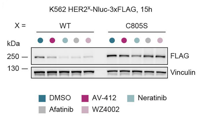
Immunoblot analysis of cell lines (K562 HER2WT or HER2C805S Nluc-3xFLAG reporter cell lines) treated for 15 h with AV-412 (HY-10346; 2.5 µM), neratinib (HY-32721; 10 µM), afatinib (HY-10261; 10 µM), WZ4002 (HY-12026; 10 µM).
Afatinib purchased from MedChemExpress. Usage Cited in: Nature. 2026 Jan;649(8098):1032-1041. [Abstract]
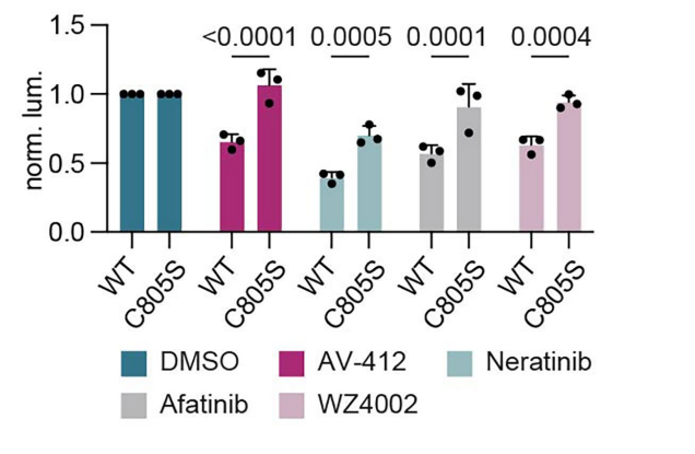
Luminescent reporter assay of K562 HER2WT or HER2C805S Nluc-3xFLAG reporter cell lines treated for 15 h with the indicated compounds (AV-412 (HY-10346; 2.5 µM), neratinib (HY-32721; 10 µM), afatinib (HY-10261; 10 µM), WZ4002 (HY-12026; 10 µM)) shown as normalized luminescence per genetic construct (two-way ANOVA, Sidak corrected) (n = 3).
-
-
Cancer Cell
Anti-tumor efficacy of HRS-4642 and its potential combination with proteasome inhibition in KRAS G12D-mutant cancer. [Abstract]2024 Jul 8;42(7):1286-1300.e8. PMID: 38942026 -
Cancer Cell
KMT2D deficiency drives lung squamous cell carcinoma and hypersensitivity to RTK-RAS inhibition. [Abstract]2023 Jan 9;41(1):88-105.e8. PMID: 36525973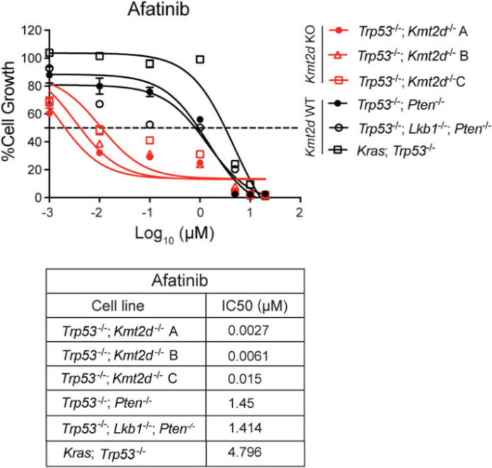
Afatinib purchased from MedChemExpress. Usage Cited in: Cancer Cell. 2023 Jan 9;41(1):88-105.e8. [Abstract]
Cell viability assays of Kmt2d KO LUSC cell lines, Kmt2d WT LUSC cell lines, and LUAD (KP) cell line treated with afatinib for 72h. Data presented as mean ± SD (n = 3). The calculated IC50 values of afatinib is shown below.
-
J Hematol Oncol
SMARCA4 controls state plasticity in small cell lung cancer through regulation of neuroendocrine transcription factors and REST splicing. [Abstract]2024 Jul 30;17(1):58. PMID: 39080761 -
Cancer Discov
Tumor-Intrinsic Kinome Landscape of Pancreatic Cancer Reveals New Therapeutic Approaches. [Abstract]2025 Feb 7;15(2):346-362. PMID: 39632628 -
Nat Genet
Loss of Kmt2c or Kmt2d primes urothelium for tumorigenesis and redistributes KMT2A-menin to bivalent promoters. [Abstract]2025 Jan;57(1):165-179. PMID: 39806204
Afatinib purchased from MedChemExpress. Usage Cited in: Nat Genet. 2025 Jan;57(1):165-179. [Abstract]
Afatinib (10 mg/kg/day; daily, 5 days per week by oral gavage) decreased the intensity of EGFR phosphorylation (pY845) in NOD-SCID mice.

Afatinib purchased from MedChemExpress. Usage Cited in: Nat Genet. 2025 Jan;57(1):165-179. [Abstract]
Afatinib (10 mg/kg/day; daily, 5 days per week by oral gavage) reduced the number of Ki-67-positive proliferating cells in NOD-SCID mice.
-
Nat Cell Biol
2025 Mar;27(3):449-463. PMID: 39984654 -
Cancer Res
2025 Dec 29. PMID: 41460723 -
Cancer Res
NF1 Loss Promotes EGFR Activation and Confers Sensitivity to EGFR Inhibition in NF1 Mutant Melanoma. [Abstract]2025 Jun 10:10.1158/0008-5472.CAN-24-3904. PMID: 40494652
Afatinib purchased from MedChemExpress. Usage Cited in: Cancer Res. 2025 Jun 10:10.1158/0008-5472.CAN-24-3904. [Abstract]
Growth curves of NF1Mut melanoma STCs treated with 0.1, 0.5, 1, 5, or 10 μM Afatinib.
-
Mol Cell
Amplified dosage of the NKX2-1 lineage transcription factor controls its oncogenic role in lung adenocarcinoma. [Abstract]2025 Mar 21:S1097-2765(25)00194-7. PMID: 40139189 -
Cancer Res
Targeting c-Myc to Overcome Acquired Resistance of EGFR Mutant NSCLC Cells to the Third-Generation EGFR Tyrosine Kinase Inhibitor, Osimertinib. [Abstract]2021 Sep 15;81(18):4822-4834. PMID: 34289988 -
Nat Commun
Human iPSC-based Modeling of Pulmonary Fibrosis Reveals p300/CBP Inhibition Suppresses Alveolar Transitional Cell State. [Abstract]2026 Feb 12;17(1):1214. PMID: 41680175 -
Nat Commun
An alternative EGFR activation by patient-derived R252C mutation promotes cancer progression. [Abstract]2026 Jan 21;17(1):1902. PMID: 41565660 -
Nat Commun
Fructose intake driven glycolysis-ROS-EGFR axis specifically promotes the generation and pathogenicity of Th17 cells. [Abstract]2025 Nov 23;16(1):11115. PMID: 41276507 -
Nat Commun
PPARα-mediated lipid metabolism reprogramming supports anti-EGFR therapy resistance in head and neck squamous cell carcinoma. [Abstract]2025 Feb 1;16(1):1237. PMID: 39890801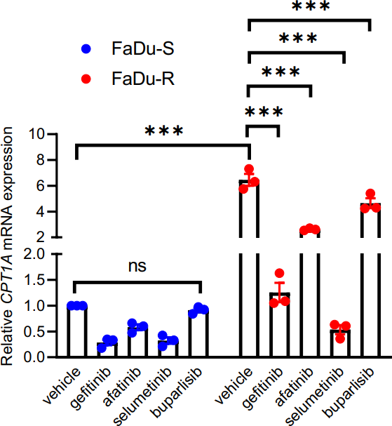
Afatinib purchased from MedChemExpress. Usage Cited in: Nat Commun. 2025 Feb 1;16(1):1237. [Abstract]
mRNA expression for CPT1A in cetuximab-sensitive (-S) and -resistant (-R) FaDu cells upon treatment with gefitinib, Afatinib, selumetinib or buparlisib (2 µM each, for 24 h) (N = 3, n = 3).
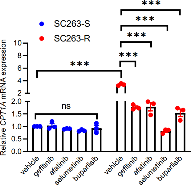
Afatinib purchased from MedChemExpress. Usage Cited in: Nat Commun. 2025 Feb 1;16(1):1237. [Abstract]
mRNA expression for CPT1A in cetuximab-sensitive (-S) and -resistant (-R) SC263 cells upon treatment with gefitinib, Afatinib, selumetinib or buparlisib (2 µM each, for 24 h) (N = 3, n = 3).
-
Nat Commun
AURKB as a target in non-small cell lung cancer with acquired resistance to anti-EGFR therapy. [Abstract]2019 Apr 18;10(1):1812 PMID: 31000705 -
Sci Transl Med
PP2A inhibition is a druggable MEK inhibitor resistance mechanism in KRAS-mutant lung cancer cells. [Abstract]2018 Jul 18;10(450):eaaq1093. PMID: 30021885 -
-
Biomaterials
2022 Oct:289:121800. PMID: 36166893 -
J Exp Clin Cancer Res
Pyrotinib targeted EGFR/GRP78 mediated cell apoptosis in high EGFR gene copy number gastric cancer. [Abstract]2025 Aug 19;44(1):245. PMID: 40830980 -
J Exp Clin Cancer Res
Dual inhibition of HERs and PD-1 counteract resistance in KRASG12C-mutant head and neck cancer. [Abstract]2024 Nov 20;43(1):308. PMID: 39567998 -
J Exp Clin Cancer Res
The potential of swine pseudorabies virus attenuated vaccine for oncolytic therapy against malignant tumors. [Abstract]2023 Oct 27;42(1):284. PMID: 37891570 -
Redox Biol
C-terminal interleukin 1 alpha (IL-1α) overexpression drives EMT and a vulnerability to ferroptosis in HNSCC. [Abstract]2026 Jun:93:104172. PMID: 42013543 -
Cell Rep Med
CAN-Scan: A multi-omic phenotype-driven precision oncology platform identifies prognostic biomarkers of therapy response for colorectal cancer. [Abstract]2025 Apr 2:102053. PMID: 40187357 -
Cell Rep Med
Using patient-derived organoids to predict locally advanced or metastatic lung cancer tumor response: A real-world study. [Abstract]2023 Feb 21;4(2):100911. PMID: 36657446 -
Pharmacol Res
Chelidonine selectively inhibits the growth of gefitinib-resistant non-small cell lung cancer cells through the EGFR-AMPK pathway. [Abstract]2020 Sep;159:104934. PMID: 32464330 -
Pharmacol Res
Cordycepin Inhibits Drug-resistance Non-small Cell Lung Cancer Progression by Activating AMPK Signaling Pathway. [Abstract]2019 Jun:144:79-89. PMID: 30974169 -
Cancer Lett
2025 Apr 10:217715. PMID: 40220852 -
Cell Death Dis
TRIB2 modulates proteasome function to reduce ubiquitin stability and protect liver cancer cells against oxidative stress. [Abstract]2021 Jan 7;12(1):42. PMID: 33414446 -
Osteoarthritis Cartilage
Synovial fluid from end-stage osteoarthritis induces proliferation and fibrosis of articular chondrocytes via MAPK and RhoGTPase signaling. [Abstract]2022 Jun;30(6):862-874. PMID: 35176481 -
J Pharm Anal
Development of a competitive enzyme-linked immunosorbent assay for therapeutic drug monitoring of afatinib. [Abstract]2019 Feb;9(1):49-54. PMID: 30740257 -
Acta Pharmacol Sin
143D, a novel selective KRASG12C inhibitor exhibits potent antitumor activity in preclinical models. [Abstract]2023 Jul;44(7):1475-1486. PMID: 36725884 -
EMBO Mol Med
Upfront admixing antibodies and EGFR inhibitors preempts sequential treatments in lung cancer models. [Abstract]2021 Apr 9;13(4):e13144. PMID: 33660397 -
NPJ Parkinsons Dis
2025 Jun 7;11(1):157. PMID: 40483356 -
ACS Appl Mater Interfaces
Intelligent Biomimetic Nanoplatform for Systemic Treatment of Metastatic Triple-Negative Breast Cancer via Enhanced EGFR-Targeted Therapy and Immunotherapy. [Abstract]2022 May 25;14(20):23152-23163. PMID: 35549005 -
NPJ Precis Oncol
High-throughput screening identifies NT-1 that synergizes with MRTX1133 against acquired resistant KRASG12D colorectal cancer. [Abstract]2026 Apr 8;10(1):211. PMID: 41951768 -
J Transl Med
Combined treatment with inhibitors of ErbB Receptors and Hh signaling pathways is more effective than single treatment in reducing the growth of malignant mesothelioma both in vitro and in vivo. [Abstract]2022 Jun 25;20(1):286. PMID: 35752861 -
Oncogene
Suppression of heparan sulfation re-sensitizes YAP1-driven melanoma to MAPK pathway inhibitors. [Abstract]2022 Aug;41(32):3953-3968. PMID: 35798875 -
Oncogene
Epigenetic silencing of miR-483-3p promotes acquired gefitinib resistance and EMT in EGFR-mutant NSCLC by targeting integrin β3. [Abstract]2018 Aug;37(31):4300-4312. PMID: 29717264 -
Clin Transl Med
Multi-omic profiling defines three distinct molecular subtypes of urothelial carcinoma with implications for precision therapy. [Abstract]2026 Mar;16(3):e70638. PMID: 41804750 -
Sci Signal
TSHZ2 is an EGF-regulated tumor suppressor that binds to the cytokinesis regulator PRC1 and inhibits metastasis. [Abstract]2021 Jun 22;14(688):eabe6156. PMID: 34158398 -
Acta Neuropathol Commun
Identifying and exploiting combinatorial synthetic lethality by characterizing adaptive kinome rewiring of EGFRvIII-driven glioblastoma. [Abstract]2025 Jun 28;13(1):143. PMID: 40581663 -
J Mater Chem B
Overcoming ABCG2-mediated multidrug resistance by a mineralized hyaluronan-drug nanocomplex. [Abstract]2016 Nov 7;4(41):6652-6661. PMID: 32263520 -
Mol Cancer Ther
Dual Inhibitors of KRASG12D and HSP90 are Effective Against KRASG12D Inhibitor Resistance. [Abstract]2025 Oct 23. PMID: 41129140 -
Pharmaceutics
Drug Repurposing for the Identification of Compounds with Anti-SARS-CoV-2 Capability via Multiple Targets. [Abstract]2022 Jan 12;14(1):176. PMID: 35057070 -
Mol Cancer Ther
Afatinib Is a New Therapeutic Approach in Chordoma with a Unique Ability to Target EGFR and Brachyury. [Abstract]2018 Mar;17(3):603-613. PMID: 29237806
Afatinib purchased from MedChemExpress. Usage Cited in: Mol Cancer Ther. 2018 Mar;17(3):603-613. [Abstract]
Immunoblot analysis of U-CH1, MUG-Chor1 and Chor-IN-1 cells treated with Afatinib for 2 h or 48 h. Protein cell extracts were resolved on SDS-PAGE gel and membranes probed with the indicated antibodies. IC50s for each cell line are reported.

Afatinib purchased from MedChemExpress. Usage Cited in: Mol Cancer Ther. 2018 Mar;17(3):603-613. [Abstract]
Immunoblot analysis of U-CH1 cells treated with the indicated doses of inhibitors (Afatinib, Erlotinib and Lapatinib) for 2 h (upper panel) or 48 h (lower panel). Protein cell extracts were resolved on SDS-PAGE gel and membranes probed with the indicated antibodies. IC50s of the different inhibitors are reported.
-
Cancer Drug Resist
Novel FAK inhibitors suppress tumor growth and reverse EGFR-TKI resistance in non-small cell lung cancer. [Abstract]2025 Nov 5:8:57. PMID: 41281943 -
Biol Direct
In vitro synergistic effect of AXL, FAK and ErbB receptors inhibitors for head and neck cancer. [Abstract]2025 Jul 2;20(1):77. PMID: 40605022 -
-
Int Immunopharmacol
Afatinib inhibits esophageal squamous cell carcinoma by regulating ferroptosis and NRF2 protein homeostasis. [Abstract]2026 Mar 1:172:116131. PMID: 41520560 -
Eur J Pharmacol
Discovery of 4-((3,4-dichlorophenyl)amino)-2-methylquinolin-6-ol derivatives as EGFR and HDAC dual inhibitors. [Abstract]2023 Dec 5:960:176114. PMID: 37863412 -
Mol Cancer Res
TAS6417/CLN-081 Is a Pan-Mutation-Selective EGFR Tyrosine Kinase Inhibitor with a Broad Spectrum of Preclinical Activity against Clinically Relevant EGFR Mutations. [Abstract]2019 Nov;17(11):2233-2243. PMID: 31467113 -
Toxicology
EGFR-TKIs induce acneiform rash and xerosis via Caspase-3/GSDME-mediated pyroptosis of keratinocytes and sebocytes. [Abstract]2024 Nov 26:154018. PMID: 39608440 -
Cancers (Basel)
Evaluation of Combined Chemotherapy and Genomic-Driven Targeted Therapy in Patient-Derived Xenografts Identifies New Therapeutic Approaches in Squamous Non-Small-Cell Lung Cancer Patients. [Abstract]2024 Aug 7;16(16):2785. PMID: 39199558 -
Cancer Sci
2018 Apr;109(4):1166-1176. PMID: 29465762
Afatinib purchased from MedChemExpress. Usage Cited in: Cancer Sci. 2018 Apr;109(4):1166-1176. [Abstract]
Influence of Afatinib or Neratinib on human epidermal growth factor receptor 2 (HER2) and the downsignal pathway in gastric cancer cell lines.
-
-
Sci Rep
A novel human acute myeloid leukemia cell line SDEY-AML1 with KMT2A: MLLT3, IKZF1: EVX1 fusions exhibits high tumorigenicity in NSG mice. [Abstract]2026 Feb 8;16(1):7792. PMID: 41656387 -
PLoS Comput Biol
2020 Feb 26;16(2):e1007701. PMID: 2101536 -
Viruses
Screening and Identification of Lujo Virus Inhibitors Using a Recombinant Reporter Virus Platform. [Abstract]2021 Jun 28;13(7):1255. PMID: 34203149 -
Exp Cell Res
Network-based analysis with primary cells reveals drug response landscape of acute myeloid leukemia. [Abstract]2020 Aug 1;393(1):112054. PMID: 32376287 -
Cell Biol Int
Sensitization of HT29 colorectal cancer cells to vemurafenib in three-dimensional collagen cultures. [Abstract]2020 Feb;44(2):621-629. PMID: 31736196 -
J Pharm Biomed Anal
Comparative studies on the human serum albumin binding of the clinically approved EGFR inhibitors gefitinib, erlotinib, afatinib, osimertinib and the investigational inhibitor KP2187. [Abstract]2018 May 30:154:321-331. PMID: 29567575 -
Genes (Basel)
The Impact of Bevacizumab and miR200c on EMT and EGFR-TKI Resistance in EGFR-Mutant Lung Cancer Organoids. [Abstract]2024 Dec 19;15(12):1624. PMID: 39766891 -
PLoS One
A novel small molecule screening assay using normal human chondrocytes toward osteoarthritis drug discovery. [Abstract]2024 Nov 1;19(11):e0308647. PMID: 39485774 -
PLoS One
2018 Jun 4;13(6):e0198364. PMID: 29864158 -
Fundam Clin Pharmacol
2021 Oct;35(5):919-929. PMID: 33523504 -
Eur J Drug Metab Pharmacokinet
Differential Inhibition of Equilibrative Nucleoside Transporter 1 (ENT1) Activity by Tyrosine Kinase Inhibitors. [Abstract]2021 Sep;46(5):625-635. PMID: 34275128 -
Int J Radiat Biol
Celecoxib and Afatinib synergistic enhance radiotherapy sensitivity on human non-small cell lung cancer A549 cells. [Abstract]2021;97(2):170-178. PMID: 33164600 -
Acta Histochem
Immunohistochemical localization of afatinib in male rat intestines and skin after its oral administration. [Abstract]2019 Nov;121(8):151439. PMID: 31500866 -
-
J Oral Biosci
Antihypertensive agent losartan promotes tongue squamous cell carcinoma cell proliferation via EGFR/ERK1/2/cyclin D1 signaling axis. [Abstract]2024 Sep 6:S1349-0079(24)00198-1. PMID: 39245205 -
Afatinib purchased from MedChemExpress. Usage Cited in: Biochem Biophys Res Commun. 2026 Feb 12:800:153165.
ActE_21 binding to A549, A431, and SK-BR-3 cells was assessed after incubation with Afatinib (Afatinib dimaleate; 50 nM) for 120 min at 37 °C. Afatinib increased the surface levels of EGF-EGFR complexes in A549 and SK-BR-3 cells.
-
Oncol Lett
EGFR and ERK activation resists flavonoid quercetin-induced anticancer activities in human cervical cancer cells in vitro. [Abstract]2021 Nov;22(5):754. PMID: 34539858 -
Cryobiology
2019 Feb:86:71-76. PMID: 30527584 -
Cell Physiol Biochem
2018;47(3):1259-1273. PMID: 29913444 -
Bioanalysis
Development and validation of an ELISA with high sensitivity for therapeutic monitoring of afatinib. [Abstract]2018 Sep 1;10(18):1511-1523. PMID: 30117333 -
Biomed Chromatogr
Simultaneous and rapid determination of gefitinib, erlotinib and afatinib plasma levels using liquid chromatography/tandem mass spectrometry in patients with non-small-cell lung cancer. [Abstract]2016 Jul;30(7):1150-4. PMID: 26525154 -
Biol Methods Protoc
Optimizing drug sensitivity assays in patient-derived tumor organoids: a comparison of IC50 estimation methods and experimental parameters. [Abstract]2025 Feb 13;10(1):bpaf012. PMID: 40060949 -
Xenobiotica
Substrate-dependent effects of molecular-targeted anticancer agents on activity of organic anion transporting polypeptide 1B1. [Abstract]2018 Oct;48(10):1059-1071. PMID: 29034773 -
-
Afatinib purchased from MedChemExpress. Usage Cited in: bioRxiv. 2026 Mar 12.
A549 cells co-expressing CFP-EGFR and YFP-GRB2 were exposed to Afatinib (Afatinib dimaleate; AFA; 25.84 μM; 4 h). FRET analysis showed that Afatinib reduced the mean donor-centric FRET efficiency from 0.27 in the control to 0.13 and produced the highest T score, indicating reduced EGFR-GRB2 interaction.
-
-
-
-
-
-
-
bioRxiv
Modeling acquired TKI resistance and effective combination therapeutic strategies in murine RET+ lung adenocarcinoma. [Abstract]2025 Jun 7:2025.06.04.657911. PMID: 40502048 -
World J Exp Med
2025 Jun 20;15(2):100443. PMID: 40546672 -
-
bioRxiv
BRAFV600E-Driven Lung Tumorigenesis Requires Ligand-Mediated Activation of ERBB Receptor Signaling. [Abstract]2025 May 9:2025.05.04.652129. PMID: 40654950
Afatinib purchased from MedChemExpress. Usage Cited in: bioRxiv. 2025 May 9:2025.05.04.652129. [Abstract]
Afatinib dimaleate (15 mg/kg; oral gavage; once daily for 8 weeks) reduced tumor burden and tumor diameter in BrafCAT mice.

Afatinib purchased from MedChemExpress. Usage Cited in: bioRxiv. 2025 May 9:2025.05.04.652129. [Abstract]
Afatinib dimaleate (Afatinib; 15 mg/kg; oral gavage; once daily for 8 weeks). Immunohistochemistry analyses of phosphorylated ERK (pT202, Y204) in BrafCAT formalin fixed paraffin embedded (FFPE) mouse lung tissue.

Afatinib purchased from MedChemExpress. Usage Cited in: bioRxiv. 2025 May 9:2025.05.04.652129. [Abstract]
Afatinib dimaleate (A; 50 nM; 2 or 24 h), alone or in combination with dabrafenib and trametinib, enhanced pathway blockade in HCC364 human BRAFV600E+ lung cancer cells, as indicated by reduced pEGFR, pHER2, and pERK levels, with a greater effect after 24 h than after 2 h.
-
-
-
-
-
-
bioRxiv
Structural dynamics of the active HER4 and HER2/HER4 complexes is finely tuned by different growth factors and glycosylation. [Abstract]2024 Jan 4:2023.10.06.561161. PMID: 38260342 -
-
-
-
-
-
-
-
Oncotarget
Ibrutinib selectively and irreversibly targets EGFR (L858R, Del19) mutant but is moderately resistant to EGFR (T790M) mutant NSCLC Cells. [Abstract]2015 Oct 13;6(31):31313-22. PMID: 26375053 -
Oncotarget
Afatinib circumvents multidrug resistance via dually inhibiting ATP binding cassette subfamily G member 2 in vitro and in vivo. [Abstract]2014 Dec 15;5(23):11971-85. PMID: 25436978
Afatinib purchased from MedChemExpress. Usage Cited in: Oncotarget. 2014 Dec 15;5(23):11971-85. [Abstract]
H460/MX20 cells are treated with varying concentrations (0–2.0 μM) of Afatinib for 48 h, or with 1.0 μM Afatinib for 24 h, 48 h and 72 h, respectively. ABCG2 protein levels are analyzed by Western blot. GAPDH is used as a loading control.


Solvent & Solubility
DMSO : 100 mg/mL (205.79 mM; Need ultrasonic; Hygroscopic DMSO has a significant impact on the solubility of product, please use newly opened DMSO)
Please refer to the solubility information to select the appropriate solvent. Once prepared, please aliquot and store the solution to prevent product inactivation from repeated freeze-thaw cycles.
Storage method and period of stock solution: -80°C, 2 years; -20°C, 1 year. When stored at -80°C, please use it within 2 years. When stored at -20°C, please use it within 1 year.
Concentration (start) × Volume (start) = Concentration (final) × Volume (final)
Select the appropriate dissolution method based on your experimental animal and administration route.
- For the following dissolution methods, please ensure to first prepare a clear stock solution using an In Vitro approach and then sequentially add co-solvents:
- To ensure reliable experimental results, the clarified stock solution can be appropriately stored based on storage conditions. As for the working solution for In Vivo experiments, it is recommended to prepare freshly and use it on the same day.
- The percentages shown for the solvents indicate their volumetric ratio in the final prepared solution. If precipitation or phase separation occurs during preparation, heat and/or sonication can be used to aid dissolution.
Add each solvent one by one: 10% DMSO 40% PEG300 5% Tween-80 45% Saline
Solubility: ≥ 2.5 mg/mL (5.14 mM); Clear solution
This protocol yields a clear solution of ≥ 2.5 mg/mL (saturation unknown).
Taking 1 mL working solution as an example, add 100 μL DMSO stock solution (25.0 mg/mL) to 400 μL PEG300, and mix evenly; then add 50 μL Tween-80 and mix evenly; then add 450 μL Saline to adjust the volume to 1 mL.
Preparation of Saline: Dissolve 0.9 g sodium chloride in ddH₂O and dilute to 100 mL to obtain a clear Saline solution.
For the following dissolution methods, please prepare the working solution directly:
It is recommended to prepare fresh solutions and use them promptly within a short period of time.
The percentages shown for the solvents indicate their volumetric ratio in the final prepared solution. If precipitation or phase separation occurs during preparation, heat and/or sonication can be used to aid dissolution.
Add each solvent one by one: 0.5% Methylcellulose/saline water
Solubility: 5 mg/mL (10.29 mM); Suspended solution; Need ultrasonic
Please enter the basic information of animal experiments:
-
-
-
-
Recommended: Prepare an additional quantity of animals to account for potential losses during experiments.
Please enter your animal formula composition:
-
%DMSO +
Recommended: Keep the proportion of DMSO in working solution below 2% if your animal is weak.
-
%+
-
+%Tween-80 + +
-
%Saline +
The co-solvents required include: DMSO, . All of co-solvents are available by MedChemExpress (MCE). , Tween 80. All of co-solvents are available by MedChemExpress (MCE).
Working solution concentration: 0.22 mg/mL
Method for preparing stock solution: mg drug dissolved in μL DMSO. Stock solution concentration: mg/mL.
1. Take μL DMSO stock solution;
2. Add μL .
μL , mix evenly;
3. Then add μL Tween 80, mix evenly;
4. Then add μL
Please ensure that the stock solution in the first step is dissolved to a clear state, and add co-solvents in sequence. You can use ultrasonic heating (ultrasonic cleaner, recommended frequency 20-40 kHz), vortexing, etc. to assist dissolution.


Purity & Documentation
-
Data Sheet (289 KB)
-
SDS (557 KB)
- English - EN (557 KB)
- Français - FR (557 KB)
- Deutsch - DE (557 KB)
- Norwegian - NO (557 KB)
- Español - ES (557 KB)
- Swedish - SV (557 KB)
- Italian - IT (557 KB)
- Korean - KR (557 KB)
- Portuguese - PT (557 KB)
-
Handling Instructions (2659 KB)
References
[1]. Li D, et al. BIBW2992, an irreversible EGFR/HER2 inhibitor highly effective in preclinical lung cancer models. Oncogene. 2008 Aug 7;27(34):4702-11. [Content Brief]
[2]. Wong CH, et al. Preclinical evaluation of afatinib (BIBW2992) in esophageal squamous cell carcinoma (ESCC). Am J Cancer Res. 2015 Nov 15;5(12):3588-99. [Content Brief]
[3]. Wang XK, et al. Afatinib circumvents multidrug resistance via dually inhibiting ATP binding cassette subfamily G member 2 in vitro and in vivo. Oncotarget. 2014 Dec 15;5(23):11971-85. [Content Brief]
[4]. Yoshioka T, et al. Antitumor activity of pan-HER inhibitors in HER2-positive gastric cancer. Cancer Sci. 2018 Apr;109(4):1166-1176. [Content Brief]


Complete Stock Solution Preparation Table
Please refer to the solubility information to select the appropriate solvent. Once prepared, please aliquot and store the solution to prevent product inactivation from repeated freeze-thaw cycles.
Storage method and period of stock solution: -80°C, 2 years; -20°C, 1 year. When stored at -80°C, please use it within 2 years. When stored at -20°C, please use it within 1 year.
| Optional Solvent | Concentration Solvent Mass | 1 mg | 5 mg | 10 mg | 25 mg |
|---|---|---|---|---|---|
| DMSO | 1 mM | 2.0579 mL | 10.2893 mL | 20.5787 mL | 51.4467 mL |
| 5 mM | 0.4116 mL | 2.0579 mL | 4.1157 mL | 10.2893 mL | |
| 10 mM | 0.2058 mL | 1.0289 mL | 2.0579 mL | 5.1447 mL | |
| 15 mM | 0.1372 mL | 0.6860 mL | 1.3719 mL | 3.4298 mL | |
| 20 mM | 0.1029 mL | 0.5145 mL | 1.0289 mL | 2.5723 mL | |
| 25 mM | 0.0823 mL | 0.4116 mL | 0.8231 mL | 2.0579 mL | |
| 30 mM | 0.0686 mL | 0.3430 mL | 0.6860 mL | 1.7149 mL | |
| 40 mM | 0.0514 mL | 0.2572 mL | 0.5145 mL | 1.2862 mL | |
| 50 mM | 0.0412 mL | 0.2058 mL | 0.4116 mL | 1.0289 mL | |
| 60 mM | 0.0343 mL | 0.1715 mL | 0.3430 mL | 0.8574 mL | |
| 80 mM | 0.0257 mL | 0.1286 mL | 0.2572 mL | 0.6431 mL | |
| 100 mM | 0.0206 mL | 0.1029 mL | 0.2058 mL | 0.5145 mL |


 Powered by Bioz