


Deguelin
Based on 15 publication(s) in Google Scholar
Deguelin, a naturally occurring rotenoid, acts as a chemopreventive agent by blocking multiple pathways like PI3K-Akt, IKK-NF-κB, and MAPK-mTOR-survivin-mediated apoptosis. Deguelin binding to Hsp90 leads to a decreased expression of numerous oncogenic proteins, including MEK1/2, Akt, HIF1α, COX-2, and NF-κB.
Nur für Forschungszwecke. Wir verkaufen nicht an Patienten.
- Reinheit: 99.20%
- CAS. Nr.: 522-17-8
- Formel: C23H22O6
- Molecular Weight:394.42
-
Speicherung:Powder -20°C, 3 years , 4°C, 2 years ; In solvent -80°C, 1 year , -20°C, 6 months

To place orders, for customer services and technical support, please contact: MedChemExpress USA
Tel: 609-228-6898 E-mail: [email protected] [email protected]


-
 Biologische Aktivität
-
Chemical Information
-
Lösungsmittel & Löslichkeit
- Protokoll
- Reinheit & Dokumentation
- Verweise
-
 Help & FAQs
-


Apoptosis Compound Library
HY-L003
-
Immunology/Inflammation Compound Library
HY-L007
-
Kinase Inhibitor Library
HY-L009
-
NF-κB Signaling Compound Library
HY-L014
-
PI3K/Akt/mTOR Compound Library
HY-L015
-
Stem Cell Signaling Compound Library
HY-L017
-
Natural Product Library
HY-L021
-
Anti-Cancer Compound Library
HY-L025
-
Autophagy Compound Library
HY-L029
-
Anti-Aging Compound Library
HY-L034
-
Differentiation Inducing Compound Library
HY-L038
-
Lipid Compound Library
HY-L043
-
Oxygen Sensing Compound Library
HY-L045
-
Glycolysis Compound Library
HY-L058
-
Cytoskeleton Compound Library
HY-L060
-
Glutamine Metabolism Compound Library
HY-L064
-
Traditional Chinese Medicine Active Compound Library
HY-L065
-
Flavonoids Library
HY-L068
-
Anti-Breast Cancer Compound Library
HY-L074
-
Anti-Lung Cancer Compound Library
HY-L075
-
Anti-Pancreatic Cancer Compound Library
HY-L077
-
Anti-Blood Cancer Compound Library
HY-L079
-
Anti-Cancer Metabolism Compound Library
HY-L083
-
Anti-Obesity Compound Library
HY-L087
-
Angiogenesis-Related Compound Library
HY-L088
-
Glucose Metabolism Compound Library
HY-L092
-
Food-Sourced Compound Library
HY-L094
-
Anti-Liver Cancer Compound Library
HY-L101
-
Anti-Colorectal Cancer Compound Library
HY-L103
-
Anti-Cancer Natural Product Library
HY-L107
-
Anti-inflammatory Traditional Chinese Medicine Active Compound Library
HY-L114
-
Plant-Sourced Natural Product Library
HY-L115
-
Human Metabolite Library
HY-L123
-
Anti-Prostate Cancer Compound Library
HY-L124
-
Anti-Pulmonary Fibrosis Compound Library
HY-L125
-
Cancer Stem Cells Compound Library
HY-L135
-
Pain-Related Compound Library
HY-L139
-
Mitochondrial Toxicity Compound Library
HY-L155
-
Cell Death Library
HY-L162
-
Serine/Threonine Kinase Inhibitor Library
HY-L164
-
Extracellular Vesicles (EVs) Compound Library
HY-L168
-
Anti-Hematopathy Compound Library
HY-L171
-
Anti-Ovarian Cancer Compound Library
HY-L173
-
Bioactive Compound Library Max
HY-L181
-
MCE Bioactive Compound Library
HY-L001V
-
Natural Product Library Plus
HY-L021P
-
Natural Product and Natural Product-Like Compound Library
HY-L021M
-
Bioactive Compound Library
HY-L001
-
Anti-Gastric Cancer Compound Library
HY-L184
-
Anti-Fibrosis Compound Library
HY-L185
-
Protein Kinase Compound Library
HY-L196
-
Non-Alcoholic Fatty Liver Disease (NAFLD) Compound Library
HY-L199
-
RO5 Drug-like Natural Product Library
HY-L200
-
Cell Proliferation Compound Library
HY-L201
-
High-Throughput Bioactive Compound Library
HY-L205
-
High-Throughput Natural Product Library
HY-L206
-
Mass Spectrometry Human Metabolite Library
HY-L215
-
Posttranslational Modification Library
HY-L226
-
Natural Product Diversity Scaffold Library
HY-L245
-
Lactylation Compound Library
HY-L249
-
Mass Spectrometry Natural Product Library
HY-L262


Publications Citing Use of MedChemExpress (MCE) Deguelin
More- Acta Pharm Sin B. 2021 Jan;11(1):143-155. [Abstract]
- Food Chem. 2025 Dec 30:497:146992. [Abstract]
- Acta Pharmacol Sin. 2025 Nov 3. [Abstract]
- Food Chem. 2025 May 31:489:144992. [Abstract]
- Int J Mol Med. 2018 Jun;41(6):3157-3166. [Abstract]
- PLoS Biol. 2024 Jun 27;22(6):e3002672. [Abstract]
- Cancer Biol Ther. 2026 Dec 31;27(1):2644788. [Abstract]
- Neuropharmacology. 2024 Aug 9:110109. [Abstract]
- J Med Virol. 2024 Sep;96(9):e29891. [Abstract]
- Toxicol Appl Pharmacol. 2025 Sep:502:117453. [Abstract]
- Arch Med Res. 2021 Jul;52(5):494-504. [Abstract]
- Int J Biochem Cell Biol. 2026 Jun-Jul:195-196:106940. [Abstract]
- J Cancer. 2021 Jan 1;12(1):270-280. [Abstract]
- Res Sq. 2025 Aug 20.
- bioRxiv. 2024 Apr 3:2023.06.02.542933. [Abstract]
 Customer Validation & Images

-
Cell Proliferation/Viability Assay
-
WB
-
Apoptosis Analysis
-
Cell Imaging/Staining
-
In Vivo Efficacy Study


Biologische Aktivität
|
Cell Line
|
Type | Value | Description | References |
|---|---|---|---|---|
| A549 | IC50 |
6.05 μM
Compound: 4
|
Antiproliferative activity against human A549 cells after 72 hrs by MTT assay
Antiproliferative activity against human A549 cells after 72 hrs by MTT assay
|
[PMID: 30784881] |
| Astrocyte | IC50 |
>80 μM
Compound: 1
|
Growth inhibition of Nf1 defective and Trp53 defective mouse primary astrocytes after 2 hrs by XTT assay
Growth inhibition of Nf1 defective and Trp53 defective mouse primary astrocytes after 2 hrs by XTT assay
|
[PMID: 26467198] |
| HeLa | IC50 |
0.22 μM
Compound: 3
|
Inhibition of TNF-alpha-induced NF-kappaB activation in human HeLa cells by SEAP reporter gene assay
Inhibition of TNF-alpha-induced NF-kappaB activation in human HeLa cells by SEAP reporter gene assay
|
[PMID: 18841906] |
| HEp-2 | CC50 |
26.5 μM
Compound: 10
|
Cytotoxicity against human HEp-2 cells measured after 3 days by CellTiter 96 aqueous one solution assay (Rvb >100 microM)
Cytotoxicity against human HEp-2 cells measured after 3 days by CellTiter 96 aqueous one solution assay (Rvb >100 microM)
|
[PMID: 38579352] |
| HEp-2 | IC50 |
0.4 μM
Compound: 10
|
Antiviral activity against Human respiratory syncytial virus infected in human HEp-2 cells assessed as plaque reduction preincubated with cells 15 mins followed by viral infection for 2.5 hrs and measured after 3 days by crystal violet staining based micr
Antiviral activity against Human respiratory syncytial virus infected in human HEp-2 cells assessed as plaque reduction preincubated with cells 15 mins followed by viral infection for 2.5 hrs and measured after 3 days by crystal violet staining based micr
|
[PMID: 38579352] |
| L5178Y | IC50 |
0.2 μM
Compound: 26
|
Cytotoxicity against mouse L5178Y cells by MTT assay
Cytotoxicity against mouse L5178Y cells by MTT assay
|
[PMID: 28075580] |
| MCF7 | IC50 |
10.59 μM
Compound: i
|
Cytotoxicity against human MCF7 cells assessed as cell growth inhibition after 24 hrs by MTT assay
Cytotoxicity against human MCF7 cells assessed as cell growth inhibition after 24 hrs by MTT assay
|
[PMID: 27376493] |
| MCF7 | IC50 |
8.17 μM
Compound: 4
|
Antiproliferative activity against human MCF7 cells after 72 hrs by MTT assay
Antiproliferative activity against human MCF7 cells after 72 hrs by MTT assay
|
[PMID: 30784881] |
| NCI-H1299 | IC50 |
0.11 μM
Compound: 1, (-)-Deguelin
|
Cytotoxicity against human H1299 cells after 3 days by MTS assay
Cytotoxicity against human H1299 cells after 3 days by MTS assay
|
[PMID: 23186287] |
| NCI-H1299 | IC50 |
5.67 μM
Compound: 4
|
Antiproliferative activity against human NCI-H1299 cells after 72 hrs by MTT assay
Antiproliferative activity against human NCI-H1299 cells after 72 hrs by MTT assay
|
[PMID: 30784881] |
| RAW264.7 | IC50 |
1.75 μM
Compound: 3
|
Inhibition of LPS-induced NF-kappaB activation in mouse RAW264.7 cells by SEAP reporter gene assay
Inhibition of LPS-induced NF-kappaB activation in mouse RAW264.7 cells by SEAP reporter gene assay
|
[PMID: 18841906] |
| U-937 | IC50 |
16 μM
Compound: 1, deguelin
|
Antiproliferative activity against human U937 cells after 48 hrs by WST-8 assay
Antiproliferative activity against human U937 cells after 48 hrs by WST-8 assay
|
[PMID: 17158054] |
| U-937 | IC50 |
42 μM
Compound: 1, deguelin
|
Antiproliferative activity against human U937 cells after 24 hrs by WST-8 assay
Antiproliferative activity against human U937 cells after 24 hrs by WST-8 assay
|
[PMID: 17158054] |
| U-937 | IC50 |
5.8 μM
Compound: 1, deguelin
|
Antiproliferative activity against human U937 cells after 72 hrs by WST-8 assay
Antiproliferative activity against human U937 cells after 72 hrs by WST-8 assay
|
[PMID: 17158054] |
Deguelin (0-500 nM) in a dose and time dependent manner inhibits the growth of MDA-MB-231, MDA-MB-468, BT-549 and BT-20 cells. Deguelin at all concentrations fails to reduce cell numbers in the presence of 1 ng EGF but in the presence of EGF 20 ng reinstated deguelin mediated growth inhibition. Deguelin treated cells show reduced expression of Survivin as determined by western blot and immunofluorescence examinations. Deguelin inhibits p-ERK and its downstream target p-STAT-3 and c-Myc expression in a dose dependent manner[1].
Deguelin down-regulates Akt signaling probably by disrupting its association with Hsp 90 in cultured HNSCC cells. Deguelin deguelin disrupts the association between Hsp 90 with survivin and Cdk4. Deguelin deguelin treatment increases cellular ceramide level through de novo synthase pathway to mediate HNSCC cell death and apoptosis[2].
Deguelin inhibits the proliferation of MPC-11 cells in a concentration- and time-dependent manner and causes the apoptotic death of MPC-11 cells. Following exposure to deguelin, the phosphorylation of Akt is decreased. Deguelin-induced apoptosis is characterized by the upregulation of Bax, downregulation of Bcl-2 and activation of caspase-3[3].
MedChemExpress (MCE) has not independently confirmed the accuracy of these methods. They are for reference only.
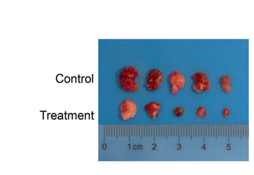
Deguelin (4 mg/kg, p.o.) treatment shows a great inhibition in tumor growth, which is demonstrated by reduced tumor size and improved mice survival and, indicating a significant anti-tumor ability by deguelin in vivo[2].
In the colon cancer xenograft model, the volume of the tumor treated with deguelin is significantly lower than that of the control, and the apoptotic index for deguelin-treated mice is much higher[4].
MedChemExpress (MCE) has not independently confirmed the accuracy of these methods. They are for reference only.


Chemical Information
-
CAS. Nr. 522-17-8
-
Appearance Solid
-
Molecular Weight 394.42
-
Formel C23H22O6
-
Color Light yellow to yellow
-
SMILES
O=C1[C@]2([H])[C@](COC3=CC(OC)=C(OC)C=C32)([H])OC4=C5C=CC(C)(C)OC5=CC=C14
-
Synonyms
(-)-Deguelin; (-)-cis-Deguelin
-
Structure Classification
-
Initial Source
-
Versand
Room temperature in continental US; may vary elsewhere.
-
Speicherung
Powder -20°C 3 years 4°C 2 years In solvent -80°C 1 year -20°C 6 months


Publications (15)
-
Journal Impact Factor
-
Most Recent
-
Acta Pharm Sin B
Chrysin serves as a novel inhibitor of DGK α/FAK interaction to suppress the malignancy of esophageal squamous cell carcinoma (ESCC). [Abstract]2021 Jan;11(1):143-155. PMID: 33532186 -
Food Chem
Effects of sun drying combined with baking processes on the flavor quality of Chongqing Tuocha raw tea. [Abstract]2025 Dec 30:497:146992. PMID: 41285060 -
Acta Pharmacol Sin
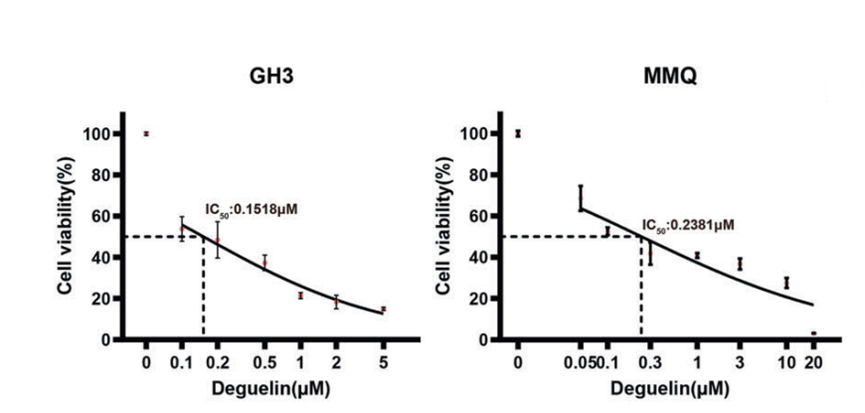
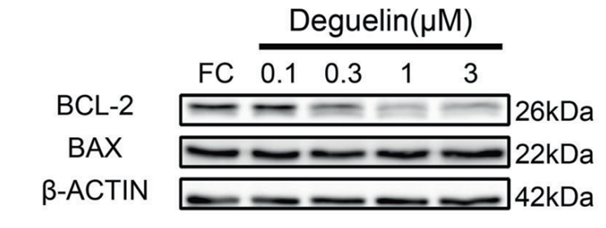
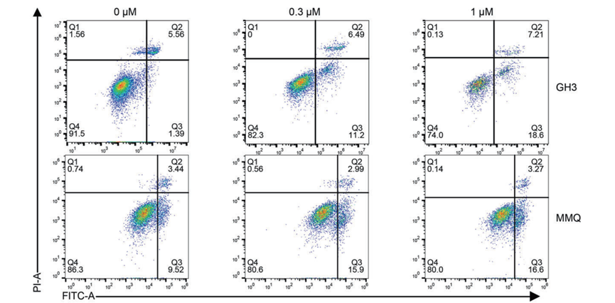
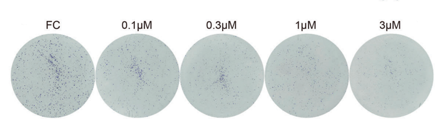
Deguelin inhibits growth and prolactin synthesis in prolactinomas by targeting the PI3K/AKT/CREB3L1 pathway and ornithine decarboxylase. [Abstract]2025 Nov 3. PMID: 41184622
Deguelin purchased from MedChemExpress. Usage Cited in: Acta Pharmacol Sin. 2025 Nov 3. [Abstract]
Deguelin concentration response curves for both cell lines, as determined by the CCK-8 assay.
Deguelin purchased from MedChemExpress. Usage Cited in: Acta Pharmacol Sin. 2025 Nov 3. [Abstract]
Western blot analysis showing the levels of BCL-2, BAX in GH3 cells treated with Deguelin (0.1, 0.3, 1, 3 μM).
Deguelin purchased from MedChemExpress. Usage Cited in: Acta Pharmacol Sin. 2025 Nov 3. [Abstract]
Apoptosis analysis based on cell flow cytometry of two cell lines treated with different doses of Deguelin (1, 0.3, 1 μM) for 48 h.
Deguelin purchased from MedChemExpress. Usage Cited in: Acta Pharmacol Sin. 2025 Nov 3. [Abstract]
Colony formation assay of GH3 cells treated with different doses of Deguelin (0.1, 0.3, 1, 3 μM).
Deguelin purchased from MedChemExpress. Usage Cited in: Acta Pharmacol Sin. 2025 Nov 3. [Abstract]
Deguelin (4 mg/kg, i.p.) significantly inhibited GH3 tumor growth in vivo.
-
Food Chem
Flavonoid-mediated metabolic underpinning quality variation in red bud-sport pear mutants. [Abstract]2025 May 31:489:144992. PMID: 40466530 -
Int J Mol Med
Deguelin exerts anticancer activity of human gastric cancer MGC-803 and MKN-45 cells in vitro. [Abstract]2018 Jun;41(6):3157-3166. PMID: 29512685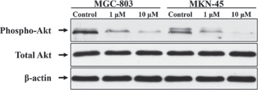
Deguelin purchased from MedChemExpress. Usage Cited in: Int J Mol Med. 2018 Jun;41(6):3157-3166. [Abstract]
MGC-803 and MKN-45 cells are treated with increasing doses of Deguelin (0, 1 and 10 μM) for 6 h and harvested for western blot (WB) analysis to assess the p-Akt signaling.
-
PLoS Biol
2024 Jun 27;22(6):e3002672. PMID: 38935621 -
Cancer Biol Ther
Deguelin inhibits perineural invasion in esophageal squamous cell carcinoma via targeting BDNF/TrkB axis. [Abstract]2026 Dec 31;27(1):2644788. PMID: 41851958 -
Neuropharmacology
Deguelin inhibits the glioblastoma progression through suppressing CCL2/NFκB signaling pathway. [Abstract]2024 Aug 9:110109. PMID: 39128581 -
J Med Virol
E3 ubiquitin ligase FBXO22 inhibits SARS-CoV-2 replication via promoting proteasome-dependent degradation of NSP5. [Abstract]2024 Sep;96(9):e29891. PMID: 39223933 -
Toxicol Appl Pharmacol
Deguelin inhibits IL-1β-induced chondrocyte inflammation in vitro and ameliorates murine osteoarthritis in vivo. [Abstract]2025 Sep:502:117453. PMID: 40582373 -
Arch Med Res
microRNA-103 Contributes to Progression of Polycystic Ovary Syndrome Through Modulating the IRS1/PI3K/AKT Signal Axis. [Abstract]2021 Jul;52(5):494-504. PMID: 33583602 -
Int J Biochem Cell Biol
Estradiol facilitates urate excretion by reducing GLUT9 expression via ERβ/TMEM106B/PI3K/AKT1 pathway in renal tubular epithelial cells. [Abstract]2026 Jun-Jul:195-196:106940. PMID: 41825791 -
J Cancer
Tephrosin induces apoptosis of human pancreatic cancer cells through the generation of reactive oxygen species. [Abstract]2021 Jan 1;12(1):270-280. PMID: 33391424 -
-
bioRxiv
An efficient behavioral screening platform classifies natural products and other chemical cues according to their chemosensory valence in C. elegans. [Abstract]2024 Apr 3:2023.06.02.542933. PMID: 37333363


Lösungsmittel & Löslichkeit
DMSO : 100 mg/mL (253.54 mM; Need ultrasonic; Hygroscopic DMSO has a significant impact on the solubility of product, please use newly opened DMSO)
Please refer to the solubility information to select the appropriate solvent. Once prepared, please aliquot and store the solution to prevent product inactivation from repeated freeze-thaw cycles.
Storage method and period of stock solution: -80°C, 1 year; -20°C, 6 months. When stored at -80°C, please use it within 1 year. When stored at -20°C, please use it within 6 months.
Konzentration (Stammlösung) × Volumen (Stammlösung) = Konzentration (Ziellösung) × Volumen (Ziellösung)
Select the appropriate dissolution method based on your experimental animal and administration route.
- For the following dissolution methods, please ensure to first prepare a clear stock solution using an In Vitro approach and then sequentially add co-solvents:
- To ensure reliable experimental results, the clarified stock solution can be appropriately stored based on storage conditions. As for the working solution for In Vivo experiments, it is recommended to prepare freshly and use it on the same day.
- The percentages shown for the solvents indicate their volumetric ratio in the final prepared solution. If precipitation or phase separation occurs during preparation, heat and/or sonication can be used to aid dissolution.
Add each solvent one by one: 10% DMSO 40% PEG300 5% Tween-80 45% Saline
Solubility: ≥ 2.5 mg/mL (6.34 mM); Clear solution
This protocol yields a clear solution of ≥ 2.5 mg/mL (saturation unknown).
Taking 1 mL working solution as an example, add 100 μL DMSO stock solution (25.0 mg/mL) to 400 μL PEG300, and mix evenly; then add 50 μL Tween-80 and mix evenly; then add 450 μL Saline to adjust the volume to 1 mL.
Preparation of Saline: Dissolve 0.9 g sodium chloride in ddH₂O and dilute to 100 mL to obtain a clear Saline solution.
Add each solvent one by one: 10% DMSO 90% (20% SBE-β-CD in Saline)
Solubility: 2.5 mg/mL (6.34 mM); Suspended solution; Need ultrasonic
This protocol yields a suspended solution of 2.5 mg/mL. Suspended solution can be used for oral and intraperitoneal injection.
Taking 1 mL working solution as an example, add 100 μL DMSO stock solution (25.0 mg/mL) to 900 μL 20% SBE-β-CD in Saline, and mix evenly.
Preparation of 20% SBE-β-CD in Saline (4°C, storage for one week): 2 g SBE-β-CD powder is dissolved in 10 mL Saline, completely dissolve until clear.
Please enter the basic information of animal experiments:
-
-
-
-
Recommended: Prepare an additional quantity of animals to account for potential losses during experiments.
Please enter your animal formula composition:
-
%DMSO +
Recommended: Keep the proportion of DMSO in working solution below 2% if your animal is weak.
-
%+
-
+%Tween-80 + +
-
%Saline +
The co-solvents required include: DMSO, . All of co-solvents are available by MedChemExpress (MCE). , Tween 80. All of co-solvents are available by MedChemExpress (MCE).
Working solution concentration: 0.22 mg/mL
Method for preparing stock solution: mg drug dissolved in μL DMSO. Stock solution concentration: mg/mL.
1. Take μL DMSO stock solution;
2. Add μL .
μL , mix evenly;
3. Then add μL Tween 80, mix evenly;
4. Then add μL
Please ensure that the stock solution in the first step is dissolved to a clear state, and add co-solvents in sequence. You can use ultrasonic heating (ultrasonic cleaner, recommended frequency 20-40 kHz), vortexing, etc. to assist dissolution.

Protokoll
Caspase 3 activity is determined using Caspase-Glo-3 assays. This assay provides luminogenic substrate in a buffer system optimized for each specific caspase activity. The caspase cleavage of the substrate is followed by generation of a luminescent signal. The signal generated is proportional to the amount of caspase activity present in the sample. Protein (10 µg) from the cell samples is diluted in water to a final volume of 50 µL and added to a white 96-well microtitre plate, followed by 50 µL of Caspase-Glo-3 reagent. The plate is sealed and gently mixed at 300-500 rpm for 30 s and incubated at room temperature for 30 min. Luminescence is measured in a microplate reader (TECAN Infinite 200).
MedChemExpress (MCE) has not independently confirmed the accuracy of these methods. They are for reference only.
Breast cancer cells are incubated with increasing concentration of Deguelin ranging from 31 nM to 500 nM for 24, 48 and 72 h. At the termination the cells are trypsinized and cell proliferation is evaluated by counting cells using Z-series Coulter counter. Data are presented as Mean±SE percent of control.
MedChemExpress (MCE) has not independently confirmed the accuracy of these methods. They are for reference only.
Six to seven weeks old female athymic mice (nu/nu) are housed in a barrier free environment under 24±2°C temperature, 50±10% relative humidity, and 12-hour light/12-hour dark cycle. Mice are provided with sterile mouse chow and water ad libitum. MDA-MB-231 cells (3.0 million cells/animal) are suspended in sterile PBS and then injected subcutaneously into the dorsal flank region using 23 g hypodermic needle. Animals are observed daily for the growth of palpable tumor at the site of injection. Once the tumor (approximately 50 mm3) appears, the mice are randomized in to three groups, animals receiving either 1) vehicle as a control 2) Deguelin treatment at 2 mg/kg bodyweight dose or 3) Deguelin at 4 mg/kg body weight. Each group consists of 10 animals. Vehicle or Deguelin is administered through i.p. injection daily for 21 days. Animals are monitored daily for the signs of drug/vehicle associated toxicity and weighed once weekly. Growth of tumor at the site of cell injection is monitored every alternate day and of tumor size is measured using calipers. Tumor volume is calculated by using the well-established formula: tumor volume (mm3)=π/6 length×width×depth. Data represent the mean tumor volume+SE (mm3) in each group. The animals are sacrificed at the indicated time unless they appear to be moribund or tumors show sign of necrosis. At termination, the tumor is excised, freed from connective tissue and other organs, a small piece is fixed in 10% buffered formalin and remaining tumor is snap frozen for future biochemical analysis. Liver, lung, kidney and spleen are excised and weighed.
MedChemExpress (MCE) has not independently confirmed the accuracy of these methods. They are for reference only.


Reinheit & Dokumentation
-
Data Sheet (283 KB)
-
SDS (393 KB)
- English - EN (393 KB)
- Français - FR (393 KB)
- Deutsch - DE (393 KB)
- Norwegian - NO (393 KB)
- Español - ES (393 KB)
- Swedish - SV (393 KB)
- Italian - IT (393 KB)
- Korean - KR (393 KB)
- Portuguese - PT (393 KB)
-
Handling Instructions (2659 KB)
Verweise
[1]. Mehta R, et al. Deguelin action involves c-Met and EGFR signaling pathways in triple negative breast cancer cells. PLoS One. 2013 Jun 10;8(6):e65113. [Content Brief]
[2]. Yang YL, et al. Deguelin induces both apoptosis and autophagy in cultured head and neck squamous cell carcinoma cells. PLoS One. 2013;8(1):e54736. [Content Brief]
[3]. Li Z, et al. Deguelin, a natural rotenoid, inhibits mouse myeloma cell growth in vitro via induction of apoptosis. Oncol Lett. 2012 Oct;4(4):677-681. [Content Brief]
[4]. Kang HW, et al. Deguelin, an Akt inhibitor, down-regulates NF-κB signaling and induces apoptosis in colon cancer cells and inhibits tumor growth in mice. Dig Dis Sci. 2012 Nov;57(11):2873-82. [Content Brief]


Complete Stock Solution Preparation Table
Please refer to the solubility information to select the appropriate solvent. Once prepared, please aliquot and store the solution to prevent product inactivation from repeated freeze-thaw cycles.
Storage method and period of stock solution: -80°C, 1 year; -20°C, 6 months. When stored at -80°C, please use it within 1 year. When stored at -20°C, please use it within 6 months.
| Optional Solvent | Concentration Solvent Mass | 1 mg | 5 mg | 10 mg | 25 mg |
|---|---|---|---|---|---|
| DMSO | 1 mM | 2.5354 mL | 12.6768 mL | 25.3537 mL | 63.3842 mL |
| 5 mM | 0.5071 mL | 2.5354 mL | 5.0707 mL | 12.6768 mL | |
| 10 mM | 0.2535 mL | 1.2677 mL | 2.5354 mL | 6.3384 mL | |
| 15 mM | 0.1690 mL | 0.8451 mL | 1.6902 mL | 4.2256 mL | |
| 20 mM | 0.1268 mL | 0.6338 mL | 1.2677 mL | 3.1692 mL | |
| 25 mM | 0.1014 mL | 0.5071 mL | 1.0141 mL | 2.5354 mL | |
| 30 mM | 0.0845 mL | 0.4226 mL | 0.8451 mL | 2.1128 mL | |
| 40 mM | 0.0634 mL | 0.3169 mL | 0.6338 mL | 1.5846 mL | |
| 50 mM | 0.0507 mL | 0.2535 mL | 0.5071 mL | 1.2677 mL | |
| 60 mM | 0.0423 mL | 0.2113 mL | 0.4226 mL | 1.0564 mL | |
| 80 mM | 0.0317 mL | 0.1585 mL | 0.3169 mL | 0.7923 mL | |
| 100 mM | 0.0254 mL | 0.1268 mL | 0.2535 mL | 0.6338 mL |


 Powered by Bioz