



MC2590
Based on 1 publication(s) in Google Scholar
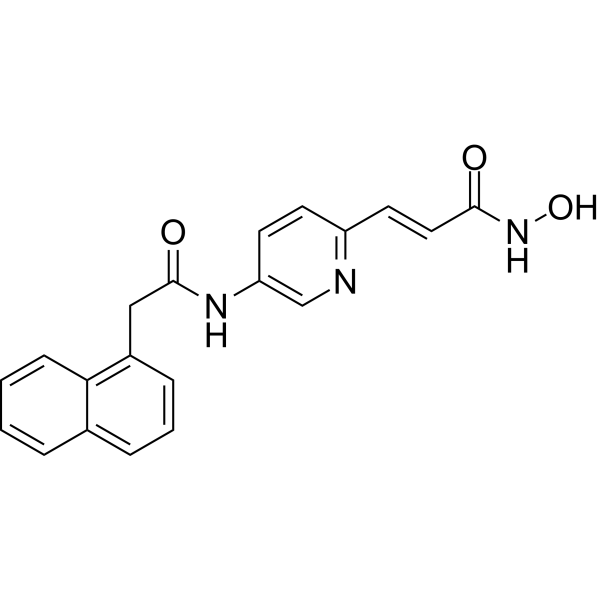
MC2590 is a potent pyridine-containing histone deacetylase (HDAC) inhibitor. MC2590 is a inhibitor of HDAC1-3, -6, -8, and -10 (class I/IIb-selective inhibitor) with IC50s of 0.015 μM-0.156 μM. MC2590 also inhibits HDAC isoforms HDAC4, HDAC5, HDAC7, HDAC9, HDAC11 with IC50s of 1.35 μM-3.98 μM. MC2625 induces G2/M cell cycle arrest and modulates pro- and anti-apoptotic microRNAs towards apoptosis induction.
Nur für Forschungszwecke. Wir verkaufen nicht an Patienten.
- Reinheit : 98.71%
- CAS. Nr.: 2284460-01-9
- Formel: C20H17N3O3
- Molecular Weight:347.37
-
Speicherung:Powder -20°C, 3 years , 4°C, 2 years ; In solvent -80°C, 6 months , -20°C, 1 month

To place orders, for customer services and technical support, please contact: MedChemExpress USA
Tel: 609-228-6898 E-mail: [email protected] [email protected]


-
 Biologische Aktivität
-
Chemical Information
-
Lösungsmittel & Löslichkeit
- Protokoll
- Reinheit & Dokumentation
- Verweise
-
 Help & FAQs


Publications Citing Use of MedChemExpress (MCE) MC2590
More

Biologische Aktivität
Beschreibung
IC50 & Target
[1]|
HDAC1 0.098 μM (IC50) |
HDAC2 0.156 μM (IC50) |
HDAC3 0.039 μM (IC50) |
HDAC6 0.015 μM (IC50) |
HDAC8 0.047 μM (IC50) |
HDAC10 0.071 μM (IC50) |
HDAC4 2.73 μM (IC50) |
HDAC5 1.35 μM (IC50) |
HDAC7 2.06 μM (IC50) |
HDAC9 2.79 μM (IC50) |
HDAC11 3.98 μM (IC50) |
Cellular Effect
|
Cell Line

|
Type | Value | Description | References |
|---|---|---|---|---|
| A549 | IC50 |
0.32 μM
Compound: 5e; MC2590
|
Antiproliferative activity against human A549 cells incubated for 72 hrs by CellTiter-Glo luminescent cell viability assay
Antiproliferative activity against human A549 cells incubated for 72 hrs by CellTiter-Glo luminescent cell viability assay
|
[PMID: 36549114] |
| HCT-116 | IC50 |
0.07 μM
Compound: 5e; MC2590
|
Antiproliferative activity against human HCT-116 cells incubated for 72 hrs by CellTiter-Glo luminescent cell viability assay
Antiproliferative activity against human HCT-116 cells incubated for 72 hrs by CellTiter-Glo luminescent cell viability assay
|
[PMID: 36549114] |
| HFF | IC50 |
>10000 nM
Compound: 6
|
Cytotoxicity against HFF
Cytotoxicity against HFF
|
[PMID: 30366254] |
| HL-60 | IC50 |
0.276 μM
Compound: 5e; MC2590
|
Antiproliferative activity against human HL-60 cells incubated for 48 hrs
Antiproliferative activity against human HL-60 cells incubated for 48 hrs
|
[PMID: 36549114] |
| K562 | IC50 |
0.05 μM
Compound: 5e; MC2590
|
Antiproliferative activity against human K562 cells incubated for 72 hrs by CellTiter-Glo luminescent cell viability assay
Antiproliferative activity against human K562 cells incubated for 72 hrs by CellTiter-Glo luminescent cell viability assay
|
[PMID: 36549114] |
| KARPAS-299 | IC50 |
0.4 μM
Compound: 5e; MC2590
|
Antiproliferative activity against human KARPAS-299 cells incubated for 48 hrs
Antiproliferative activity against human KARPAS-299 cells incubated for 48 hrs
|
[PMID: 36549114] |
| Kasumi 1 | IC50 |
0.066 μM
Compound: 5e; MC2590
|
Antiproliferative activity against human Kasumi 1 cells incubated for 48 hrs
Antiproliferative activity against human Kasumi 1 cells incubated for 48 hrs
|
[PMID: 36549114] |
| MV4-11 | IC50 |
0.059 μM
Compound: 5e; MC2590
|
Antiproliferative activity against human MV4-11 cells incubated for 48 hrs
Antiproliferative activity against human MV4-11 cells incubated for 48 hrs
|
[PMID: 36549114] |
| NB-4 | IC50 |
0.084 μM
Compound: 5e; MC2590
|
Antiproliferative activity against human NB4 cells incubated for 48 hrs
Antiproliferative activity against human NB4 cells incubated for 48 hrs
|
[PMID: 36549114] |
| OCI-AML2 | IC50 |
0.379 μM
Compound: 5e; MC2590
|
Antiproliferative activity against human OCI-AML2 cells incubated for 48 hrs
Antiproliferative activity against human OCI-AML2 cells incubated for 48 hrs
|
[PMID: 36549114] |
| OCI-AML-3 | IC50 |
0.529 μM
Compound: 5e; MC2590
|
Antiproliferative activity against human OCI-AML-3 cells incubated for 48 hrs
Antiproliferative activity against human OCI-AML-3 cells incubated for 48 hrs
|
[PMID: 36549114] |
| U-937 | IC50 |
0.258 μM
Compound: 5e; MC2590
|
Antiproliferative activity against human U-937 cells incubated for 48 hrs
Antiproliferative activity against human U-937 cells incubated for 48 hrs
|
[PMID: 36549114] |
In Vitro
MC2625 (compound 5e) has antiproliferative activity with Colorectal carcinoma HCT116 (IC50=0.07 μM), Lung adenocarcinoma A549 (IC50=0.32 μM), Chronic myelogenous leukaemia K562 (IC50=0.05 μM) for 72 h[1].
MC2625 (1, 5 μM; 24, 48 h) displays mainly G2/M cell cycle arrest[1].
MC2625 (1, 5 μM; 24, 48 h) reveals H3K9/14 hyperacetylation activity, increases the acetyl-α-tubulin level, markedly upregulates the p21 protein[1].
MC2625 (1, 5 μM; 48 h) increases mRNA expression of p21, BAX and BAK, downregulates cyclin D1 and BCL-2 and modulates pro- and anti-apoptotic microRNAs towards apoptosis induction[1].
MedChemExpress (MCE) has not independently confirmed the accuracy of these methods. They are for reference only.
-
Cell Line:Human acute myeloid leukaemia U937 cells
-
Concentration:1, 5 μM
-
Incubation Time:24, 48 h
-
Result:At 24 h, showed very low increase of the pre-G1 peak and led to a G2/M phase arrest at 1 μM; induced a 10% pre-G1 increase and displayed a block at the G2/M phase at 5 μM.
At 48 h, induced a 70-85% block of the cell cycle at the G1 phase.
-
Cell Line:Human acute myeloid leukaemia U937 cells
-
Concentration:1, 5 μM
-
Incubation Time:24, 48 h
-
Result:At 1 μM revealed H3K9/14 hyperacetylation activity, increased the acetyl-α-tubulin level, markedly upregulated the p21 protein.
-
Cell Line:Human acute myeloid leukaemia U937 cells
-
Concentration:1, 5 μM
-
Incubation Time:48 h
-
Result:At 1 μM significantly induced the expression of BAX and BAK, dose-dependently downregulated the antiapoptotic factor BCL-2.
Downregulated miRNAs with antiapoptotic activity (miR-17-5p, miR-18-5p, miR-19b-3p, miR-20a-5p, miR-21-5p); induced the proapoptotic miRNAs (miR-let7a-5p, miR-125b-5p, miR-181a-5p, miR-181b-5p, miR-769-5p, miR-122-5p).


Chemical Information
-
CAS. Nr. 2284460-01-9
-
Appearance Solid
-
Molecular Weight 347.37
-
Formel C20H17N3O3
-
Color Off-white to light yellow
-
SMILES
O=C(NO)/C=C/C1=NC=C(C=C1)NC(CC2=C3C=CC=CC3=CC=C2)=O
-
Versand
Room temperature in continental US; may vary elsewhere.
-
Speicherung
Powder -20°C 3 years 4°C 2 years In solvent -80°C 6 months -20°C 1 month


Publications (1)
-
Journal Impact Factor
-
Most Recent


Lösungsmittel & Löslichkeit
In Vitro:
DMSO : 100 mg/mL (287.88 mM; Need ultrasonic; Hygroscopic DMSO has a significant impact on the solubility of product, please use newly opened DMSO)
Please refer to the solubility information to select the appropriate solvent. Once prepared, please aliquot and store the solution to prevent product inactivation from repeated freeze-thaw cycles.
Storage method and period of stock solution: -80°C, 6 months; -20°C, 1 month. When stored at -80°C, please use it within 6 months. When stored at -20°C, please use it within 1 month.
Please refer to the solubility information to select the appropriate solvent. Once prepared, please aliquot and store the solution to prevent product inactivation from repeated freeze-thaw cycles.
Storage method and period of stock solution: -80°C, 6 months; -20°C, 1 month. When stored at -80°C, please use it within 6 months. When stored at -20°C, please use it within 1 month.
Konzentration (Stammlösung) × Volumen (Stammlösung) = Konzentration (Ziellösung) × Volumen (Ziellösung)


Reinheit & Dokumentation
-
Data Sheet (273 KB)
-
SDS (251 KB)
- English - EN (251 KB)
- Français - FR (251 KB)
- Deutsch - DE (251 KB)
- Norwegian - NO (251 KB)
- Español - ES (251 KB)
- Swedish - SV (251 KB)
- Italian - IT (251 KB)
- Korean - KR (251 KB)
- Portuguese - PT (251 KB)
-
Handling Instructions (2659 KB)
Verweise


Complete Stock Solution Preparation Table
Please refer to the solubility information to select the appropriate solvent. Once prepared, please aliquot and store the solution to prevent product inactivation from repeated freeze-thaw cycles.
Storage method and period of stock solution: -80°C, 6 months; -20°C, 1 month. When stored at -80°C, please use it within 6 months. When stored at -20°C, please use it within 1 month.
| Optional Solvent | Concentration Solvent Mass | 1 mg | 5 mg | 10 mg | 25 mg |
|---|---|---|---|---|---|
| DMSO | 1 mM | 2.8788 mL | 14.3939 mL | 28.7877 mL | 71.9694 mL |
| 5 mM | 0.5758 mL | 2.8788 mL | 5.7575 mL | 14.3939 mL | |
| 10 mM | 0.2879 mL | 1.4394 mL | 2.8788 mL | 7.1969 mL | |
| 15 mM | 0.1919 mL | 0.9596 mL | 1.9192 mL | 4.7980 mL | |
| 20 mM | 0.1439 mL | 0.7197 mL | 1.4394 mL | 3.5985 mL | |
| 25 mM | 0.1152 mL | 0.5758 mL | 1.1515 mL | 2.8788 mL | |
| 30 mM | 0.0960 mL | 0.4798 mL | 0.9596 mL | 2.3990 mL | |
| 40 mM | 0.0720 mL | 0.3598 mL | 0.7197 mL | 1.7992 mL | |
| 50 mM | 0.0576 mL | 0.2879 mL | 0.5758 mL | 1.4394 mL | |
| 60 mM | 0.0480 mL | 0.2399 mL | 0.4798 mL | 1.1995 mL | |
| 80 mM | 0.0360 mL | 0.1799 mL | 0.3598 mL | 0.8996 mL | |
| 100 mM | 0.0288 mL | 0.1439 mL | 0.2879 mL | 0.7197 mL |


MC2590 Related Classifications
HY-152226 Related Classifications
 Powered by Bioz