



KP372-1
Based on 3 publication(s) in Google Scholar
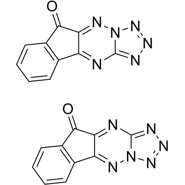
KP372-1 is an Akt inhibitor that inhibits proliferation and induces apoptosis and anoikis. KP372-1 is also an NQO1 redox cycling agent that causes DNA damage (including DNA breakage) by generating ROS. KP372-1 can be used in cancer research (such as head and neck squamous cell carcinoma (HNSCC) and pancreatic cancer).
For research use only. We do not sell to patients.
- Purity : 99.5%
- CAS No.: 1374996-60-7
- Formula: C20H8N12O2
- Molecular Weight:448.36
-
Storage:Powder -20°C, 3 years , 4°C, 2 years ; In solvent -80°C, 2 years , -20°C, 1 year

To place orders, for customer services and technical support, please contact: MedChemExpress USA
Tel: 609-228-6898 E-mail: [email protected] [email protected]


-
 Biological Activity
-
Chemical Information
-
Solvent & Solubility
- Protocols
- Purity & Documentation
- References
-
 Help & FAQs


-
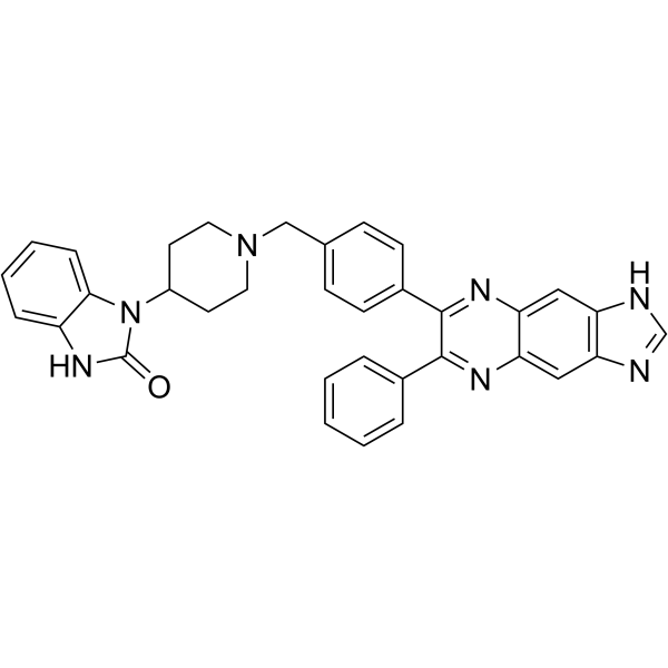
HY-10355
AKT inhibitor VIIIAKT Inhibitor -
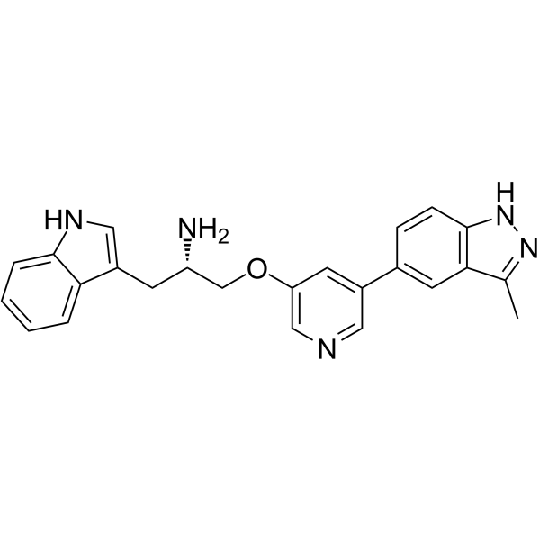
HY-10425
Rizavasertibpan-Akt Inhibitor -
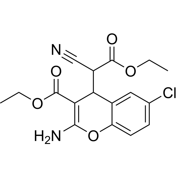
HY-18749
SC79Akt Activator -
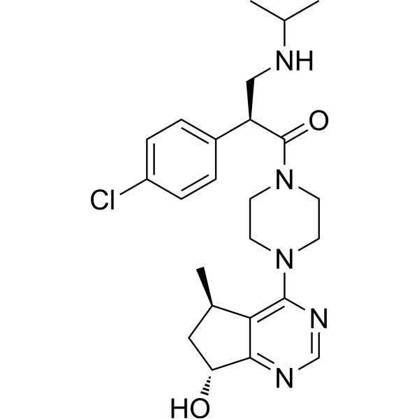
HY-15186
IpatasertibPan-Akt Inhibitor -
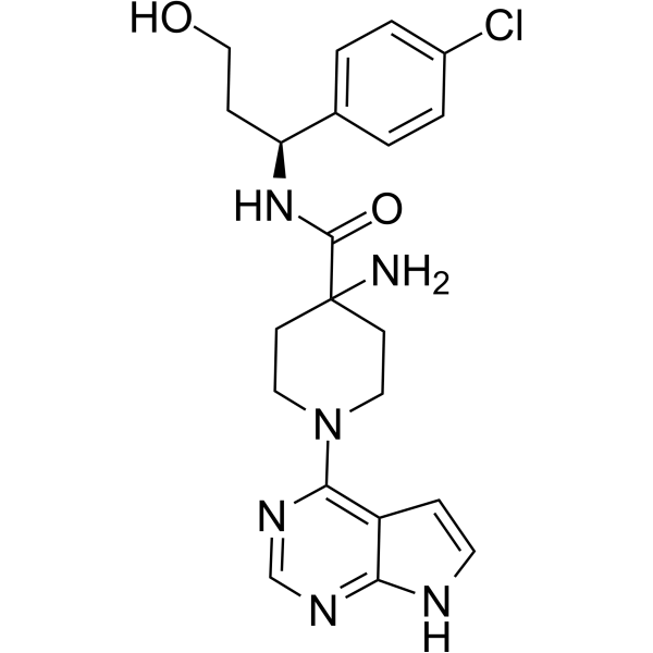
HY-15431
CapivasertibAkt Inhibitor -
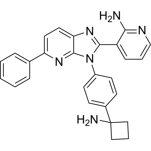
HY-19719
MiransertibAKT Inhibitor -
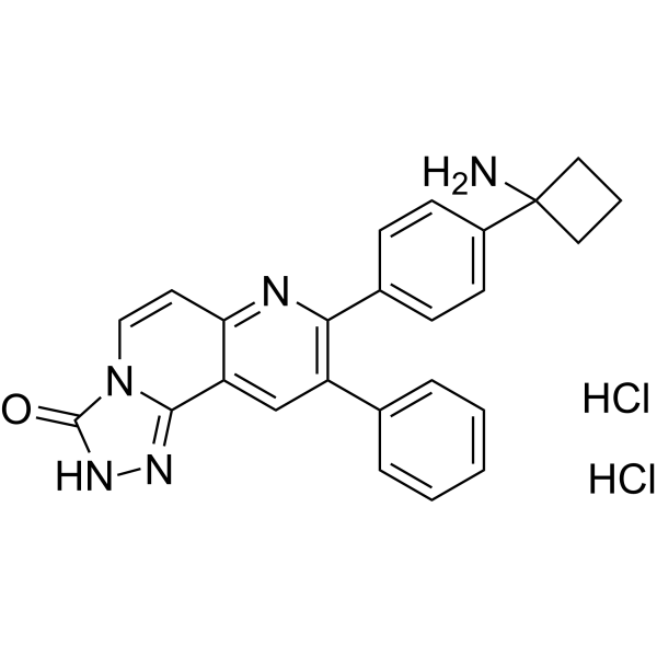
HY-10358
MK-2206 dihydrochlorideAllosteric Akt Inhibitor -
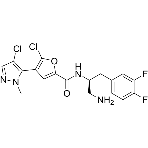
HY-15965
UprosertibAkt Inhibitor -
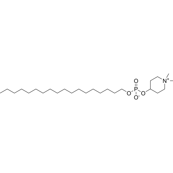
HY-50909
PerifosineAkt Inhibitor -
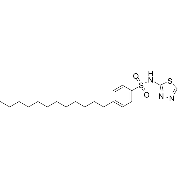
HY-12063
PHT-427Akt Inhibitor -
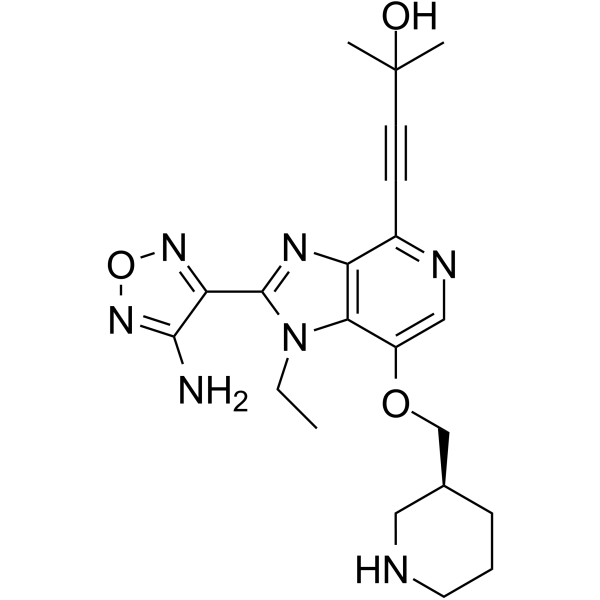
HY-10249
GSK-690693Akt Inhibitor -
HY-100018
BAY1125976AKT1/2 Inhibitor
Publications Citing Use of MedChemExpress (MCE) KP372-1
More

Biological Activity
Description
In Vitro
KP372-1 (0.0625, 0.125, 0.25, 0.5, 1.0 μM; 48 h) inhibits growth of JMARc42 and Tu167c2 cells with IC50s of 200 and 100 nM, respectively[1].
KP372-1 (125 nM; 24 h) induces Tu167c2 cells apoptosis and induces anoikis in the JMARc42 cells[1].
KP372-1 (125 nM; 30 min) blocks Akt, thereby decreasing the phosphorylation of the S6 ribosomal protein in both Tu167 and JMAR cells[1].
KP372-1 (0.250, 0.5, 1.0 μM; 30 min) inhibits Akt kinase activity with an IC50 of 250 nM in JMAR cells[1].
MedChemExpress (MCE) has not independently confirmed the accuracy of these methods. They are for reference only. Further protocols information, click here.
-
Cell Line:JMARc42 and Tu167c2 cells
-
Concentration:0.0625, 0.125, 0.25, 0.5, 1.0 µM
-
Incubation Time:48 h
-
Result:Showed antiproliferative activity.
-
Cell Line:Tu167c2 and JMARc42 cells
-
Concentration:125 nM
-
Incubation Time:24 h
-
Result:Induced approximately 90% of cells apoptosis.
-
Cell Line:Tu167 and JMAR cells
-
Concentration:125 nM
-
Incubation Time:30 min
-
Result:Induced a small but consistent decrease in Akt phosphorylation with a concomitant marked decrease in S6 phosphorylation.
Inhibited the EGF induced phosphorylation of Aktser473 in Tu167 and AktThr308 in JMAR.
-
Cell Line:JMAR cells
-
Concentration:0.250, 0.5, 1.0 µM
-
Incubation Time:30 min
-
Result:Significantly blocked Akt kinase activity in a dose-dependent fashion, with an IC50 of 250 nM.
In Vivo
MedChemExpress (MCE) has not independently confirmed the accuracy of these methods. They are for reference only.
-
Animal Model:Nude mice (H1299 xenografts model)[2].
-
Dosage:10, 20 mg/kg
-
Administration:intragastric injection; single daily for 33 days
-
Result:Affected tumor metabolism and suppressed tumor growth.


Chemical Information
-
CAS No. 1374996-60-7
-
Appearance Solid
-
Molecular Weight 448.36
-
Formula C20H8N12O2
-
Color Light yellow to yellow
-
SMILES
O=C1C2=C(C3=NC4=NN=NN4N=C31)C=CC=C2.O=C5C6=NC7=NN=NN7N=C6C8=C5C=CC=C8
-
Shipping
Room temperature in continental US; may vary elsewhere.
-
Storage
Powder -20°C 3 years 4°C 2 years In solvent -80°C 2 years -20°C 1 year


Publications (3)
-
Journal Impact Factor
-
Most Recent
-
Biomaterials
Nano-sensitizer with self-amplified drug release and hypoxia normalization properties potentiates efficient chemoradiotherapy of pancreatic cancer. [Abstract]2024 Oct:310:122634. PMID: 38823195 -
J Control Release
Bladder cancer selective chemotherapy with potent NQO1 substrate co-loaded prodrug nanoparticles. [Abstract]2022 May 31;347:632-648. PMID: 35618186 -
J Biochem Mol Toxicol
Heme Oxygenase-1 Regulates the JAK/STAT Pathway to Inhibit Ferroptosis in Metabolic Dysfunction-Associated Steatotic Liver Disease. [Abstract]2026 Jan;40(1):e70661. PMID: 41486516


Solvent & Solubility
In Vitro:
DMSO : 17.86 mg/mL (39.83 mM; Need ultrasonic; Hygroscopic DMSO has a significant impact on the solubility of product, please use newly opened DMSO)
Please refer to the solubility information to select the appropriate solvent. Once prepared, please aliquot and store the solution to prevent product inactivation from repeated freeze-thaw cycles.
Storage method and period of stock solution: -80°C, 2 years; -20°C, 1 year. When stored at -80°C, please use it within 2 years. When stored at -20°C, please use it within 1 year.
Please refer to the solubility information to select the appropriate solvent. Once prepared, please aliquot and store the solution to prevent product inactivation from repeated freeze-thaw cycles.
Storage method and period of stock solution: -80°C, 2 years; -20°C, 1 year. When stored at -80°C, please use it within 2 years. When stored at -20°C, please use it within 1 year.
Concentration (start) × Volume (start) = Concentration (final) × Volume (final)

Protocols
-
Apoptosis
Apoptosis, also called programmed cell death, is generally characterized by distinct morphological characteristics.
-
TUNEL staining for apoptotic DNA fragmentation
TUNEL staining detects DNA strand breaks by using terminal deoxynucleotidyl transferase to add labeled nucleotides to exposed 3′-OH DNA termini, generating either microscopic staining in fixed cells or tissue sections, or fluorescence/cytometric signal in cell suspensions. TUNEL positivity reflects DNA fragmentation but should not be interpreted alone as definitive apoptosis, because TUNEL can also label necrotic, autolytic, mechanically damaged, or DNA-repair-associated DNA breaks.
-
Annexin V plus membrane-impermeant dye apoptosis staining
Annexin V-based apoptosis assays rely on the detection of phosphatidylserine (PS) externalization from the inner leaflet of the plasma membrane to the outer leaflet, an early biochemical hallmark of apoptosis. Fluorescently labeled Annexin V binds PS in a calcium-dependent manner, enabling identification of early apoptotic cells by flow cytometry or fluorescence microscopy. When combined with a membrane-impermeant DNA-binding dye (e. g. , propidium iodide), this approach allows discrimination between viable (Annexin V−/dye−), early apoptotic (Annexin V+/dye−), and late apoptotic or necrotic (Annexin V+/dye+) cell populations by assessing membrane integrity and PS exposure.
-
ROS/oxidative-stress fluorescent staining
ROS/oxidative-stress fluorescent staining uses cell-permeant fluorogenic probes that become fluorescent after oxidation inside cells or tissues; commonly used examples include DCFH-DA/DCFDA for broad cellular oxidant detection, DHE for superoxide-related signal detection, MitoSOX for mitochondrial superoxide-related signal detection, and CellROX probes for oxidative-stress-associated fluorescence readouts. The assay detects probe oxidation rather than a single ROS species unless the probe and analysis method have been chemically validated for that species. DCFH-DA enters cells, is deacetylated by intracellular esterases to DCFH, and produces fluorescent DCF after oxidation, so the readout is used as an operational measure of total cellular oxidative stress rather than a species-specific ROS measurement. DHE and MitoSOX can report superoxide-related oxidation, but red fluorescence alone can include non-specific ethidium-like oxidation products; HPLC or optimized spectral approaches are
-
Apoptosis Solutions
Apoptosis is a regulated, generally non-lytic cell-death pathway that removes unwanted, damaged, infected, or abnormal cells through coordinated morphological changes, caspase activation, DNA fragmentation, and membrane remodeling. The intrinsic apoptosis pathway is controlled mainly by mitochondrial outer membrane permeabilization, BCL-2 family proteins, cytochrome c release, apoptosome formation, caspase-9 activation, and downstream executioner caspase-3/7 activation. The extrinsic apoptosis pathway is initiated by death receptors such as Fas, TNFR, and TRAIL receptors, which recruit adaptor proteins and activate caspase-8 before engaging executioner caspases or mitochondrial amplification through BID cleavage. Apoptosis is linked to many phenotypes, including cancer cell killing, tissue homeostasis, immune regulation, neurodegeneration, infection response, and treatment-induced cytotoxicity; unresolved questions include how apoptosis interacts with necroptosis, pyroptosis, ferroptos
-
Genotoxicity/Mutagenicity Study
The bacterial reverse mutation assay detects point mutations that restore amino-acid prototrophy in auxotrophic Salmonella typhimurium or Escherichia coli tester strains; after exposure to a test article, mutagenic activity is read out as an increased number of revertant colonies on minimal agar compared with the vehicle control. The assay uses tester strains with different mutation targets so that base-substitution and frameshift mutagens can be detected, and testing is performed with and without exogenous mammalian metabolic activation because some chemicals require biotransformation to become mutagenic.
-
Cell Viability Determination by MTT Colorimetric Assay
The following protocol uses the MTT colorimetric assay as a classic literature-established method for assessing cell viability/metabolic activity in cultured mammalian cells. MTT[3-(4,5-dimethylthiazol-2-yl)-2,5-diphenyltetrazolium bromide] is reduced by metabolically active cells to a colored formazan product; the amount of formazan is quantified spectrophotometrically and provides an indirect measure of metabolically active viable cells. Importantly, MTT reduction reflects cellular oxidoreductase/metabolic activity rather than an absolute direct count of living cells, so changes in cellular metabolism can alter the signal independently of cell number.


Purity & Documentation
-
Data Sheet (288 KB)
-
SDS (392 KB)
- English - EN (392 KB)
- Français - FR (392 KB)
- Deutsch - DE (392 KB)
- Norwegian - NO (392 KB)
- Español - ES (392 KB)
- Swedish - SV (392 KB)
- Italian - IT (392 KB)
- Korean - KR (392 KB)
- Portuguese - PT (392 KB)
-
Handling Instructions (2659 KB)
References
[1]. Mandal M, et al. The Akt inhibitor KP372-1 inhibits proliferation and induces apoptosis and anoikis in squamous cell carcinoma of the head and neck. Oral Oncol. 2006 Apr;42(4):430-9. [Content Brief]
[2]. Zhao Y, et al. SoNar, a Highly Responsive NAD+/NADH Sensor, Allows High-Throughput Metabolic Screening of Anti-tumor Agents. Cell Metab. 2015 May 5;21(5):777-89. [Content Brief]
[3]. Viera T, et al. DNA damage induced by KP372-1 hyperactivates PARP1 and enhances lethality of pancreatic cancer cells with PARP inhibition. Sci Rep. 2020 Nov 19;10(1):20210. [Content Brief]


Complete Stock Solution Preparation Table
Please refer to the solubility information to select the appropriate solvent. Once prepared, please aliquot and store the solution to prevent product inactivation from repeated freeze-thaw cycles.
Storage method and period of stock solution: -80°C, 2 years; -20°C, 1 year. When stored at -80°C, please use it within 2 years. When stored at -20°C, please use it within 1 year.
| Optional Solvent | Concentration Solvent Mass | 1 mg | 5 mg | 10 mg | 25 mg |
|---|---|---|---|---|---|
| DMSO | 1 mM | 2.2304 mL | 11.1518 mL | 22.3035 mL | 55.7588 mL |
| 5 mM | 0.4461 mL | 2.2304 mL | 4.4607 mL | 11.1518 mL | |
| 10 mM | 0.2230 mL | 1.1152 mL | 2.2304 mL | 5.5759 mL | |
| 15 mM | 0.1487 mL | 0.7435 mL | 1.4869 mL | 3.7173 mL | |
| 20 mM | 0.1115 mL | 0.5576 mL | 1.1152 mL | 2.7879 mL | |
| 25 mM | 0.0892 mL | 0.4461 mL | 0.8921 mL | 2.2304 mL | |
| 30 mM | 0.0743 mL | 0.3717 mL | 0.7435 mL | 1.8586 mL |


KP372-1 Related Classifications
HY-15673 Related Classifications
 Powered by Bioz