From 11:00 pm to 12:00 pm EST ( 8:00 pm to 9:00 pm PST ) on January 6th, the website will be under maintenance. We are sorry for the inconvenience. Please arrange your schedule properly.
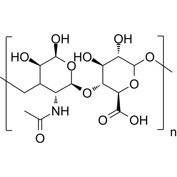
Hyaluronic acid is a biopolymer composed of repeating units of disaccharides with various applications. Hyaluronic acid is a major component of the extracellular matrix (ECM). Hyaluronic acid is synthesized at the plasma membrane. Increased hyaluronic acid levels are associated with tumor cell growth, adhesion, migration, invasion and angiogenesis in digestive cancers. Hyaluronic acid participates in tissue remodeling and rapid cell proliferation in some physiological processes including embryonic morphogenesis and wound-healing. Hyaluronic acid activates the PI3K-Akt signaling. Hyaluronic acid acts as a regulator of cancer-associated lymphangiogenesis. Hyaluronic acid also enhances cell invasion and angiogenesis by promoting proteolytic MMP-9 binding to cell surface or stimulating MMP-9 binding to cell surface. Hyaluronic acid can be used as drug delivery for sodium butyrate to improve the anti-proliferative activity on breast cancer cell line. Hyaluronic acid can be studied in joint diseases, wound healing and cancer .
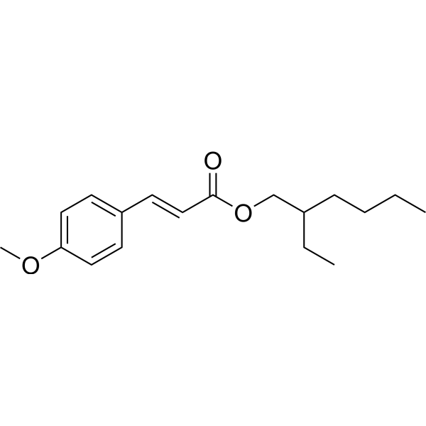
Octinoxate (Octyl methoxycinnamate) is a thyroid hormone receptor agonist, reducing the levels of triiodothyronine (T3) and thyroxine (T4) and transcription levels of genes related to type II deiodinase (deio2) in Japanese Medaka. Octinoxate is commonly used as a safe ultraviolet (UV) filter used in the aquatic environment. Octinoxate inhibits CYP1A1 and CYP1B1 to regulate hyaluronan (HA) (HY-B0633A) metabolism in a PI3K pathway-dependent manner in human keratinocytes. Octinoxate also exhibits an anti-estrogenic and anti-androgenic effect in vitro and in vivo .
Hyaluronic acid, low endotoxin (Hyaluronan, low endotoxin) is a biopolymer composed of repeating disaccharide units containing low levels of endotoxin. Hyaluronic acid is a major component of the extracellular matrix (ECM). It is synthesized on the plasma membrane. Hyaluronic acid exerts its effects by binding to receptors CD44 and RHAMM. Hyaluronic acid activates PI3K-Akt signaling. Hyaluronic acid also enhances cell invasion and angiogenesis by promoting or stimulating the binding of proteolytic MMP-9 to the cell surface. Elevated hyaluronic acid levels are associated with tumor cell growth, adhesion, migration, invasion, and angiogenesis in digestive system cancers. Hyaluronic acid is involved in tissue remodeling and rapid cell proliferation in several physiological processes, including embryonic morphogenesis and wound healing. Hyaluronic acid can be used as a regulator of cancer-associated lymphangiogenesis. Hyaluronic acid can be used as a drug delivery carrier for sodium butyrate, enhancing its anti-proliferative activity against breast cancer cell lines. Hyaluronic acid can lubricate the corneal endothelium. Hyaluronic acid can improve tissue hydration and enhance the resistance of cells to mechanical damage. Hyaluronic acid has been conjugated with antibodies to ensure that the active compound continues to exert its effects at the site of inflammation. Hyaluronic acid can be used in research in the fields of osteoarthritis, ophthalmology, cosmetic dermatology, oncology, and liver diseases .
Hyaluronic acid (MW 50000-100000) (Hyaluronan (MW 50000-100000)) is a biopolymer composed of repeating units of disaccharides with a molecular weight of 50000-100000 Da. Hyaluronic acid (MW 50000-100000) can be used as a drug carrier for drug delivery .
Tetrapeptide-21 is a bioactive peptide composed of four amino acids. Tetrapeptide-21 effectively enhances the vitality of human dermal fibroblasts. Tetrapeptide-21 upregulates the expression of key extracellular matrix (ECM) genes and promotes the synthesis of ECM proteins (such as type I collagen, hyaluronic acid synthase 1, and fibronectin). Tetrapeptide-21 has the efficacy of anti-wrinkle and improving skin elasticity, and has been reported to be used as a cosmetic ingredient .
Hyaluronan-binding peptide, biotin labeled TFA is a biological active peptide. (This peptide is a hyaluronan-binding peptide biotinylated through a C-terminal GGGSK linker. Hyaluronan (HA) is a nonsulfated glycosaminoglycan expressed in the extracellular matrix and on cell surfaces. HA plays a role in fertilization, embryonic development, wound healing, angiogenesis, leukocyte trafficking to inflamed tissues, and cancer metastasis. This peptide has been shown to block HA binding to CD44 receptors and inhibit T cell proliferation.) .
Anti-CD44 Antibody (Hermes-1) is a kind of rat IgG2a κ chimeric antibody, targeting to human CD44. Anti-CD44 Antibody (Hermes-1) blocks the binding of hyaluronan to CD44. Anti-CD44 Antibody (Hermes-1) restores (Platelet-derived Growth Factor-BB)-induced β-receptor activation and motility in fibroblasts. Anti-CD44 Antibody (Hermes-1) causes partial loss of the anti-aging effect of hyaluronic .
Hyaluronan-IN-1 (Pep-1) is a Hyaluronan inhibitor with a Kd value of 1.65 μM. Hyaluronan-IN-1 blocks CD44-dependent cell adhesion. Hyaluronan-IN-1 inhibits cell adhesion to hyaluronan substrates. Hyaluronan-IN-1 suppresses the development of contact hypersensitivity in mice by blocking the homing process of inflammatory cells to the skin. Hyaluronan-IN-1 also inhibits responses during the sensitization phase. Hyaluronan-IN-1 reduces lung metastasis of melanoma and prolongs the survival of mice. Hyaluronan-IN-1 can be used in research related to contact hypersensitivity, chronic skin inflammation, and melanoma .
4-Methylumbelliferone (sodium salt) is a coumarin derivative. 4-Methylumbelliferone (sodium salt) can be used as a standard to quantify the free 4-methylumbelliferone released as a result of enzyme-substrate action. 4-Methylumbelliferone (sodium salt) inhibits hyaluronan production in vitro. 4-Methylumbelliferone (sodium salt) can be studied in ovarian cancer research .
IGFBP-3 peptide is a 18-amino acid insulin-like growth factor binding protein-3. IGFBP-3 peptide binds Humanin (HY-P1928) and Hyaluronan (HY-B0633A), blocks the interaction of CD44 and hyaluronan .
Octinoxate-13C,d3 is the deuterium labeled Octinoxate (HY-W245806). Octinoxate (Octyl methoxycinnamate) is a thyroid hormone receptor agonist, reducing the levels of triiodothyronine (T3) and thyroxine (T4) and transcription levels of genes related to type II deiodinase (deio2) in Japanese Medaka. Octinoxate is commonly used as a safe ultraviolet (UV) filter used in the aquatic environment. Octinoxate inhibits CYP1A1 and CYP1B1 to regulate hyaluronan (HA) (HY-B0633A) metabolism in a PI3K pathway-dependent manner in human keratinocytes. Octinoxate also exhibits an anti-estrogenic and anti-androgenic effect in vitro and in vivo .
2-Ethylhexyl (E)-3-(4-(methoxy-d3)phenyl)acrylate is the deuterium labeled Octinoxate (HY-B1234). Octinoxate (Octyl methoxycinnamate) is a thyroid hormone receptor agonist, reducing the levels of triiodothyronine (T3) and thyroxine (T4) and transcription levels of genes related to type II deiodinase (deio2) in Japanese Medaka. Octinoxate is commonly used as a safe ultraviolet (UV) filter used in the aquatic environment. Octinoxate inhibits CYP1A1 and CYP1B1 to regulate hyaluronan (HA) (HY-B0633A) metabolism in a PI3K pathway-dependent manner in human keratinocytes. Octinoxate also exhibits an anti-estrogenic and anti-androgenic effect in vitro and in vivo .
Hyaluronidase, Streptomyces hyalurolyticus (EC 3.2.1.35) degrades hyaluronan and has been found to be inappropriately regulated during cancer progression. Hyaluronidase randomly cleaves β-N-acetylhexosamine-[1→4] glycosidic bonds in hyaluronic acid, chondroitin, and chondroitin sulfates.
Anti-Mouse CD44 Antibody (KM703) recognizes mouse CD44. CD44 is the main receptor for hyaluronan (HA). Anti-Mouse CD44 Antibody (KM703) does not recognize the HA binding site of CD44. Anti-Mouse CD44 Antibody (KM703) is a non-HA-blocking antibody. Recommend Isotype Controls: Rat IgG2a kappa, Isotype Control (HY-P990679) .
Hyaluronic acid is a biopolymer composed of repeating units of disaccharides with various applications. Hyaluronic acid is a major component of the extracellular matrix (ECM). Hyaluronic acid is synthesized at the plasma membrane. Increased hyaluronic acid levels are associated with tumor cell growth, adhesion, migration, invasion and angiogenesis in digestive cancers. Hyaluronic acid participates in tissue remodeling and rapid cell proliferation in some physiological processes including embryonic morphogenesis and wound-healing. Hyaluronic acid activates the PI3K-Akt signaling. Hyaluronic acid acts as a regulator of cancer-associated lymphangiogenesis. Hyaluronic acid also enhances cell invasion and angiogenesis by promoting proteolytic MMP-9 binding to cell surface or stimulating MMP-9 binding to cell surface. Hyaluronic acid can be used as drug delivery for sodium butyrate to improve the anti-proliferative activity on breast cancer cell line. Hyaluronic acid can be studied in joint diseases, wound healing and cancer .
Hyaluronic acid, low endotoxin (Hyaluronan, low endotoxin) is a biopolymer composed of repeating disaccharide units containing low levels of endotoxin. Hyaluronic acid is a major component of the extracellular matrix (ECM). It is synthesized on the plasma membrane. Hyaluronic acid exerts its effects by binding to receptors CD44 and RHAMM. Hyaluronic acid activates PI3K-Akt signaling. Hyaluronic acid also enhances cell invasion and angiogenesis by promoting or stimulating the binding of proteolytic MMP-9 to the cell surface. Elevated hyaluronic acid levels are associated with tumor cell growth, adhesion, migration, invasion, and angiogenesis in digestive system cancers. Hyaluronic acid is involved in tissue remodeling and rapid cell proliferation in several physiological processes, including embryonic morphogenesis and wound healing. Hyaluronic acid can be used as a regulator of cancer-associated lymphangiogenesis. Hyaluronic acid can be used as a drug delivery carrier for sodium butyrate, enhancing its anti-proliferative activity against breast cancer cell lines. Hyaluronic acid can lubricate the corneal endothelium. Hyaluronic acid can improve tissue hydration and enhance the resistance of cells to mechanical damage. Hyaluronic acid has been conjugated with antibodies to ensure that the active compound continues to exert its effects at the site of inflammation. Hyaluronic acid can be used in research in the fields of osteoarthritis, ophthalmology, cosmetic dermatology, oncology, and liver diseases .
Hyaluronic acid (MW 50000-100000) (Hyaluronan (MW 50000-100000)) is a biopolymer composed of repeating units of disaccharides with a molecular weight of 50000-100000 Da. Hyaluronic acid (MW 50000-100000) can be used as a drug carrier for drug delivery .
Tetrapeptide-21 is a bioactive peptide composed of four amino acids. Tetrapeptide-21 effectively enhances the vitality of human dermal fibroblasts. Tetrapeptide-21 upregulates the expression of key extracellular matrix (ECM) genes and promotes the synthesis of ECM proteins (such as type I collagen, hyaluronic acid synthase 1, and fibronectin). Tetrapeptide-21 has the efficacy of anti-wrinkle and improving skin elasticity, and has been reported to be used as a cosmetic ingredient .
Hyaluronan-binding peptide, biotin labeled TFA is a biological active peptide. (This peptide is a hyaluronan-binding peptide biotinylated through a C-terminal GGGSK linker. Hyaluronan (HA) is a nonsulfated glycosaminoglycan expressed in the extracellular matrix and on cell surfaces. HA plays a role in fertilization, embryonic development, wound healing, angiogenesis, leukocyte trafficking to inflamed tissues, and cancer metastasis. This peptide has been shown to block HA binding to CD44 receptors and inhibit T cell proliferation.) .
Hyaluronan-IN-1 (Pep-1) is a Hyaluronan inhibitor with a Kd value of 1.65 μM. Hyaluronan-IN-1 blocks CD44-dependent cell adhesion. Hyaluronan-IN-1 inhibits cell adhesion to hyaluronan substrates. Hyaluronan-IN-1 suppresses the development of contact hypersensitivity in mice by blocking the homing process of inflammatory cells to the skin. Hyaluronan-IN-1 also inhibits responses during the sensitization phase. Hyaluronan-IN-1 reduces lung metastasis of melanoma and prolongs the survival of mice. Hyaluronan-IN-1 can be used in research related to contact hypersensitivity, chronic skin inflammation, and melanoma .
Hyaluronan-binding peptide, biotin labeled is a biological active peptide. (This peptide is a hyaluronan-binding peptide biotinylated through a C-terminal GGGSK linker. Hyaluronan (HA) is a nonsulfated glycosaminoglycan expressed in the extracellular matrix and on cell surfaces. HA plays a role in fertilization, embryonic development, wound healing, angiogenesis, leukocyte trafficking to inflamed tissues, and cancer metastasis. This peptide has been shown to block HA binding to CD44 receptors and inhibit T cell proliferation.)
IGFBP-3 peptide is a 18-amino acid insulin-like growth factor binding protein-3. IGFBP-3 peptide binds Humanin (HY-P1928) and Hyaluronan (HY-B0633A), blocks the interaction of CD44 and hyaluronan .
Anti-CD44 Antibody (Hermes-1) is a kind of rat IgG2a κ chimeric antibody, targeting to human CD44. Anti-CD44 Antibody (Hermes-1) blocks the binding of hyaluronan to CD44. Anti-CD44 Antibody (Hermes-1) restores (Platelet-derived Growth Factor-BB)-induced β-receptor activation and motility in fibroblasts. Anti-CD44 Antibody (Hermes-1) causes partial loss of the anti-aging effect of hyaluronic .
Anti-Mouse CD44 Antibody (KM703) recognizes mouse CD44. CD44 is the main receptor for hyaluronan (HA). Anti-Mouse CD44 Antibody (KM703) does not recognize the HA binding site of CD44. Anti-Mouse CD44 Antibody (KM703) is a non-HA-blocking antibody. Recommend Isotype Controls: Rat IgG2a kappa, Isotype Control (HY-P990679) .
Hyaluronic acid is a biopolymer composed of repeating units of disaccharides with various applications. Hyaluronic acid is a major component of the extracellular matrix (ECM). Hyaluronic acid is synthesized at the plasma membrane. Increased hyaluronic acid levels are associated with tumor cell growth, adhesion, migration, invasion and angiogenesis in digestive cancers. Hyaluronic acid participates in tissue remodeling and rapid cell proliferation in some physiological processes including embryonic morphogenesis and wound-healing. Hyaluronic acid activates the PI3K-Akt signaling. Hyaluronic acid acts as a regulator of cancer-associated lymphangiogenesis. Hyaluronic acid also enhances cell invasion and angiogenesis by promoting proteolytic MMP-9 binding to cell surface or stimulating MMP-9 binding to cell surface. Hyaluronic acid can be used as drug delivery for sodium butyrate to improve the anti-proliferative activity on breast cancer cell line. Hyaluronic acid can be studied in joint diseases, wound healing and cancer .
Octinoxate (Octyl methoxycinnamate) is a thyroid hormone receptor agonist, reducing the levels of triiodothyronine (T3) and thyroxine (T4) and transcription levels of genes related to type II deiodinase (deio2) in Japanese Medaka. Octinoxate is commonly used as a safe ultraviolet (UV) filter used in the aquatic environment. Octinoxate inhibits CYP1A1 and CYP1B1 to regulate hyaluronan (HA) (HY-B0633A) metabolism in a PI3K pathway-dependent manner in human keratinocytes. Octinoxate also exhibits an anti-estrogenic and anti-androgenic effect in vitro and in vivo .
Hyaluronate lyase is a polysaccharide-derived carboxylase, also known as a diffusion factor or mucin. This enzyme can cleave hyaluronic acid salts at the β-D-GalNAc-(1→4)-β-D-GlcA glycosidic bond site through a β-elimination reaction, and the resulting unsaturated disaccharide can initiate downstream reaction pathways. Hyaluronan Lyase Protein, S. agActiae (His) is a recombinant hyaluronic acid lyase protein with the His tag.
HABP2 is a protein that cleaves the alpha chain of fibrinogen as well as the beta chain between "Lys-53" and "Lys-54" at multiple sites, preventing direct formation of a fibrin clot. It activates coagulation factor VII and converts prourokinase into its active form. HABP2 Protein, Human (HEK293, His) is the recombinant human-derived HABP2 protein, expressed by HEK293 , with C-6*His labeled tag.
The HAS2 protein adds GlcNAc or GlcUA monosaccharides to hyaluronic acid, playing a crucial role in its synthesis. HAS2 Protein, Mouse (His) is the recombinant mouse-derived HAS2 protein, expressed by E. coli , with N-6*His labeled tag.
The ITIH3 protein has a dual role: as a carrier of hyaluronic acid in serum and as a binding protein that facilitates interactions with various matrix proteins. These interactions regulate the localization, synthesis, and degradation of hyaluronic acid, which is critical for multiple cellular activities. ITIH3 Protein, Human (HEK293, His) is the recombinant human-derived ITIH3 protein, expressed by HEK293 , with C-6*His labeled tag.
Stabilin-2 protein is a multifunctional phosphatidylserine and hyaluronic acid receptor that engulfs apoptotic cells and mediates hyaluronic acid endocytosis. As a systemic scavenger receptor, it binds various ligands and supports extracellular matrix turnover and body mobility. Stabilin-2 Protein, Human (His) is the recombinant human-derived Stabilin-2 protein, expressed by E. coli , with C-His labeled tag.
Octinoxate-13C,d3 is the deuterium labeled Octinoxate (HY-W245806). Octinoxate (Octyl methoxycinnamate) is a thyroid hormone receptor agonist, reducing the levels of triiodothyronine (T3) and thyroxine (T4) and transcription levels of genes related to type II deiodinase (deio2) in Japanese Medaka. Octinoxate is commonly used as a safe ultraviolet (UV) filter used in the aquatic environment. Octinoxate inhibits CYP1A1 and CYP1B1 to regulate hyaluronan (HA) (HY-B0633A) metabolism in a PI3K pathway-dependent manner in human keratinocytes. Octinoxate also exhibits an anti-estrogenic and anti-androgenic effect in vitro and in vivo .
2-Ethylhexyl (E)-3-(4-(methoxy-d3)phenyl)acrylate is the deuterium labeled Octinoxate (HY-B1234). Octinoxate (Octyl methoxycinnamate) is a thyroid hormone receptor agonist, reducing the levels of triiodothyronine (T3) and thyroxine (T4) and transcription levels of genes related to type II deiodinase (deio2) in Japanese Medaka. Octinoxate is commonly used as a safe ultraviolet (UV) filter used in the aquatic environment. Octinoxate inhibits CYP1A1 and CYP1B1 to regulate hyaluronan (HA) (HY-B0633A) metabolism in a PI3K pathway-dependent manner in human keratinocytes. Octinoxate also exhibits an anti-estrogenic and anti-androgenic effect in vitro and in vivo .
Hyaluronic acid is a biopolymer composed of repeating units of disaccharides with various applications. Hyaluronic acid is a major component of the extracellular matrix (ECM). Hyaluronic acid is synthesized at the plasma membrane. Increased hyaluronic acid levels are associated with tumor cell growth, adhesion, migration, invasion and angiogenesis in digestive cancers. Hyaluronic acid participates in tissue remodeling and rapid cell proliferation in some physiological processes including embryonic morphogenesis and wound-healing. Hyaluronic acid activates the PI3K-Akt signaling. Hyaluronic acid acts as a regulator of cancer-associated lymphangiogenesis. Hyaluronic acid also enhances cell invasion and angiogenesis by promoting proteolytic MMP-9 binding to cell surface or stimulating MMP-9 binding to cell surface. Hyaluronic acid can be used as drug delivery for sodium butyrate to improve the anti-proliferative activity on breast cancer cell line. Hyaluronic acid can be studied in joint diseases, wound healing and cancer .
Inquiry Online
Your information is safe with us. * Required Fields.
Western blot analysis of extracts from THP-1(lane 2(20μg), Jurkat (lane 3(20μg) and NIH3T3(lane 4(20μg) using FOXO1A (HY-P80132) Rabbit mAb. Proteins were transferred
to a PVDF membrane and blocked with 5% non-fat milk in TBST for 2 hour at room temperature. The primary antibody (1/1000) and Loading control antibody (Beta Actin, HY-P80438, 1/10000) was
used in 5% non-fat milk in TBST at 4°C overnight. Goat Anti-Mouse/Rabbit IgG-HRP Secondary Antibody (1/10000) was used for 1 hour at room temperature.
Western blot analysis of extracts from THP-1(lane 2(20μg), Jurkat (lane 3(20μg) and NIH3T3(lane 4(20μg) using FOXO1A (HY-P80132) Rabbit mAb. Proteins were transferred
to a PVDF membrane and blocked with 5% non-fat milk in TBST for 2 hour at room temperature. The primary antibody (1/1000) and Loading control antibody (Beta Actin, HY-P80438, 1/10000) was
MedchemExpress Validation 03
Western blot analysis of extracts from THP-1(lane 2(20μg), Jurkat (lane 3(20μg) and NIH3T3(lane 4(20μg) using FOXO1A (HY-P80132) Rabbit mAb. Proteins were transferred
MedchemExpress Validation 04
Western blot analysis of extracts from THP-1(lane 2(20μg), Jurkat (lane 3(20μg) and NIH3T3(lane 4(20μg) using FOXO1A (HY-P80132) Rabbit mAb. Proteins were transferred
to a PVDF membrane and blocked with 5% non-fat milk in TBST for 2 hour at room temperature. The primary antibody (1/1000) and Loading control antibody (Beta Actin, HY-P80438, 1/10000) was
used in 5% non-fat milk in TBST at 4°C overnight. Goat Anti-Mouse/Rabbit IgG-HRP Secondary Antibody (1/10000) was used for 1 hour at room temperature.
MedchemExpress Validation
Western blot analysis of extracts from THP-1(lane 2(20μg), Jurkat (lane 3(20μg) and NIH3T3(lane 4(20μg) using FOXO1A (HY-P80132) Rabbit mAb. Proteins were transferred
to a PVDF membrane and blocked with 5% non-fat milk in TBST for 2 hour at room temperature. The primary antibody (1/1000) and Loading control antibody (Beta Actin, HY-P80438, 1/10000) was
used in 5% non-fat milk in TBST at 4°C overnight. Goat Anti-Mouse/Rabbit IgG-HRP Secondary Antibody (1/10000) was used for 1 hour at room temperature.
MedchemExpress Validation
Western blot analysis of extracts from THP-1(lane 2(20μg), Jurkat (lane 3(20μg) and NIH3T3(lane 4(20μg) using FOXO1A (HY-P80132) Rabbit mAb. Proteins were transferred
to a PVDF membrane and blocked with 5% non-fat milk in TBST for 2 hour at room temperature. The primary antibody (1/1000) and Loading control antibody (Beta Actin, HY-P80438, 1/10000) was
used in 5% non-fat milk in TBST at 4°C overnight. Goat Anti-Mouse/Rabbit IgG-HRP Secondary Antibody (1/10000) was used for 1 hour at room temperature.
MedChemExpress values your privacy and your trust is important to us. We use cookies to enhance your website experience. Some cookies are necessary to run the website.
Privacy and Cookie Policy