Delivery of temperature sensitive items including proteins and kits will be paused on 6/19 for the Juneteenth holiday. For urgent orders please contact customer service.
Sphingomyelin is a eukaryotic sphingolipid and one of the major constituents of cell membranes and particularly abundant in the myelin sheath that surrounds neuronal axons. Sphingomyelin plays an important role in cell processes, the regulation of inflammatory responses, and signal transduction. Sphingomyelin metabolism is associated with various central nervous system diseases and Niemann–Pick disease .
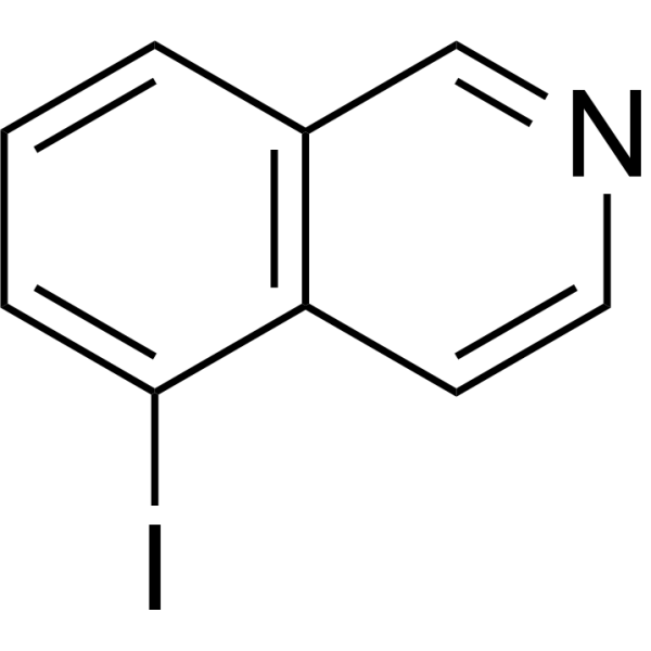
DSRM-3716 (5-Iodoisoquinoline) is a potent and selective SARM1 NADase inhibitor with an IC50 of 75 nM. DSRM-3716 is selective against other NAD +-processing enzymes, receptors, and transporters. DSRM-3716 provides robust axon protection .
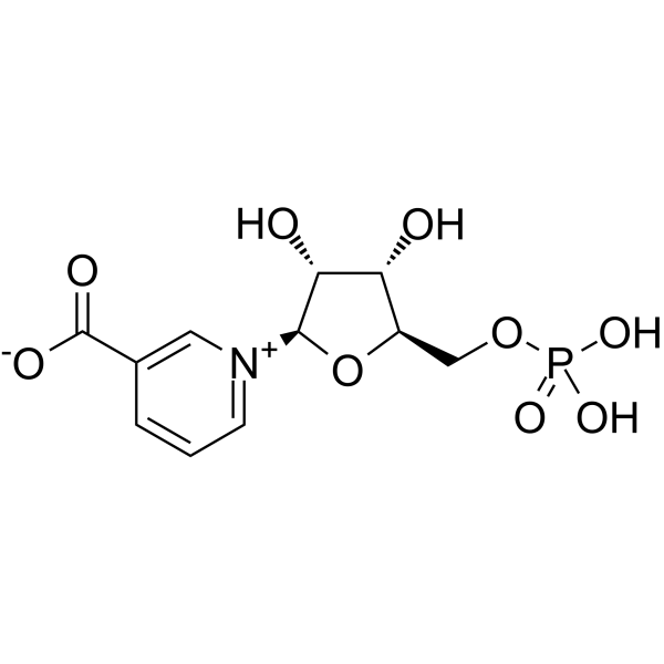
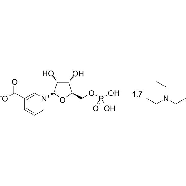
Nicotinic acid mononucleotide acts as a SARM1 inhibitor and a NAD + biosynthesis intermediate, with an IC50 value of 93.3 μM against SARM1. Nicotinic acid mononucleotide exerts axon-protective effects, delays axonal degeneration, elevates NAD + levels, enhances Sirt1 activity, improves myocardial capillary density and alleviates myocardial fibrosis. Nicotinic acid mononucleotide reverses diabetic cardiomyopathy in diabetic mice by increasing myocardial NAD + levels. Nicotinic acid mononucleotide is applicable to research related to cancer, multiple sclerosis, diabetic cardiomyopathy, neurodegenerative diseases and Huntington's disease .
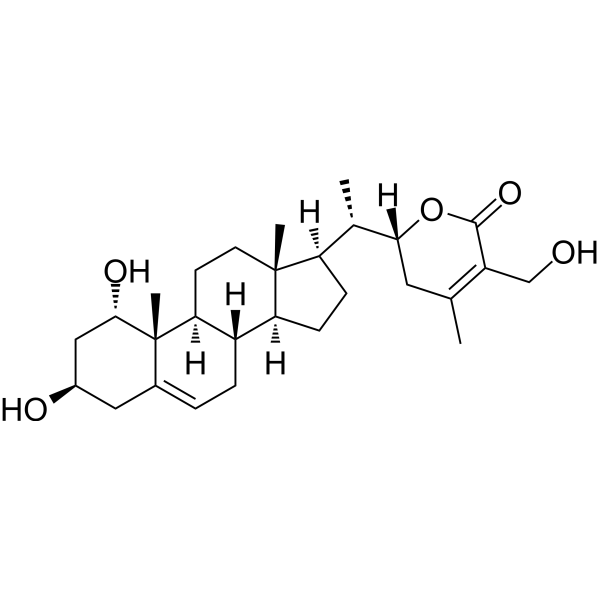
Sominone is the active metabolite of Withanoside IV (HY-N8693). Sominone enhances neuronal morphological plasticity by activating the RET pathway. Sominone can also induce axon/dendrite regeneration and synaptic reconstruction, thereby improving spatial memory. Sominone can be used in the research of neurodegenerative diseases such as Alzheimer's disease .
Dextran T3 (Dextran 3; Dextran T3(MW 2400-3600)) is a neural tracer and intestinal permeability probe that can move anterogradely and retrogradely in neuronal axons by passive diffusion. Dextran T3 (MW 3,000) is able to permeate across the intestinal epithelial cell membrane in the presence of cholera toxin-induced cytoskeletal disturbance. Dextran T3 (MW 3,000) is used as a fluorescent marker to rapidly label developing neurons (such as Xenopus retinal ganglion cells) and to assess intestinal barrier function. It can be used to study axonal transport in neuroanatomy and permeability changes in intestinal pathophysiology. The Dextran series of compounds are also natural polysaccharide drug carriers that can be connected to drugs through covalent bonding methods such as ester bonds, amide bonds or click chemistry, or self-assembled to form carriers such as nanoparticles and hydrogels. Dextran is biodegradable and biocompatible, and can achieve targeted delivery and controlled release of drugs. Dextran derivatives can prolong the half-life of drugs, increase local concentrations, and reduce the activity of immune clearance .
GNE-8505 is an orally active, blood-brain barrier-permeable selective dual leucine zipper kinase (DLK) inhibitor. GNE-8505 has an IC50 of 0.144 μM for pJNK, and EC50 of 0.457 μM for DRG. GNE-8505 inhibits the DLK/JNK pathway, reduces stress-induced c-Jun phosphorylation levels, decreases neuronal death and suppresses axonal degeneration. GNE-8505 reduces phosphorylated c-Jun levels in the retina, spinal cord and brain tissues of mice. GNE-8505 is applicable to research related to Alzheimer's disease and amyotrophic lateral sclerosis (ALS) .
PI3K/Akt/CREB activator 1 (compound AE-18) is a potent, orally active PI3K/Akt/CREB activator. PI3K/Akt/CREB activator 1 promotes neuronal proliferation, induced differentiation of Neuro-2a cells into a neuron-like morphology, and accelerated the establishment of axon-dendrite polarization of primary hippocampal neurons through upregulating brain-derived neurotrophic factor via the PI3K/Akt/CREB pathway. PI3K/Akt/CREB activator 1 can be used in research of vascular dementia (VaD) .
Diadenosine pentaphosphate pentasodium is an agonist and negative modulator of the P2X1 receptor, an endogenous vasoactive purine dinucleotide that can be isolated from platelets. Diadenosine pentaphosphate pentasodium mediates negative regulation of dendrite growth and number by activating homologous and heterologous P2X1 receptors, which triggers a transient and moderate increase in intracellular calcium levels within dendritic growth cones. Diadenosine pentaphosphate pentasodium is widely present in secretory vesicles such as platelets, chromaffin cells and brain synaptosomes, and exhibits selective activity on dendrite growth of cultured hippocampal neurons, inhibiting only dendrite growth without affecting axon growth. Diadenosine pentaphosphate pentasodium has a weaker ability to compete with RcCHAD for binding to polyP than short-chain polyPs .
SARM1-IN-2 (Example 82) is a SARM1 inhibitor (IC50 <1 μM) that inhibits axon regeneration. Axon regeneration refers to the process by which neuronal axons attempt to restore their structure and function after axonal degeneration. SARM1-IN-2 inhibits axon regeneration by reducing or inhibiting the binding of SARM1 to NAD+. SARM1-IN-2 can be used to study axonal degeneration .
Phaseolus vulgaris leucoagglutinin (PHA-L) is a lectin, that can be extracted from red kidney beans (Phaseolus vulgaris). Phaseolus vulgaris leucoagglutinin can be used as an anterograde axonal tracer in neuroanatomical research to study the morphology of neurons, axons, and terminal structures in the nervous system .
BN201 promotes neuronal differentiation, the differentiation of precursor cells to mature oligodendrocytes (EC50 of 6.3 μM) in vitro, and the myelination of new axons (EC50 of 16.6 μM). BN201 is able to cross the blood-brain barrier by active transport and activate pathways (IGF-1 pathway) associated with the response to stress and neuron survival. BN201 has potently neuroprotective effects .
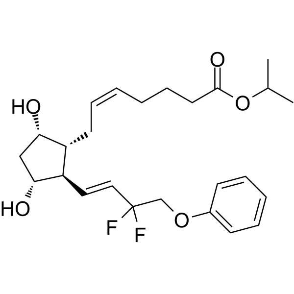
Tafluprost (AFP-168) is an anti-glaucoma prostaglandin (PG) analog. Tafluprost can inhibit the apoptosis of retinal ganglion cells (RGCs) and rat RGCs cells. Tafluprost promotes axon regeneration by regulating Zn 2+-mTORpathway, inhibits intracellular lipid accumulation in human preorbital adipocytes. Tafluprost can be used in the study of optic nerve injury in glaucoma .
Dehydronitrosonisoldipine, a derivative of Nisoldipine (HY-17402), is an irreversible and cell-permeant sterile alpha and TIR motif-containing 1 (SARM1) inhibitor. Dehydronitrosonisoldipine acts mainly by blocking SARM1 activation but not its enzymatic activities. Dehydronitrosonisoldipine inhibits SARM1 and axon degenration (AxD) by covalently modifying cysteines, also inhibits the Vincristine-activated cADPR production in neurons. Dehydronitrosonisoldipine can be used for researching neurodegenerative disorders .
ESI1 is a small molecule epigenetic silencing inhibitor. ESI1 can trigger the formation of nuclear condensates of key lipid metabolism regulators SREBP1/2, concentrating transcriptional co-activators to drive lipid/cholesterol biosynthesis. ESI1 can promote myelin regeneration in demyelinated animal models and facilitate de novo myelination on regenerating CNS axons, reversing age-related declines in cognitive abilities .
Xelafaslatide (ONL-1204) is a Fas receptor antagonist. Xelafaslatide blocks the Fas receptor signaling pathway and inhibits downstream apoptosis and inflammatory pathways. Xelafaslatide suppresses neuroinflammation and microglial activation in glaucoma models, protects retinal ganglion cells and prevents axonal degeneration. Xelafaslatide is applicable to relevant research on glaucoma .
Hexane-2,5-dione (2,5-HD) is a cytotoxic agent. Hexane-2,5-dione causes an accumulation of neurofilaments within axons in rats that may lead to their degeneration. Hexane-2,5-dione is promising for research of neurodegenerative diseases (e.g., Alzheimer's, Parkinson's) .
D-threo-PDMP is a potent glucoceramide synthase (GCS) inhibitor, which reduces the glycosphingolipids (such as GM3 and GD3) on the cell surface by inhibiting glycosylation, reduces the total length of the axon plexus and the number of axon branch points, and inhibits neurite growth. D-threo-PDMP inhibits the synthesis of GM3, thereby reducing the adhesion ability of B16 melanoma cells and mimicking the pathological effects of hyperglycemia/TGF-β1. D-threo-PDMP inhibits the synthesis of GD3, thereby protecting liver cells from apoptosis induced by TNF-α. D-threo-PDMP can be used to study diseases related to targeted glycosphingolipid metabolism .
D-threo-PDMP hydrochloride is a potent glucoceramide synthase (GCS) inhibitor, which reduces the glycosphingolipids (such as GM3 and GD3) on the cell surface by inhibiting glycosylation, reduces the total length of the axon plexus and the number of axon branch points, and inhibits neurite growth. D-threo-PDMP hydrochloride inhibits the synthesis of GM3, thereby reducing the adhesion ability of B16 melanoma cells and mimicking the pathological effects of hyperglycemia/TGF-β1. D-threo-PDMP hydrochloride inhibits the synthesis of GD3, thereby protecting liver cells from apoptosis induced by TNF-α. D-threo-PDMP hydrochloride can be used to study diseases related to targeted glycosphingolipid metabolism .
Sob-AM2 is a potent substrate (Km=1.3 μM) targeting fatty acid amide hydrolase (FAAH) expressed in the brain and has blood-brain barrier permeability. Sob-AM2 delivers high concentrations of Sobetirome (HY-14823) to the central nervous system with minimal peripheral systemic dose, thereby stimulating central thyroid hormone receptor β (TRβ). In addition, Sob-AM2 can prevent myelin and axon degeneration in experimental autoimmune encephalomyelitis (EAE) mice .
GDC-0134 (RG6000) is a modulator targeting dual leucine zipper kinase (DLK) that can cross the blood-brain barrier. By inhibiting the kinase activity of DLK, GDC-0134 blocks the activation of the downstream JNK signaling pathway, suppresses DLK-dependent retrograde signal transduction of axon-to-soma degeneration, and exerts neuroprotective activity. GDC-0134 reduces TDP-43 protein aggregation and decreases the degree of neuromuscular junction denervation in motor neurons. GDC-0134 can be used in the research of amyotrophic lateral sclerosis (ALS), Alzheimer's disease and other DLK-related neurodegenerative diseases .
Nicotinic acid mononucleotide triethylamine acts as a SARM1 inhibitor and a NAD + biosynthesis intermediate, with an IC50 value of 93.3 μM against SARM1. Nicotinic acid mononucleotide triethylamine exerts axon-protective effects, delays axonal degeneration, elevates NAD + levels, enhances Sirt1 activity, improves myocardial capillary density and alleviates myocardial fibrosis. Nicotinic acid mononucleotide triethylamine reverses diabetic cardiomyopathy in diabetic mice by increasing myocardial NAD + levels. Nicotinic acid mononucleotide triethylamine is applicable to research related to cancer, multiple sclerosis, diabetic cardiomyopathy, neurodegenerative diseases and Huntington's disease .
GNE-5152 is an orthosteric SARM1 base-exchange (BE) inhibitor. GNE-5152 sustainably activates SARM1 at subinhibitory concentrations under mildly activating conditions, and this synergistic adverse effect increases NAD consumption, induces axon degradation and neurodegeneration and releases neurofilament-light (NfL) in cortical neurons. GNE-5152 can be used for neurodegenerative diseases research .
5-Chloroisoquinoline (compound 42) is an inhibitor of SARM1 (Sterile alpha and toll/interleukin receptor (TIR) motif containing protein 1), an enzyme involved in axon degeneration that catalyzes multiple activities through a ternary complex mechanism. 5-Chloroisoquinoline can be used in the study of neurodegenerative diseases or axon degeneration .
Withanoside IV is an orally active, blood-brain barrier-permeable withanolide derivative. Withanoside IV specifically binds to the Sudlow I site of HSA, induces secondary structural changes in HSA, and forms stable HSA complexes. Withanoside IV inhibits the enzymatic activity of COX-2. Withanoside IV induces axonal regeneration, peripheral nervous system myelination and increased axonal density in spinal cord tissue, reduces reactive gliosis-related changes, and improves hindlimb motor function. Withanoside IV binds to amyloid-β 1-42 to inhibit its aggregation, induces neurite outgrowth and synapse reconstruction, repairs damaged axons and dendrites, enhances mitochondrial biogenesis, exerts neuroprotective effects via the BDNF and SIRT1 signaling pathways, reduces ROS production and neuronal apoptosis, and ameliorates memory deficits. Withanoside IV inhibits the activity of the SARS-CoV-2 main protease. Withanoside IV can be used in research related to spinal cord injury, Alzheimer's disease, and coronavirus disease 2019 (COVID-19) .
FFN-102 trifluoroacetate is an analogue of biogenic neurotransmitters. FFN-102 trifluoroacetate is a pH-dependent fluorescent probe that labels dopamine cell bodies, axons, and presynaptic terminals .
Tafluprost (Standard) is the analytical standard of Tafluprost. This product is intended for research and analytical applications. Tafluprost (AFP-168) is an anti-glaucoma prostaglandin (PG) analog. Tafluprost can inhibit the apoptosis of retinal ganglion cells (RGCs) and rat RGCs cells. Tafluprost promotes axon regeneration by regulating Zn2+-mTORpathway, inhibits intracellular lipid accumulation in human preorbital adipocytes. Tafluprost can be used in the study of optic nerve injury in glaucoma [4] .
TRITC-lysine-dextran (MW 70kDa) is a fluorescent label prepared by the conjugation of TRITC (HY-D0791), lysine and dextran. TRITC-lysine-dextran (MW 70kDa) serves multiple functions as an axonal tracer, non-viral nanocarrier and fixable fluorescent clonal marker. TRITC-lysine-dextran (MW 70kDa) undergoes anterograde and retrograde transport within axons of sensory neurons, and acts as a non-viral delivery system to precisely deliver biomolecules to neurons. TRITC-lysine-dextran (MW 70kDa) remains stably retained during histological preparation, thereby supporting continuous observation in live or fixed samples .
DLK-IN-2 is a selective inhibitor of DLK and neuroprotective agent. DLK-IN-2 shows no significant inhibition against CYPs 3A4, 2D6 and 2C9. DLK-IN-2 inhibits acute axonal palmitoylation of DLK, blocks DLK-dependent pro-degenerative axon-to-soma retrograde signaling and suppresses c-Jun phosphorylation. DLK-IN-2 can be used for the mechanistic study of neurodegenerative diseases .
SARM1-IN-9 (Compound MY-13B) is a stereoselective SARM1 inhibitor. SARM1-IN-9 is applicable to research related to axon degeneration-dependent neurological diseases .
(S,S)-SARM1-IN-9 (Compound MY-13A) is a stereoselective SARM1 inhibitor with covalent binding properties. (S,S)-SARM1-IN-9 covalently modifies Cys311 in the autoregulatory ARM domain of wild-type SARM1, thereby blocking NADase activity, without inhibiting the SARM1C311A or SARM1C311S mutants. (S,S)-SARM1-IN-9 blocks vacor- and vincristine-induced axon degeneration in primary rodent dorsal root ganglion neurons. (S,S)-SARM1-IN-9 can be used for research on axon degeneration-dependent neurological disorders, including chemotherapy-induced peripheral neuropathy .
SARM1-IN-10 is an orally active SARM1 inhibitor with a pIC50 of 7.1 and a pKd of 8.3. As a base-exchange inhibitor, SARM1-IN-10 forms a NAD + adduct at the active site of the TIR domain of SARM1, blocks enzymatic function, and induces a unique rotameric state of W662 at the catalytic site of SARM1. SARM1-IN-10 acts as a paradoxical neurodegeneration inducer at low doses and an inhibitor at high doses, and it can exacerbate or protect against SARM1-mediated neurodegeneration depending on concentration. SARM1-IN-10 can be used in studies of peripheral neurodegeneration .
Spindlactone B (SPL-B) is a TACC3 inhibitor. Spindlactone B reduces the level of acetylated Microtubules. Spindlactone B can be used in the research of neurological diseases .
Dextran T3 (Dextran 3; Dextran T3(MW 2400-3600)) is a neural tracer and intestinal permeability probe that can move anterogradely and retrogradely in neuronal axons by passive diffusion. Dextran T3 (MW 3,000) is able to permeate across the intestinal epithelial cell membrane in the presence of cholera toxin-induced cytoskeletal disturbance. Dextran T3 (MW 3,000) is used as a fluorescent marker to rapidly label developing neurons (such as Xenopus retinal ganglion cells) and to assess intestinal barrier function. It can be used to study axonal transport in neuroanatomy and permeability changes in intestinal pathophysiology. The Dextran series of compounds are also natural polysaccharide drug carriers that can be connected to drugs through covalent bonding methods such as ester bonds, amide bonds or click chemistry, or self-assembled to form carriers such as nanoparticles and hydrogels. Dextran is biodegradable and biocompatible, and can achieve targeted delivery and controlled release of drugs. Dextran derivatives can prolong the half-life of drugs, increase local concentrations, and reduce the activity of immune clearance .
Phaseolus vulgaris leucoagglutinin (PHA-L) is a lectin, that can be extracted from red kidney beans (Phaseolus vulgaris). Phaseolus vulgaris leucoagglutinin can be used as an anterograde axonal tracer in neuroanatomical research to study the morphology of neurons, axons, and terminal structures in the nervous system .
TRITC-lysine-dextran (MW 70kDa) is a fluorescent label prepared by the conjugation of TRITC (HY-D0791), lysine and dextran. TRITC-lysine-dextran (MW 70kDa) serves multiple functions as an axonal tracer, non-viral nanocarrier and fixable fluorescent clonal marker. TRITC-lysine-dextran (MW 70kDa) undergoes anterograde and retrograde transport within axons of sensory neurons, and acts as a non-viral delivery system to precisely deliver biomolecules to neurons. TRITC-lysine-dextran (MW 70kDa) remains stably retained during histological preparation, thereby supporting continuous observation in live or fixed samples .
Xelafaslatide (ONL-1204) is a Fas receptor antagonist. Xelafaslatide blocks the Fas receptor signaling pathway and inhibits downstream apoptosis and inflammatory pathways. Xelafaslatide suppresses neuroinflammation and microglial activation in glaucoma models, protects retinal ganglion cells and prevents axonal degeneration. Xelafaslatide is applicable to relevant research on glaucoma .
Tat-peptide 190-208 TFA is a cell-permeable and Tat-labeled fusion peptide, corresponding to residues 190-208 of rat G3BP1. Tat sequence from HIV, is placed at the least conserved end of the sequence, for cell permeability. Tat-peptide 190-208 TFA increases axon growth and increases the number of neurites per neuron. Tat-peptide 190-208 TFA likely exhibits an axon intrinsic mechanism. Tat-peptide 190-208 TFA can be used for ischemic protection during endovascular repair for intracranial aneurysms .
Tat-peptide 190-208 is a cell-permeable and Tat-labeled fusion peptide, corresponding to residues 190-208 of rat G3BP1. Tat sequence from HIV, is placed at the least conserved end of the sequence, for cell permeability. Tat-peptide 190-208 increases axon growth and increases the number of neurites per neuron. Tat-peptide 190-208 likely exhibits an axon intrinsic mechanism. Tat-peptide 190-208 can be used for ischemic protection during endovascular repair for intracranial aneurysms .
Tat-peptide 168-189 is a cell-permeable and Tat-labeled fusion peptide, corresponding to residues 168-189 of rat G3BP1. Tat sequence from HIV, is placed at the least conserved end of the sequence, for cell permeability. Tat-peptide 168-189 is the negtive control of Tat-peptide 190-208 (HY-P5118), as Tat-peptide 190-208 increases axon growth and increases the number of neurites per neuron .
Tat-peptide 168-189 is a cell-permeable and Tat-labeled fusion peptide, corresponding to residues 168-189 of rat G3BP1. Tat sequence from HIV, is placed at the least conserved end of the sequence, for cell permeability. Tat-peptide 168-189 is the negtive control of Tat-peptide 168-189 TFA (HY-P5118A), as Tat-peptide 168-189 TFA increases axon growth and increases the number of neurites per neuron .
Sphingomyelin is a eukaryotic sphingolipid and one of the major constituents of cell membranes and particularly abundant in the myelin sheath that surrounds neuronal axons. Sphingomyelin plays an important role in cell processes, the regulation of inflammatory responses, and signal transduction. Sphingomyelin metabolism is associated with various central nervous system diseases and Niemann–Pick disease .
Nicotinic acid mononucleotide acts as a SARM1 inhibitor and a NAD + biosynthesis intermediate, with an IC50 value of 93.3 μM against SARM1. Nicotinic acid mononucleotide exerts axon-protective effects, delays axonal degeneration, elevates NAD + levels, enhances Sirt1 activity, improves myocardial capillary density and alleviates myocardial fibrosis. Nicotinic acid mononucleotide reverses diabetic cardiomyopathy in diabetic mice by increasing myocardial NAD + levels. Nicotinic acid mononucleotide is applicable to research related to cancer, multiple sclerosis, diabetic cardiomyopathy, neurodegenerative diseases and Huntington's disease .
Sominone is the active metabolite of Withanoside IV (HY-N8693). Sominone enhances neuronal morphological plasticity by activating the RET pathway. Sominone can also induce axon/dendrite regeneration and synaptic reconstruction, thereby improving spatial memory. Sominone can be used in the research of neurodegenerative diseases such as Alzheimer's disease .
Withanoside IV is an orally active, blood-brain barrier-permeable withanolide derivative. Withanoside IV specifically binds to the Sudlow I site of HSA, induces secondary structural changes in HSA, and forms stable HSA complexes. Withanoside IV inhibits the enzymatic activity of COX-2. Withanoside IV induces axonal regeneration, peripheral nervous system myelination and increased axonal density in spinal cord tissue, reduces reactive gliosis-related changes, and improves hindlimb motor function. Withanoside IV binds to amyloid-β 1-42 to inhibit its aggregation, induces neurite outgrowth and synapse reconstruction, repairs damaged axons and dendrites, enhances mitochondrial biogenesis, exerts neuroprotective effects via the BDNF and SIRT1 signaling pathways, reduces ROS production and neuronal apoptosis, and ameliorates memory deficits. Withanoside IV inhibits the activity of the SARS-CoV-2 main protease. Withanoside IV can be used in research related to spinal cord injury, Alzheimer's disease, and coronavirus disease 2019 (COVID-19) .
The Draxin protein is critical in commissural development in the spinal cord and forebrain, serving as a chemical pulse guidance protein for commissural axons. It inhibits dorsal spinal cord neurite outgrowth and acts as a Wnt signaling antagonist by interacting with LRP6, inhibiting cytosolic β-catenin stabilization. Draxin Protein, Human (HEK293, His) is the recombinant human-derived Draxin protein, expressed by HEK293 , with C-His labeled tag.
The ROBO1 protein is a receptor for SLIT1 and SLIT2 and critically mediates cellular responses during migration and axonal navigation. Interaction with FLRT3 promotes axonal attraction to NTN1-expressing cells. ROBO1 Protein, Rat (HEK293, His) is the recombinant rat-derived ROBO1 protein, expressed by HEK293 , with C-10*His labeled tag.
Diadenosine pentaphosphate pentasodium is an agonist and negative modulator of the P2X1 receptor, an endogenous vasoactive purine dinucleotide that can be isolated from platelets. Diadenosine pentaphosphate pentasodium mediates negative regulation of dendrite growth and number by activating homologous and heterologous P2X1 receptors, which triggers a transient and moderate increase in intracellular calcium levels within dendritic growth cones. Diadenosine pentaphosphate pentasodium is widely present in secretory vesicles such as platelets, chromaffin cells and brain synaptosomes, and exhibits selective activity on dendrite growth of cultured hippocampal neurons, inhibiting only dendrite growth without affecting axon growth. Diadenosine pentaphosphate pentasodium has a weaker ability to compete with RcCHAD for binding to polyP than short-chain polyPs .
Inquiry Online
Your information is safe with us. * Required Fields.
Western blot analysis of extracts from THP-1(lane 2(20μg), Jurkat (lane 3(20μg) and NIH3T3(lane 4(20μg) using FOXO1A (HY-P80132) Rabbit mAb. Proteins were transferred
to a PVDF membrane and blocked with 5% non-fat milk in TBST for 2 hour at room temperature. The primary antibody (1/1000) and Loading control antibody (Beta Actin, HY-P80438, 1/10000) was
used in 5% non-fat milk in TBST at 4°C overnight. Goat Anti-Mouse/Rabbit IgG-HRP Secondary Antibody (1/10000) was used for 1 hour at room temperature.
Western blot analysis of extracts from THP-1(lane 2(20μg), Jurkat (lane 3(20μg) and NIH3T3(lane 4(20μg) using FOXO1A (HY-P80132) Rabbit mAb. Proteins were transferred
to a PVDF membrane and blocked with 5% non-fat milk in TBST for 2 hour at room temperature. The primary antibody (1/1000) and Loading control antibody (Beta Actin, HY-P80438, 1/10000) was
MedchemExpress Validation 03
Western blot analysis of extracts from THP-1(lane 2(20μg), Jurkat (lane 3(20μg) and NIH3T3(lane 4(20μg) using FOXO1A (HY-P80132) Rabbit mAb. Proteins were transferred
MedchemExpress Validation 04
Western blot analysis of extracts from THP-1(lane 2(20μg), Jurkat (lane 3(20μg) and NIH3T3(lane 4(20μg) using FOXO1A (HY-P80132) Rabbit mAb. Proteins were transferred
to a PVDF membrane and blocked with 5% non-fat milk in TBST for 2 hour at room temperature. The primary antibody (1/1000) and Loading control antibody (Beta Actin, HY-P80438, 1/10000) was
used in 5% non-fat milk in TBST at 4°C overnight. Goat Anti-Mouse/Rabbit IgG-HRP Secondary Antibody (1/10000) was used for 1 hour at room temperature.
MedchemExpress Validation
Western blot analysis of extracts from THP-1(lane 2(20μg), Jurkat (lane 3(20μg) and NIH3T3(lane 4(20μg) using FOXO1A (HY-P80132) Rabbit mAb. Proteins were transferred
to a PVDF membrane and blocked with 5% non-fat milk in TBST for 2 hour at room temperature. The primary antibody (1/1000) and Loading control antibody (Beta Actin, HY-P80438, 1/10000) was
used in 5% non-fat milk in TBST at 4°C overnight. Goat Anti-Mouse/Rabbit IgG-HRP Secondary Antibody (1/10000) was used for 1 hour at room temperature.
MedchemExpress Validation
Western blot analysis of extracts from THP-1(lane 2(20μg), Jurkat (lane 3(20μg) and NIH3T3(lane 4(20μg) using FOXO1A (HY-P80132) Rabbit mAb. Proteins were transferred
to a PVDF membrane and blocked with 5% non-fat milk in TBST for 2 hour at room temperature. The primary antibody (1/1000) and Loading control antibody (Beta Actin, HY-P80438, 1/10000) was
used in 5% non-fat milk in TBST at 4°C overnight. Goat Anti-Mouse/Rabbit IgG-HRP Secondary Antibody (1/10000) was used for 1 hour at room temperature.
MedChemExpress values your privacy and your trust is important to us. We use cookies to enhance your website experience. Some cookies are necessary to run the website.
Privacy and Cookie Policy