



ProTx-I

Based on 1 Customer Validation
ProTx-I is a toxin derived from Thrixopelma pruriens and a peptide inhibitor targeting TTX-resistant sodium channels. ProTx-I interacts with voltage sensors of multiple domains such as NaV1.7, reduces neuronal excitability through allosteric modulation of channel gating and alteration of voltage dependence. The IC50 values of ProTx-I against human NaV1.7, NaV1.2, NaV1.6, and NaV1.5 are 95 nM, 104 nM, 21 nM, and 358 nM, respectively; ProTx-I also potently inhibits Ba2+ currents of hCav3.1, while its inhibitory potency against hCav3.2 is approximately 160-fold lower. ProTx-I is applicable to the research of chronic pain.
For research use only. We do not sell to patients.
- Purity : 99.80%
- CAS No.: 484598-35-8
- Formula: C171H245N53O47S6
- Molecular Weight:3987.49
-
Storage:
Sealed storage, away from moisture.
Powder -80°C, 2 years , -20°C, 1 year* In solvent : -80°C, 6 months; -20°C, 1 month (sealed storage, away from moisture)

To place orders, for customer services and technical support, please contact: MedChemExpress USA
Tel: 609-228-6898 E-mail: [email protected] [email protected]


-
 Biological Activity
-
Chemical Information
-
Solvent & Solubility
- Protocols
- Purity & Documentation
- References
-
 Help & FAQs


-
HY-P1074
SNX-482CaV2.3 Channel Blocker -

HY-P1080
ω-Agatoxin IVAP/Q Type Ca2+ Channel Blocker -
HY-P1075
CALP3Ca2+ Channel Blocker -
HY-P1079
ω-Agatoxin TKP/Q Type Ca2+ Channel Blocker -
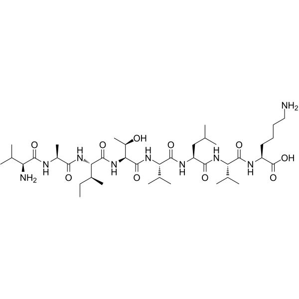
HY-P1077
CALP1Calmodulin Agonist -
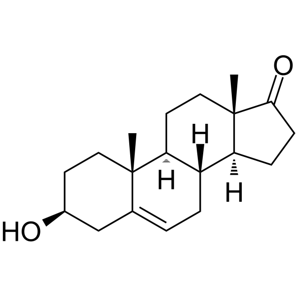
HY-14650
DHEASteroid Hormone -
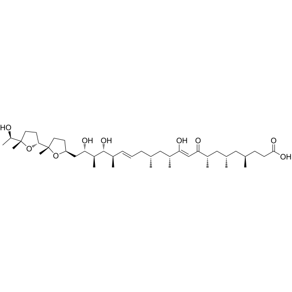
HY-13434
IonomycinCalcium Ionophore -
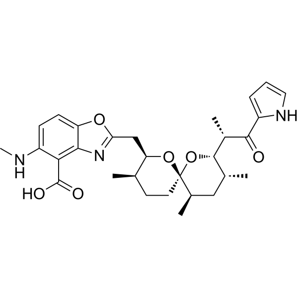
HY-N6687
CalcimycinCation Ionophore -
HY-W009724
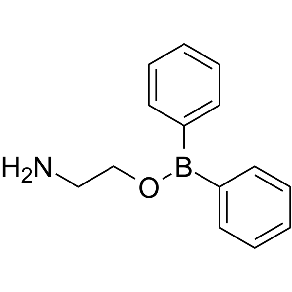
2-Aminoethyl diphenylborinateIP3R Inhibitor -
HY-13434A
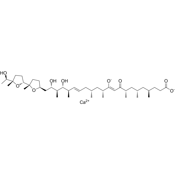
Ionomycin calciumCalcium Ionophore -
HY-B0317
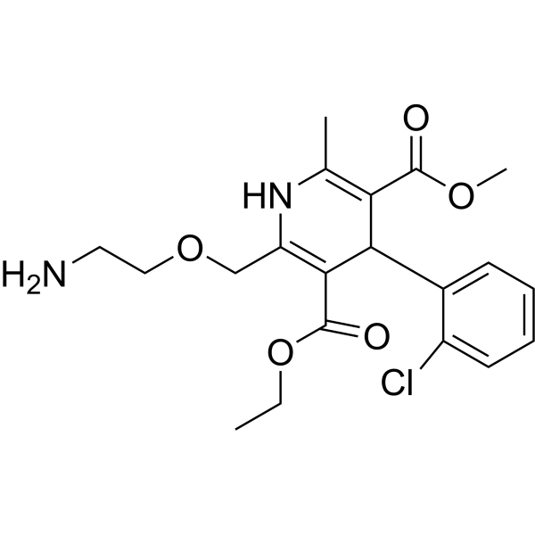
AmlodipineCalcium Channel Blocker
All Calcium Channel Isoforms
More

Biological Activity
Description
IC50 & Target
|
T-type calcium channel |
Nav1.2 104 nM (IC50) |
Nav1.6 21 nM (IC50) |
Nav1.7 95 nM (IC50) |
KV2.1 411 nM (IC50) |
TRPA1 389 nM (IC50) |
hNav1.5 358 nM (IC50) |
In Vitro
ProTx-I (0.01-10 μM; ~7-30 min) potently inhibits Ba2+ currents through hCav3.1-expressing Xenopus laevis oocytes with an IC50 of 0.20 μM, causes voltage-dependent inhibition and a small positive shift in activation potential, and exhibits partial to nearly irreversible binding[2].
ProTx-I (0.1-10 μM; ~2-7 min) weakly inhibits Ba2+ currents through hCav3.2-expressing Xenopus laevis oocytes with an IC50 of 31.8 μM, does not alter channel activation properties, and exhibits fully reversible binding[2].
ProTx-I (multiple concentrations) shows drastically reduced sensitivity when the domain IV S3-S4 linker of hCav3.1 is replaced with that of hCav3.2, resulting in an IC50 of 17.8 μM in hCav3.1/S3S4-expressing Xenopus laevis oocytes[2].
ProTx-I (multiple concentrations) shows increased sensitivity when the domain IV S3-S4 linker of hCav3.2 is replaced with that of hCav3.1, resulting in an IC50 of 15.8 μM in hCav3.2/S3S4-expressing Xenopus laevis oocytes[2].
ProTx-I (70 nM) inhibits heterologously expressed Cav3.1 voltage-gated calcium channels by shifting their voltage dependence of activation to more positive potentials, with an apparent IC50 of 70 nM during weak depolarizations[3].
MedChemExpress (MCE) has not independently confirmed the accuracy of these methods. They are for reference only.


Chemical Information
-
CAS No. 484598-35-8
-
Appearance Solid
-
Molecular Weight 3987.49
-
Formula C171H245N53O47S6
-
Color White to off-white
-
Sequence
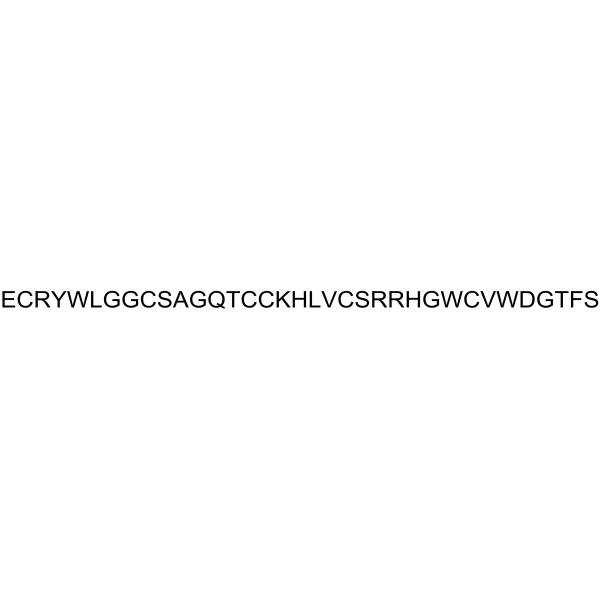
Glu-Cys-Arg-Tyr-Trp-Leu-Gly-Gly-Cys-Ser-Ala-Gly-Gln-Thr-Cys-Cys-Lys-His-Leu-Val-Cys-Ser-Arg-Arg-His-Gly-Trp-Cys-Val-Trp-Asp-Gly-Thr-Phe-Ser (Disulfide bridge:Cys2-Cys16;Cys9-Cys21;Cys15-Cys28)
-
Sequence Shortening
ECRYWLGGCSAGQTCCKHLVCSRRHGWCVWDGTFS (Disulfide bridge:Cys2-Cys16;Cys9-Cys21;Cys15-Cys28)
-
Shipping
Room temperature in continental US; may vary elsewhere.
-
Storage
Sealed storage, away from moisture
Powder -80°C 2 years -20°C 1 year * In solvent : -80°C, 6 months; -20°C, 1 month (sealed storage, away from moisture)


Solvent & Solubility
In Vitro:
DMSO : 25 mg/mL (6.27 mM; Need ultrasonic; Hygroscopic DMSO has a significant impact on the solubility of product, please use newly opened DMSO)
Please refer to the solubility information to select the appropriate solvent. Once prepared, please aliquot and store the solution to prevent product inactivation from repeated freeze-thaw cycles.
Storage method and period of stock solution: -80°C, 6 months; -20°C, 1 month (sealed storage, away from moisture). When stored at -80°C, please use it within 6 months. When stored at -20°C, please use it within 1 month.
Please refer to the solubility information to select the appropriate solvent. Once prepared, please aliquot and store the solution to prevent product inactivation from repeated freeze-thaw cycles.
Storage method and period of stock solution: -80°C, 6 months; -20°C, 1 month (sealed storage, away from moisture). When stored at -80°C, please use it within 6 months. When stored at -20°C, please use it within 1 month.
Concentration (start) × Volume (start) = Concentration (final) × Volume (final)


Purity & Documentation
-
Data Sheet (297 KB)
-
SDS (254 KB)
- English - EN (254 KB)
- Français - FR (254 KB)
- Deutsch - DE (254 KB)
- Norwegian - NO (254 KB)
- Español - ES (254 KB)
- Swedish - SV (254 KB)
- Italian - IT (254 KB)
- Korean - KR (254 KB)
- Portuguese - PT (254 KB)
-
Handling Instructions (2659 KB)
References
[1]. Rupasinghe DB, et al. Mutational analysis of ProTx-I and the novel venom peptide Pe1b provide insight into residues responsible for selective inhibition of the analgesic drug target NaV1.7. Biochem Pharmacol. 2020;181:114080. [Content Brief]
[2]. Ohkubo T, et al. Tarantula toxin ProTx-I differentiates between human T-type voltage-gated Ca2+ Channels Cav3.1 and Cav3.2. J Pharmacol Sci. 2010;112(4):452-458. [Content Brief]
[3]. Priest BT, et al. ProTx-I and ProTx-II: gating modifiers of voltage-gated sodium channels. Toxicon. 2007;49(2):194-201. [Content Brief]


Complete Stock Solution Preparation Table
Please refer to the solubility information to select the appropriate solvent. Once prepared, please aliquot and store the solution to prevent product inactivation from repeated freeze-thaw cycles.
Storage method and period of stock solution: -80°C, 6 months; -20°C, 1 month (sealed storage, away from moisture). When stored at -80°C, please use it within 6 months. When stored at -20°C, please use it within 1 month.
| Optional Solvent | Concentration Solvent Mass | 1 mg | 5 mg | 10 mg | 25 mg |
|---|---|---|---|---|---|
| DMSO | 1 mM | 0.2508 mL | 1.2539 mL | 2.5078 mL | 6.2696 mL |
| 5 mM | 0.0502 mL | 0.2508 mL | 0.5016 mL | 1.2539 mL |


ProTx-I Related Classifications
HY-P1073 Related Classifications
 Powered by Bioz