From 11:00 pm to 12:00 pm EST ( 8:00 pm to 9:00 pm PST ) on January 6th, the website will be under maintenance. We are sorry for the inconvenience. Please arrange your schedule properly.
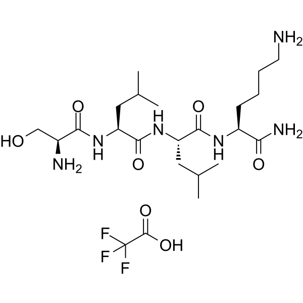
LSKL, Inhibitor of Thrombospondin (TSP-1) is a latency-associated protein (LAP)-TGFβ derived tetrapeptide and a competitive TGF-β1 antagonist. LSKL, Inhibitor of Thrombospondin (TSP-1) inhibits the binding of TSP-1 to LAP and alleviates renal interstitial fibrosis and hepatic fibrosis. LSKL, Inhibitor of Thrombospondin (TSP-1) suppresses subarachnoid fibrosis via inhibition of TSP-1-mediated TGF-β1 activity, prevents the development of chronic hydrocephalus and improves long-term neurocognitive defects following subarachnoid hemorrhage (SAH). LSKL, Inhibitor of Thrombospondin (TSP-1) can readily crosse the blood-brain barrier .
Thrombospondin (TSP-1)-derived CD36 binding motif is a bioactive hexapeptide. Thrombospondin (TSP-1)-derived CD36 binding motif interferes with the interaction between cells and the extracellular matrix by binding to CD36 and angiostatin, thereby affecting the cell adhesion and migration process. Thrombospondin (TSP-1)-derived CD36 binding motif inhibits platelet aggregation. Thrombospondin (TSP-1)-derived CD36 binding motif exerts an anti-tumor effect against colon cancer .
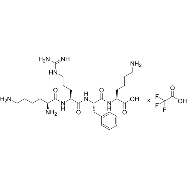
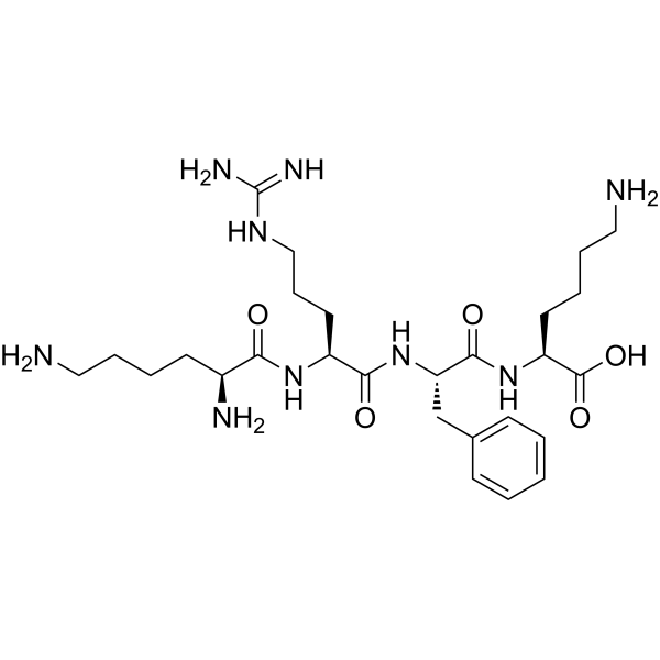
Thrombospondin-1 (1016-1023) (human, bovine, mouse), is the C-terminal end of the native sequence of Thrombospondin-1 (TSP-1), is a CD47 agonist peptide .
LSKL, Inhibitor of Thrombospondin (TSP-1) TFA is a latency-associated protein (LAP)-TGFβ derived tetrapeptide and a competitive TGF-β1 antagonist. LSKL, Inhibitor of Thrombospondin (TSP-1) TFA inhibits the binding of TSP-1 to LAP and alleviates renal interstitial fibrosis and hepatic fibrosis. LSKL, Inhibitor of Thrombospondin (TSP-1) TFA suppresses subarachnoid fibrosis via inhibition of TSP-1-mediated TGF-β1 activity, prevents the development of chronic hydrocephalus and improves long-term neurocognitive defects following subarachnoid hemorrhage (SAH). LSKL, Inhibitor of Thrombospondin (TSP-1) TFA can readily crosse the blood-brain barrier .
ABT-510 is an anti-angiogenic TSP peptide (Thrombospondin-1 analogue) that induces apoptosis and inhibits ovarian tumour growth in an orthotopic, syngeneic model of epithelial ovarian cancer. ABT-510 also reduces angiogenesis and inflammatory responses in a murine model of inflammatory bowel disease. ABT-510 can be used in studies of cancer (particularly epithelial ovarian cancer) and inflammatory bowel disease (IBD) .
KRFK TFA, a peptide derived from TSP-1, can activate TGF-β. KRFK TFA promotes TGF-β-mediated signaling and its downstream role, independent of thrombospondin (TSP) receptors such as CD47 and CD36. KRFK TFA can be used for chronic ocular surface inflammatory disorders reseach .
ABT-510 acetate is an anti-angiogenic TSP peptide (Thrombospondin-1 analogue) that induces apoptosis and inhibits ovarian tumour growth in an orthotopic, syngeneic model of epithelial ovarian cancer. ABT-510 acetate also reduces angiogenesis and inflammatory responses in a murine model of inflammatory bowel disease. ABT-510 acetate can be used in studies of cancer (particularly epithelial ovarian cancer) and inflammatory bowel disease (IBD) .
KRFK, a peptide derived from TSP-1, can activate TGF-β. KRFK promotes TGF-β-mediated signaling and its downstream role, independent of thrombospondin (TSP) receptors such as CD47 and CD36. KRFK can be used for chronic ocular surface inflammatory disorders reseach .
WAAG-3R TFA is a biological active peptide. (Aggrecanases belong to the ADAMTS (A disintegrin and metalloprotease with thrombospondin motif) family of proteases. Aggrecanases cleave aggrecan, the major structural component of cartilage. Aggrecanase-1 (ADAMTS-4) is a major aggrecanase in human osteoarthritic cartilage. This FRET peptide was used in an ADAMTS-4 (Aggrecanase-1) and ADAMTS-5 (Aggrecanase-2) assay . (Ex/Em = 340/420 nm)
Adamtsostatin 18 is an anti-angiogenic peptide derived from proteins containing type I thrombospondin motifs. Adamtsostatin 18 inhibits cell migration and proliferation .
Cys-PKHB1 (Cys-txCD47) TFA is a peptide conjugated to PKHB1, a CD47 agonist peptide and a thrombospondin-1 peptide mimic with antitumor effects. PKHB1 induces mitochondrial alterations, ROS generation, intracellular Ca + accumulation, and calcium-dependent cell death in breast cancer cells. PKHB1 induces immune system activation in breast cancer cells through immunogenic cell death .
CD36 Peptide P (93-110), Cys conjugated is a Cys labelled CD36 Peptide, and can block binding of CD36 to immobilized thrombospondin and partially inhibited collagen-induced platelet aggregation .
PKHB1 (txCD47) is a CD47 agonist and Thrombospondin-1 peptide mimetic. PKHB1 activates CD47 and triggers Caspase-independent, calcium-dependent cell death via mitochondrial alterations, ROS production, endoplasmic reticulum morphological changes, and dissipation of mitochondrial membrane potential. PKHB1 induces the exposure of Calreticulin, HSP70, and HSP90, thereby driving immunogenic cell death. PKHB1 promotes intratumoral CD8+ T cell infiltration and inhibits breast tumorigenesis. PKHB1 reduces HSV-1 levels and alleviates the severity of herpes simplex keratitis. PKHB1 can be used in research related to breast cancer, herpes simplex keratitis, and T-cell acute lymphoblastic leukemia .
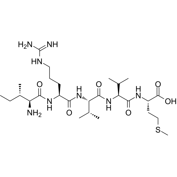
H-ILE-ARG-VAL-VAL-MET-OH is a pentapeptide from C7 with a domain that supports cell attachment. H-ILE-ARG-VAL-VAL-MET-OH is also the sequence fragment that binds to the thrombospondin-1 (TS1) receptor .
Cys-PKHB1 (Cys-txCD47) is a peptide conjugated to PKHB1, a CD47 agonist peptide and a thrombospondin-1 peptide mimic with antitumor effects. PKHB1 induces mitochondrial alterations, ROS generation, intracellular Ca + accumulation, and calcium-dependent cell death in breast cancer cells. PKHB1 induces immune system activation in breast cancer cells through immunogenic cell death .
SRI-31277 is a thrombospondin 1 (TSP-1) inhibitor with a human target pIC50 of 8.28 nM and in vivo activity, and exhibits a very short plasma half-life with intravenous administration.SRI-31277 inhibits interaction between TSP-1 and the latency-associated peptide (LAP) of latent TGF-β, blocking TSP-1-mediated TGF-β activation.SRI-31277 can be used for the research of multiple myeloma and fibrotic conditions .
SRI-35241 is a thrombospondinTSP-1 inhibitor with a pIC50 of 8.12 nM against human targets. SRI-35241 inhibits the binding of TSP-1 to latent transforming growth factor-β-associated peptide (LAP) and blocks the activation of TGF-β. SRI-35241 shows a certain time-dependent property, with a plasma half-life of 1.8 h after intravenous administration. SRI-35241 can be used for the research of multiple myeloma and fibrotic diseases .
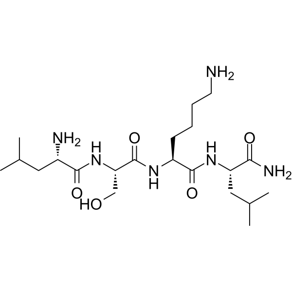
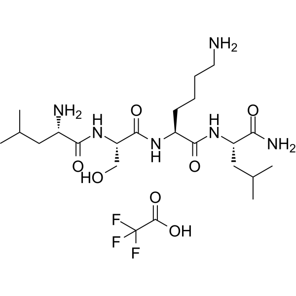
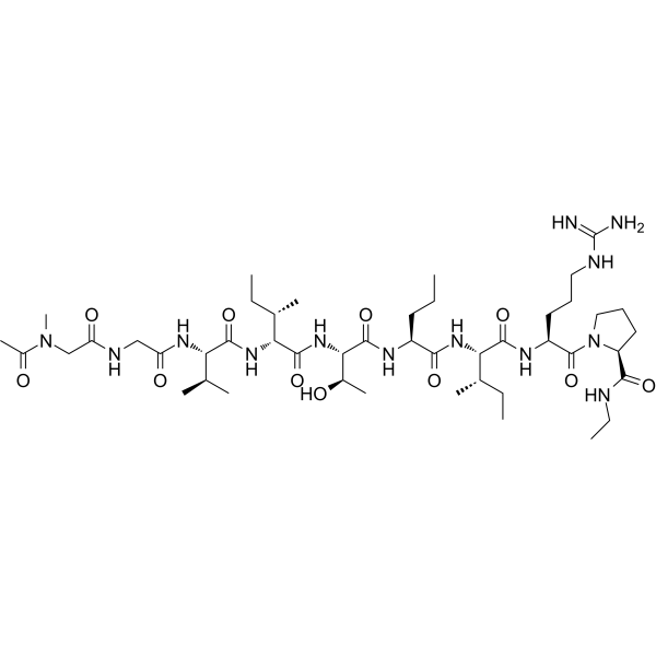
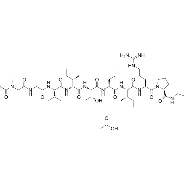
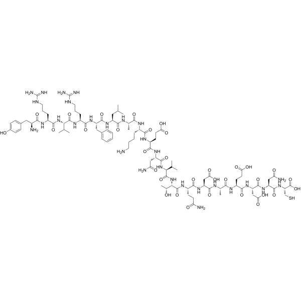
Lysyl-phenylalanyl-lysine (Compound KFK) is a tripeptide and belongs to the peptide segments related to thrombospondin-1 (TSP-1) (HY-P0299). Lysyl-phenylalanyl-lysine can activate LAP-TGF-β1 and release active TGF-β1, thereby inhibiting abnormal expression of MMP. Lysyl-phenylalanyl-lysine can be used for research on skin aging-related diseases and poor wound healing .
LSKL, Inhibitor of Thrombospondin (TSP-1) is a latency-associated protein (LAP)-TGFβ derived tetrapeptide and a competitive TGF-β1 antagonist. LSKL, Inhibitor of Thrombospondin (TSP-1) inhibits the binding of TSP-1 to LAP and alleviates renal interstitial fibrosis and hepatic fibrosis. LSKL, Inhibitor of Thrombospondin (TSP-1) suppresses subarachnoid fibrosis via inhibition of TSP-1-mediated TGF-β1 activity, prevents the development of chronic hydrocephalus and improves long-term neurocognitive defects following subarachnoid hemorrhage (SAH). LSKL, Inhibitor of Thrombospondin (TSP-1) can readily crosse the blood-brain barrier .
Thrombospondin (TSP-1)-derived CD36 binding motif is a bioactive hexapeptide. Thrombospondin (TSP-1)-derived CD36 binding motif interferes with the interaction between cells and the extracellular matrix by binding to CD36 and angiostatin, thereby affecting the cell adhesion and migration process. Thrombospondin (TSP-1)-derived CD36 binding motif inhibits platelet aggregation. Thrombospondin (TSP-1)-derived CD36 binding motif exerts an anti-tumor effect against colon cancer .
Thrombospondin-1 (1016-1023) (human, bovine, mouse), is the C-terminal end of the native sequence of Thrombospondin-1 (TSP-1), is a CD47 agonist peptide .
LSKL, Inhibitor of Thrombospondin (TSP-1) TFA is a latency-associated protein (LAP)-TGFβ derived tetrapeptide and a competitive TGF-β1 antagonist. LSKL, Inhibitor of Thrombospondin (TSP-1) TFA inhibits the binding of TSP-1 to LAP and alleviates renal interstitial fibrosis and hepatic fibrosis. LSKL, Inhibitor of Thrombospondin (TSP-1) TFA suppresses subarachnoid fibrosis via inhibition of TSP-1-mediated TGF-β1 activity, prevents the development of chronic hydrocephalus and improves long-term neurocognitive defects following subarachnoid hemorrhage (SAH). LSKL, Inhibitor of Thrombospondin (TSP-1) TFA can readily crosse the blood-brain barrier .
ABT-510 is an anti-angiogenic TSP peptide (Thrombospondin-1 analogue) that induces apoptosis and inhibits ovarian tumour growth in an orthotopic, syngeneic model of epithelial ovarian cancer. ABT-510 also reduces angiogenesis and inflammatory responses in a murine model of inflammatory bowel disease. ABT-510 can be used in studies of cancer (particularly epithelial ovarian cancer) and inflammatory bowel disease (IBD) .
KRFK TFA, a peptide derived from TSP-1, can activate TGF-β. KRFK TFA promotes TGF-β-mediated signaling and its downstream role, independent of thrombospondin (TSP) receptors such as CD47 and CD36. KRFK TFA can be used for chronic ocular surface inflammatory disorders reseach .
ABT-510 acetate is an anti-angiogenic TSP peptide (Thrombospondin-1 analogue) that induces apoptosis and inhibits ovarian tumour growth in an orthotopic, syngeneic model of epithelial ovarian cancer. ABT-510 acetate also reduces angiogenesis and inflammatory responses in a murine model of inflammatory bowel disease. ABT-510 acetate can be used in studies of cancer (particularly epithelial ovarian cancer) and inflammatory bowel disease (IBD) .
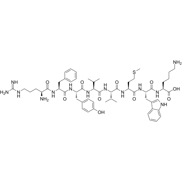
4N1K peptide is a biological active peptide. (4N1K-peptide (KRFYVVMWKK), which is derived from thrombospondins has anti-cancer activities in several cancers)
KRFK, a peptide derived from TSP-1, can activate TGF-β. KRFK promotes TGF-β-mediated signaling and its downstream role, independent of thrombospondin (TSP) receptors such as CD47 and CD36. KRFK can be used for chronic ocular surface inflammatory disorders reseach .
WAAG-3R TFA is a biological active peptide. (Aggrecanases belong to the ADAMTS (A disintegrin and metalloprotease with thrombospondin motif) family of proteases. Aggrecanases cleave aggrecan, the major structural component of cartilage. Aggrecanase-1 (ADAMTS-4) is a major aggrecanase in human osteoarthritic cartilage. This FRET peptide was used in an ADAMTS-4 (Aggrecanase-1) and ADAMTS-5 (Aggrecanase-2) assay . (Ex/Em = 340/420 nm)
Adamtsostatin 18 is an anti-angiogenic peptide derived from proteins containing type I thrombospondin motifs. Adamtsostatin 18 inhibits cell migration and proliferation .
Cys-PKHB1 (Cys-txCD47) TFA is a peptide conjugated to PKHB1, a CD47 agonist peptide and a thrombospondin-1 peptide mimic with antitumor effects. PKHB1 induces mitochondrial alterations, ROS generation, intracellular Ca + accumulation, and calcium-dependent cell death in breast cancer cells. PKHB1 induces immune system activation in breast cancer cells through immunogenic cell death .
Properdistatin is a peptide derived from the thrombospondin 1 (TSP-1) domain of properdin. Properdistatin inhibits angiogenesis and improves vascular function. Properdistatin has the potential for the research of melanoma .
CD36 Peptide P (93-110), Cys conjugated is a Cys labelled CD36 Peptide, and can block binding of CD36 to immobilized thrombospondin and partially inhibited collagen-induced platelet aggregation .
PKHB1 (txCD47) is a CD47 agonist and Thrombospondin-1 peptide mimetic. PKHB1 activates CD47 and triggers Caspase-independent, calcium-dependent cell death via mitochondrial alterations, ROS production, endoplasmic reticulum morphological changes, and dissipation of mitochondrial membrane potential. PKHB1 induces the exposure of Calreticulin, HSP70, and HSP90, thereby driving immunogenic cell death. PKHB1 promotes intratumoral CD8+ T cell infiltration and inhibits breast tumorigenesis. PKHB1 reduces HSV-1 levels and alleviates the severity of herpes simplex keratitis. PKHB1 can be used in research related to breast cancer, herpes simplex keratitis, and T-cell acute lymphoblastic leukemia .
H-ILE-ARG-VAL-VAL-MET-OH is a pentapeptide from C7 with a domain that supports cell attachment. H-ILE-ARG-VAL-VAL-MET-OH is also the sequence fragment that binds to the thrombospondin-1 (TS1) receptor .
Cys-PKHB1 (Cys-txCD47) is a peptide conjugated to PKHB1, a CD47 agonist peptide and a thrombospondin-1 peptide mimic with antitumor effects. PKHB1 induces mitochondrial alterations, ROS generation, intracellular Ca + accumulation, and calcium-dependent cell death in breast cancer cells. PKHB1 induces immune system activation in breast cancer cells through immunogenic cell death .
WAAG-3R is a biological active peptide. (Aggrecanases belong to the ADAMTS (A disintegrin and metalloprotease with thrombospondin motif) family of proteases. Aggrecanases cleave aggrecan, the major structural component of cartilage. Aggrecanase-1 (ADAMTS-4) is a major aggrecanase in human osteoarthritic cartilage. This FRET peptide was used in an ADAMTS-4 (Aggrecanase-1) and ADAMTS-5 (Aggrecanase-2) assay . (Ex/Em = 340/420 nm)
Thrombospondin-2 protein, also known as CD36 ligand, mediates anti-angiogenic properties. Thrombospondin-2 may regulate cell surface properties of mesenchymal cells and be involved in cell adhesion and migration. Thrombospondin-2 Protein, Mouse (His) is the recombinant mouse-derived Thrombospondin-2 protein, expressed by E. coli , with N-6*His labeled tag.
THBS2 protein is an extracellular matrix protein involved in multiple biological processes, including cell adhesion, migration, and angiogenesis. It plays a vital role in tissue remodeling and wound healing. Thrombospondin-2 Protein, Human (HEK293, His) is the recombinant human-derived Thrombospondin-2 protein, expressed by HEK293 , with C-10*His labeled tag. The total length of Thrombospondin-2 Protein, Human (HEK293, His) is 1154 a.a., with molecular weight of ~129.7 kDa.
Thrombospondin-2 protein, also known as CD36 ligand, mediates anti-angiogenic properties. Thrombospondin-2 may regulate cell surface properties of mesenchymal cells and be involved in cell adhesion and migration. Thrombospondin-2 Protein, Mouse (P.pastoris, His) is the recombinant mouse-derived Thrombospondin-2 protein, expressed by P. pastoris , with N-6*His labeled tag.
THSD1 Protein positively regulates nascent focal adhesion assembly, influencing endothelial cell attachment to the extracellular matrix. It forms a complex with PTK2/FAK1, TLN1, and VCL, interacting specifically with TLN1. THSD1 Protein, Human (HEK293, His) is the recombinant human-derived THSD1 protein, expressed by HEK293 , with C-6*His labeled tag.
THSD7A Protein orchestrates actin cytoskeleton rearrangement, actively influencing cellular dynamics. Its soluble form facilitates endothelial cell migration and filopodia formation in sprouting angiogenesis, relying on Focal Adhesion Kinase (FAK) activation. This emphasizes THSD7A's role in modulating key cellular processes associated with angiogenesis, highlighting its potential as a regulator of vascular development. THSD7A Protein, Human (HEK293, His) is the recombinant human-derived THSD7A protein, expressed by HEK293 , with C-His labeled tag.
The THBS1 protein is a member of the thrombospondin family and plays a key role in cell-matrix interactions, angiogenesis, and tissue remodeling.THBS1 is known for its multi-receptor binding, including integrins and CD36, regulating signaling pathways related to cell adhesion, migration and survival.THBS1 Protein, Mouse (His) is the recombinant mouse-derived THBS1 protein, expressed by E.coli , with N-6*His labeled tag.
The THBS1 protein is an adhesion glycoprotein that controls multiple cellular processes and mediates cell-cell and cell-matrix interactions. It plays multifunctional roles in inflammation, angiogenesis, wound healing, ROS and NO signaling, apoptosis, senescence, senescence, self-renewal, stemness, and cardiovascular/metabolic homeostasis. THBS1 Protein, Human (HEK293, His) is the recombinant human-derived THBS1 protein, expressed by HEK293 , with C-10*His labeled tag.
The COMP protein is critical for cartilage integrity and interacts with extracellular matrix proteins to promote the connection of chondrocytes to the cartilage matrix through integrin receptors. Its role in the pathogenesis of osteoarthritis emphasizes the significance of joint health. COMP Protein, Human (HEK293, His) is the recombinant human-derived COMP protein, expressed by HEK293 , with C-His labeled tag.
THSD1 protein actively regulates the assembly of nascent focal adhesions and affects the attachment of endothelial cells to the extracellular matrix. As a key player, THSD1 forms a complex with PTK2/FAK1, TLN1 and VCL and contributes to focal adhesion dynamics. THSD1 Protein, Mouse (HEK293, His) is the recombinant mouse-derived THSD1 protein, expressed by HEK293 , with C-His labeled tag.
CD36 is a multifunctional glycoprotein that serves as a receptor for a variety of ligands, including thrombospondin and oxidized low-density lipoprotein. Ligand induces CD36 clusters, initiating signal transduction and internalization. CD36 Protein, Human (HEK293, Fc ) is the recombinant human-derived CD36 protein, expressed by HEK293 , with C-hFc labeled tag.
The ADAMTS13 protein, a metalloproteinase, has a metalloproteinase domain, a disintegrin-like domain, and a thrombospondin type 1 motif. It cleaves von Willebrand Factor and defects in this gene cause thrombotic thrombocytopenic purpura. Alternative splicing generates multiple transcript variants. ADAMTS13 is broadly expressed in various tissues, including the liver, testis, and 24 other tissues. ADAMTS13 Protein, Human (CHO, His) is the recombinant human-derived ADAMTS13, expressed by CHO, with C-10*His labeled tag. The total length of ADAMTS13 Protein, Human (CHO, His) is 655 a.a..
RSPO3 is a potent activator of the canonical Wnt pathway and can bind to LGR4-6 receptors to initiate a complex with phosphorylated LRP6 and Frizzled receptors. This interaction activates the canonical Wnt pathway and upregulates target genes. RSPO3/R-spondin-3 Protein, Human (HEK293, Fc-His) is the recombinant human-derived RSPO3/R-spondin-3 protein, expressed by HEK293 , with C-hFc, C-6*His labeled tag.
CD36 is a multifunctional glycoprotein that serves as a receptor for a variety of ligands, including thrombospondin and oxidized low-density lipoprotein. Ligand induces CD36 clusters, initiating signal transduction and internalization. CD36 Protein, Human (HEK293, His-Avi) is the recombinant human-derived CD36 protein, expressed by HEK293 , with C-Avi, C-His labeled tag.
A disintegrin and metalloproteinase with thrombospondin motifs 5; A disintegrin like and metalloprotease (reprolysin type) with thrombospondin type 1 motif 5; A Disintigrin And Metalloproteinase with thrombospondin motif-5; ADAM metallopeptidase with thrombospondin type 1 motif 5; ADAM TS 11; ADAM TS 5; ADAM TS5; ADAMTS 11; ADAMTS 5; ADAMTS11; ADMP 2; ADMP2; Aggrecanase 2; aggrecanase-2; FLJ36738; Implantin; thrombospondin motif-5.
WB, ICC/IF, FC
Human, Mouse, Rat
ADAMTS5 Antibody (YA3480) is a Rabbit-derived and non-conjugated IgG monoclonal antibody, targeting to ADAMTS5.
Western blot analysis of extracts from THP-1(lane 2(20μg), Jurkat (lane 3(20μg) and NIH3T3(lane 4(20μg) using FOXO1A (HY-P80132) Rabbit mAb. Proteins were transferred
to a PVDF membrane and blocked with 5% non-fat milk in TBST for 2 hour at room temperature. The primary antibody (1/1000) and Loading control antibody (Beta Actin, HY-P80438, 1/10000) was
used in 5% non-fat milk in TBST at 4°C overnight. Goat Anti-Mouse/Rabbit IgG-HRP Secondary Antibody (1/10000) was used for 1 hour at room temperature.
Western blot analysis of extracts from THP-1(lane 2(20μg), Jurkat (lane 3(20μg) and NIH3T3(lane 4(20μg) using FOXO1A (HY-P80132) Rabbit mAb. Proteins were transferred
to a PVDF membrane and blocked with 5% non-fat milk in TBST for 2 hour at room temperature. The primary antibody (1/1000) and Loading control antibody (Beta Actin, HY-P80438, 1/10000) was
MedchemExpress Validation 03
Western blot analysis of extracts from THP-1(lane 2(20μg), Jurkat (lane 3(20μg) and NIH3T3(lane 4(20μg) using FOXO1A (HY-P80132) Rabbit mAb. Proteins were transferred
MedchemExpress Validation 04
Western blot analysis of extracts from THP-1(lane 2(20μg), Jurkat (lane 3(20μg) and NIH3T3(lane 4(20μg) using FOXO1A (HY-P80132) Rabbit mAb. Proteins were transferred
to a PVDF membrane and blocked with 5% non-fat milk in TBST for 2 hour at room temperature. The primary antibody (1/1000) and Loading control antibody (Beta Actin, HY-P80438, 1/10000) was
used in 5% non-fat milk in TBST at 4°C overnight. Goat Anti-Mouse/Rabbit IgG-HRP Secondary Antibody (1/10000) was used for 1 hour at room temperature.
MedchemExpress Validation
Western blot analysis of extracts from THP-1(lane 2(20μg), Jurkat (lane 3(20μg) and NIH3T3(lane 4(20μg) using FOXO1A (HY-P80132) Rabbit mAb. Proteins were transferred
to a PVDF membrane and blocked with 5% non-fat milk in TBST for 2 hour at room temperature. The primary antibody (1/1000) and Loading control antibody (Beta Actin, HY-P80438, 1/10000) was
used in 5% non-fat milk in TBST at 4°C overnight. Goat Anti-Mouse/Rabbit IgG-HRP Secondary Antibody (1/10000) was used for 1 hour at room temperature.
MedchemExpress Validation
Western blot analysis of extracts from THP-1(lane 2(20μg), Jurkat (lane 3(20μg) and NIH3T3(lane 4(20μg) using FOXO1A (HY-P80132) Rabbit mAb. Proteins were transferred
to a PVDF membrane and blocked with 5% non-fat milk in TBST for 2 hour at room temperature. The primary antibody (1/1000) and Loading control antibody (Beta Actin, HY-P80438, 1/10000) was
used in 5% non-fat milk in TBST at 4°C overnight. Goat Anti-Mouse/Rabbit IgG-HRP Secondary Antibody (1/10000) was used for 1 hour at room temperature.
MedChemExpress values your privacy and your trust is important to us. We use cookies to enhance your website experience. Some cookies are necessary to run the website.
Privacy and Cookie Policy