



COMP Protein, Human (HEK293, His)
Based on 2 publication(s) in Google Scholar
The COMP protein is critical for cartilage integrity and interacts with extracellular matrix proteins to promote the connection of chondrocytes to the cartilage matrix through integrin receptors. Its role in the pathogenesis of osteoarthritis emphasizes the significance of joint health. COMP Protein, Human (HEK293, His) is the recombinant human-derived COMP protein, expressed by HEK293 , with C-His labeled tag.
- Species: Human
- Source: HEK293
-
Storage:Stored at -20°C for 2 years from date of receipt. After reconstitution, it is stable at 4°C for 1 week or -20°C for longer (with carrier protein). It is recommended to freeze aliquots at -20°C or -80°C for extended storage.


To place orders, for customer services and technical support, please contact: MedChemExpress USA
Tel: 609-228-6898 E-mail: [email protected] [email protected]


-
 Biological Activity
-
Technical Parameters
-
Product Properties
- Documentation
- Help & FAQs


-

HY-P70030
CHI3L1 Protein, Human (HEK293, His) -
HY-P70780A
GAS6 Protein, Human (HEK293, His) -
HY-P75951
GPNMB/Osteoactivin Protein, Human (Isoform 2, HEK293, Fc) -
HY-P73414
SLPI Protein, Human (sf9, His) -
HY-P70726
IGFBP-7 Protein, Human (HEK293, His) -
HY-P702847
MGP Protein, Human (His) -
HY-P70055
CFHR1 Protein, Human (HEK293, His) -
HY-P71067
Pentraxin 2/SAP Protein, Human (HEK293, His) -
HY-P78009
Notch 3 Protein, Human (HEK293, Fc)


Biological Activity
Description
The COMP protein is critical for cartilage integrity and interacts with extracellular matrix proteins to promote the connection of chondrocytes to the cartilage matrix through integrin receptors. Its role in the pathogenesis of osteoarthritis emphasizes the significance of joint health. COMP Protein, Human (HEK293, His) is the recombinant human-derived COMP protein, expressed by HEK293 , with C-His labeled tag.
Background
The COMP protein assumes a crucial role in preserving the structural integrity of cartilage by interacting with other extracellular matrix proteins, including collagens and fibronectin. It facilitates the connection between chondrocytes and the cartilage extracellular matrix by engaging with cell surface integrin receptors. Additionally, COMP's involvement in the pathogenesis of osteoarthritis underscores its potential significance in joint health. Beyond its structural role, COMP emerges as a potent suppressor of apoptosis in both primary chondrocytes and transformed cells. Its anti-apoptotic effects are mediated by inhibiting caspase-3 activation and inducing the IAP family of survival proteins (BIRC3, BIRC2, BIRC5, and XIAP). Furthermore, COMP plays an essential role in maintaining the contractile and differentiated phenotype of vascular smooth muscle cells under both physiological and pathological stimuli, achieved through its interaction with ITGA7. This multifaceted functionality positions COMP as a key player in the maintenance of cartilage structure, joint health, and cellular survival processes.
Verified Bioactivity
1. Measured by its ability to induce adhesion of ATDC5 mouse chondrogenic cells.When cells are added to COMP-coated plates (0.625 μg/mL and 100 μL/well), approximately ≥30% cells will adhere specifically after 60 minutes at 37°C.
2. Measured by the ability of the immobilized protein to support the adhesion of ATDC5 cells. When 5 x 10⁴ cells/well are added to Recombinant Human COMP coated plates (0.625µg/mL, 100 µL/well), 48-60% cells will adhere after 1 hour incubation.
MCE Validation Data
-
Purity - SDS-PAGE
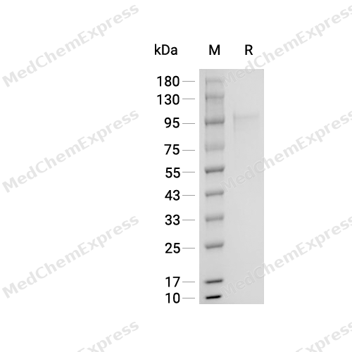
Purity - SDS-PAGE


Publications (2)
-
Journal Impact Factor
-
Most Recent
-
Cell
Comprehensive human proteome profiles across a 50-year lifespan reveal aging trajectories and signatures. [Abstract]2025 Oct 2;188(20):5763-5784.e26. PMID: 40713952 -
Cell Signal
COMP promotes pancreatic fibrosis by activating pancreatic stellate cells through CD36-ERK/AKT signaling pathways. [Abstract]2024 Jun:118:111135. PMID: 38479555


Technical Parameters
-
Species Human
-
Source HEK293
-
Tag C-His
-
Accession
P49747-1 (Q21-A757)
-
Molecular Construction
-
N-term
-
COMP (Q21-A757)
Accession # P49747-1 -
His
-
C-term
-
-
Protein Length
Full Length of Isoform-1
-
Synonyms
COMP; TSP-5; Prev. PSACH; Cartilage Oligomeric Matrix Protein (Pseudoachondroplasia, Epiphyseal Dysplasia 1, Multiple); Prev. EDM1; Multiple Epiphyseal Dysplasia; Prev. EPD1; Pseudoachondroplasia (Epiphyseal Dysplasia 1, Multiple); TSP5; Alternative Prote
-
AA Sequence
QGQSPLGSDLGPQMLRELQETNAALQDVRELLRQQVREITFLKNTVMECDACGMQQSVRTGLPSVRPLLHCAPGFCFPGVACIQTESGARCGPCPAGFTGNGSHCTDVNECNAHPCFPRVRCINTSPGFRCEACPPGYSGPTHQGVGLAFAKANKQVCTDINECETGQHNCVPNSVCINTRGSFQCGPCQPGFVGDQASGCQRRAQRFCPDGSPSECHEHADCVLERDGSRSCVCAVGWAGNGILCGRDTDLDGFPDEKLRCPERQCRKDNCVTVPNSGQEDVDRDGIGDACDPDADGDGVPNEKDNCPLVRNPDQRNTDEDKWGDACDNCRSQKNDDQKDTDQDGRGDACDDDIDGDRIRNQADNCPRVPNSDQKDSDGDGIGDACDNCPQKSNPDQADVDHDFVGDACDSDQDQDGDGHQDSRDNCPTVPNSAQEDSDHDGQGDACDDDDDNDGVPDSRDNCRLVPNPGQEDADRDGVGDVCQDDFDADKVVDKIDVCPENAEVTLTDFRAFQTVVLDPEGDAQIDPNWVVLNQGREIVQTMNSDPGLAVGYTAFNGVDFEGTFHVNTVTDDDYAGFIFGYQDSSSFYVVMWKQMEQTYWQANPFRAVAEPGIQLKAVKSSTGPGEQLRNALWHTGDTESQVRLLWKDPRNVGWKDKKSYRWFLQHRPQVGYIRVRFYEGPELVADSNVVLDTTMRGGRLGVFCFSQENIIWANLRYRCNDTIPEDYETHQLRQA
-
Molecular Weight
Approximately 120-130 kDa, based on SDS-PAGE under reducing conditions, due to the glycosylation.
-
Glycosylation
Yes
-
Purity
≥ 90%, as determined by reducing SDS-PAGE.


Product Properties
Lyophilized powder
1.Lyophilized from a 0.22 μm filtered solution of PBS, pH 7.4, 5% trehalose, 5% mannitol and 0.01% Tween 80.
2.Lyophilized from a 0.22 μm filtered solution of PBS, pH 7.4, 8% trehalose.
Please refer to the lot-specific COA for specific buffer information.
<1 EU/μg, determined by LAL method.
It is not recommended to reconstitute to a concentration less than 100 μg/mL in ddH2O.
Stored at -20°C for 2 years from date of receipt. After reconstitution, it is stable at 4°C for 1 week or -20°C for longer (with carrier protein). It is recommended to freeze aliquots at -20°C or -80°C for extended storage.
Room temperature in continental US; may vary elsewhere.


Documentation
-
Data Sheet (240 KB)
-
SDS (252 KB)
- English - EN (252 KB)
- Français - FR (252 KB)
- Deutsch - DE (252 KB)
- Norwegian - NO (252 KB)
- Español - ES (252 KB)
- Swedish - SV (252 KB)
- Italian - IT (252 KB)
- Korean - KR (252 KB)
- Portuguese - PT (252 KB)
-
Handling Instructions (2659 KB)

Calculators
Concentration (start) × Volume (start) = Concentration (final) × Volume (final)


 Powered by Bioz

