



CD36 Antibody (YA1538)
(Synonyms: CD36; GP3B; GP4; Platelet glycoprotein 4; Fatty acid translocase; FAT; Glycoprotein IIIb; GPIIIB; Leukocyte differentiation antigen CD36; PAS IV; PAS-4; Platelet collagen receptor; Platelet glycoprotein IV; GPIV; Thrombospondin receptor; CD36)Based on 2 publication(s) in Google Scholar
CD36 Antibody (YA1538) is a Rabbit-derived and non-conjugated IgG monoclonal antibody, targeting to CD36.
-
Host:
Rabbit
-
Isotype:
IgG
-
Application:
WB, IHC-P, IP
-
Reactivity :
Human, Mouse, Rat
-
Formulation:
Supplied in Rabbit IgG in PBS(pH 7.4), 150mM NaCl, 50% Glycerol, 0.02% Sodium azide and 0.05% BSA.
-
Conjugation:
Non-conjugated


To place orders, for customer services and technical support, please contact: MedChemExpress USA
Tel: 609-228-6898 E-mail: [email protected] [email protected]



Publications Citing Use of MedChemExpress (MCE) CD36 Antibody (YA1538)
More
 2 Publications Citing Use of MCE CD36 Antibody (YA1538) (1)
-
WB


Applications
| Application |
WB

WB: Western Blot
|
IP
IP: Immunoprecipitation
|
IHC
|
|---|---|---|---|
| Dilution Ratio | 1:500-1:1000 | 1:50 | 1:50-1:1000 |


Product Details
CD36 Antibody (YA1538) is a Rabbit-derived and non-conjugated IgG monoclonal antibody, targeting to CD36.
-
Host Rabbit

-
Clonality Recombinant,Monoclonal

-
Species ReactivityHuman, Mouse, Rat
-
Observed Molecular WeightObserved band size: 70-100 kDa
Note: Due to possible protein modifications or aggregation, the molecular weight should be confirmed by actual measurement, and the predicted value is for reference only.

-
Calculated Molecular Weight Predicted band size: 53 kDa

Entrez Gene: 948 Human ; 12491 Mouse
SwissProt: P16671 Human ; Q08857 Mouse ; Q07969 Rat
OMIM: 608404 Human
A synthesized peptide derived from human CD36 aa350-400.
Endogenous
Affinity Chromatography
Non-conjugated
Unmodified
IgG


Product Properties
-
Appearance
Solution
-
Formulation
Supplied in Rabbit IgG in PBS(pH 7.4), 150mM NaCl, 50% Glycerol, 0.02% Sodium azide and 0.05% BSA.
-
Concentration
Batch-dependent, Please check the COA for the concentration of each lot. Check Lot Concentration
-
Storage & Stability
Stored at -20°C for 1 year. Avoid repeated freeze / thaw cycles.
-
Shipping
Shipping with blue ice.


Publications (2)
-
Journal Impact Factor
-
Most Recent
-
Adipocyte
2025 Dec;14(1):2549456. PMID: 41381093
CD36 Antibody (YA1538) purchased from MedChemExpress. Usage Cited in: Adipocyte. 2025 Dec;14(1):2549456. [Abstract]
Western blot results of CD36, ATGL, CAPG, and GLUT4 protein levels in sh-Ctrl and sh-Capg cells treated with vehicle control (NC) or 400 μM linoleic acid (LA).


Verification Images
-
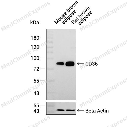 Western blot analysis was performed on extracts from Mouse brown adipose (lane 1, 15 μg), and Rat brown adipose (lane 2, 15 μg) using CD36 Rabbit mAb.Proteins were transferred to a PVDF membrane and blocked with 5% non-fat milk in TBST at 4°C overnight.The primary antibody (1:1000 dilution) and the loading control antibody (beta-Actin, HY-P83730, 1:20000 dilution) were incubated in 5% non-fat milk in TBST for 1 hour at 37°C.Goat Anti-Rabbit IgG-HRP Secondary Antibody (1:20000 dilution) was then applied for 40 minutes at 37°C.
 -
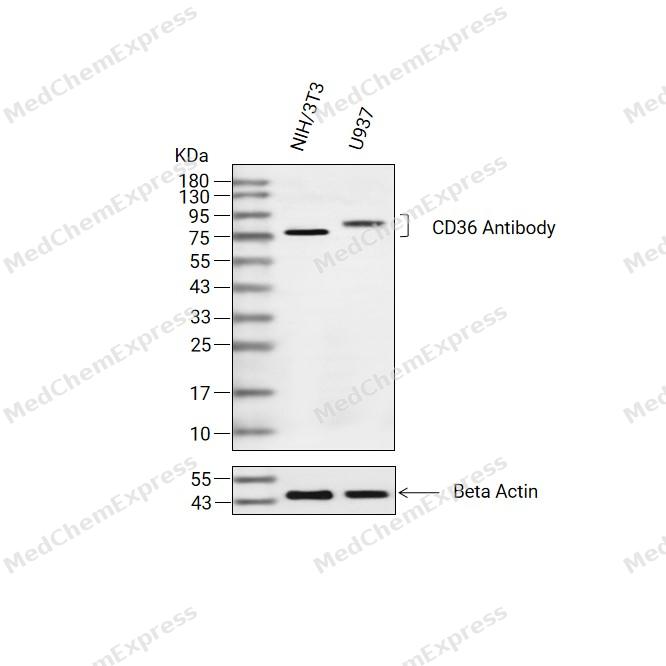 Western blot analysis of extracts from NIH/3T3(lane 2(20ug) and U937(lane 3(20ug) using CD36 Antibody (HY-P81793) Rabbit mAb. Proteins were transferred to a PVDF membrane and blocked with 5% non-fat milk in TBST for 2 hour at room temperature. The primary antibody (1/1000) and Loading control antibody (Beta Actin, HY-P83730, 1/10000) was used in 5% non-fat milk in TBST at 4°C overnight. Goat Anti-Mouse/Rabbit IgG-HRP Secondary Antibody (1/10000) was used for 1 hour at room temperature.
 -
 Immunohistochemical analysis of paraffin-embedded mouse small intestine tissue using CD36 Antibody (HY-P81793, 1/500). The section was pretreated using heat mediated antigen retrieval with sodium citrate buffer (pH 6.0) for 8 minutes. The tissues were blocked with quick block buffer for 0.5 hours at room temperature, washed with PBS and PBST, and then incubated with the primary antibody overnight at 4℃. The detection was performed using an HRP conjugated compact polymer system. DAB was used as the chromogen. Tissues were counterstained with hematoxylin and mounted with DPX.
 -
 Immunohistochemical analysis of paraffin-embedded mouse duodenum tissue using CD36 Antibody (HY-P81793, 1/500). The section was pretreated using heat mediated antigen retrieval with sodium citrate buffer (pH 6.0) for 8 minutes. The tissues were blocked with quick block buffer for 0.5 hours at room temperature, washed with PBS and PBST, and then incubated with the primary antibody overnight at 4℃. The detection was performed using an HRP conjugated compact polymer system. DAB was used as the chromogen. Tissues were counterstained with hematoxylin and mounted with DPX.
 -
 Immunohistochemical analysis of paraffin-embedded mouse epididymis tissue using CD36 Antibody (HY-P81793, 1/500). The section was pretreated using heat mediated antigen retrieval with sodium citrate buffer (pH 6.0) for 8 minutes. The tissues were blocked with quick block buffer for 0.5 hours at room temperature, washed with PBS and PBST, and then incubated with the primary antibody overnight at 4℃. The detection was performed using an HRP conjugated compact polymer system. DAB was used as the chromogen. Tissues were counterstained with hematoxylin and mounted with DPX.
 -
 Immunohistochemical analysis of paraffin-embedded mouse colon tissue using CD36 Antibody (HY-P81793, 1/500). The section was pretreated using heat mediated antigen retrieval with sodium citrate buffer (pH 6.0) for 8 minutes. The tissues were blocked with quick block buffer for 0.5 hours at room temperature, washed with PBS and PBST, and then incubated with the primary antibody overnight at 4℃. The detection was performed using an HRP conjugated compact polymer system. DAB was used as the chromogen. Tissues were counterstained with hematoxylin and mounted with DPX.
 -
 Immunohistochemical analysis of paraffin-embedded mouse rectum tissue using CD36 Antibody (HY-P81793, 1/500). The section was pretreated using heat mediated antigen retrieval with sodium citrate buffer (pH 6.0) for 8 minutes. The tissues were blocked with quick block buffer for 0.5 hours at room temperature, washed with PBS and PBST, and then incubated with the primary antibody overnight at 4℃. The detection was performed using an HRP conjugated compact polymer system. DAB was used as the chromogen. Tissues were counterstained with hematoxylin and mounted with DPX.
 -
 Immunohistochemical analysis of paraffin-embedded rat small intestine tissue using CD36 Antibody (HY-P81793, 1/500). The section was pretreated using heat mediated antigen retrieval with sodium citrate buffer (pH 6.0) for 8 minutes. The tissues were blocked with quick block buffer for 0.5 hours at room temperature, washed with PBS and PBST, and then incubated with the primary antibody overnight at 4℃. The detection was performed using an HRP conjugated compact polymer system. DAB was used as the chromogen. Tissues were counterstained with hematoxylin and mounted with DPX.


Background
-
Function
CD36 multifunctional glycoprotein that acts as a receptor for a broad range of ligands. Ligands can be of proteinaceous nature like thrombospondin, fibronectin, collagen or amyloid-beta as well as of lipidic nature such as oxidized low-density lipoprotein (oxLDL), anionic phospholipids, long-chain fatty acids and bacterial diacylated lipopeptides. They are generally multivalent and can therefore engage multiple receptors simultaneously, the resulting formation of CD36 clusters initiates signal transduction and internalization of receptor-ligand complexes. The dependency on coreceptor signaling is strongly ligand specific. Cellular responses to these ligands are involved in angiogenesis, inflammatory response, fatty acid metabolism, taste and dietary fat processing in the intestine (Probable). Binds long-chain fatty acids and facilitates their transport into cells, thus participating in muscle lipid utilization, adipose energy storage, and gut fat absorption. Mechanistically, binding of fatty acids activates downstream kinase LYN, which phosphorylates the palmitoyltransferase ZDHHC5 and inactivates it, resulting in the subsequent depalmitoylation of CD36 and caveolar endocytosis. In the small intestine, plays a role in proximal absorption of dietary fatty acid and cholesterol for optimal chylomicron formation, possibly through the activation of MAPK1/3 (ERK1/2) signaling pathway. Involved in oral fat perception and preferences. Detection into the tongue of long-chain fatty acids leads to a rapid and sustained rise in flux and protein content of pancreatobiliary secretions. In taste receptor cells, mediates the induction of an increase in intracellular calcium levels by long-chain fatty acids, leading to the activation of the gustatory neurons in the nucleus of the solitary tract. Important factor in both ventromedial hypothalamus neuronal sensing of long-chain fatty acid and the regulation of energy and glucose homeostasis. Receptor for thrombospondins, THBS1 and THBS2, mediating their antiangiogenic effects. Involved in inducing apoptosis in podocytes in response to elevated free fatty acids, acting together with THBS1. As a coreceptor for TLR4:TLR6 heterodimer, promotes inflammation in monocytes/macrophages. Upon ligand binding, such as oxLDL or amyloid-beta 42, interacts with the heterodimer TLR4:TLR6, the complex is internalized and triggers inflammatory response, leading to NF-kappa-B-dependent production of CXCL1, CXCL2 and CCL9 cytokines, via MYD88 signaling pathway, and CCL5 cytokine, via TICAM1 signaling pathway, as well as IL1B secretion, through the priming and activation of the NLRP3 inflammasome. Selective and nonredundant sensor of microbial diacylated lipopeptide that signal via TLR2:TLR6 heterodimer, this cluster triggers signaling from the cell surface, leading to the NF-kappa-B-dependent production of TNF, via MYD88 signaling pathway and subsequently is targeted to the Golgi in a lipid-raft dependent pathway; (Microbial infection) Directly mediates cytoadherence of Plasmodium falciparum parasitized erythrocytes and the internalization of particles independently of TLR signaling[1][2][3][4][5][6][7][8].
-
Subcellular Localization
Cell membrane; Multi-pass membrane protein; Membrane raft; Golgi apparatus; Apical cell membrane
-
Isoforms & Post-Translational Modification
P16671 has 4 isomers: P16671-1: 53053 Da (predicted); P16671-2: 32265 Da (predicted); P16671-3: 48600 Da (predicted); P16671-4: 46090 Da (predicted).
N-glycosylated and O-glycosylated with a ratio of 2:1;Palmitoylated by ZDHHC5 (PubMed:32958780). Palmitoylation is required for proper localization at the plasma membrane (PubMed:32958780, PubMed:37461827);Ubiquitinated at Lys-469 and Lys-472. Ubiquitination is induced by fatty acids such as oleic acid and leads to degradation by the proteasome (PubMed:18353783, PubMed:21610069). Ubiquitination and degradation are inhibited by insulin which blocks the effect of fatty acids (PubMed:18353783) -
Subunit
Interacts with THBS1 and THBS2; the interactions mediate the THBS antiangiogenic activity (PubMed:10613822, PubMed:1371676). Upon interaction with a ligand, such as oxidized low-density lipoprotein (oxLDL) or amyloid-beta 42, rapidly forms a complex with TLR4 and TLR6; the complex is internalized and triggers an inflammatory signal. Through its C-terminus, interacts with PTK2, PXN and LYN, but not with SRC. LYN kinase activity is required for facilitating TLR4:TLR6 heterodimerization and signal initiation (PubMed:1371676, PubMed:20037584). Upon interaction with ligands such as diacylated lipopeptides, interacts with the TLR2:TLR6 heterodimer (PubMed:16880211). Interacts with CD9, CD81, FCER1G, ITGB2 and/or ITGB2; forming a membrane heteromeric complex required for the internalization of CD36 and its ligands (By similarity). Interacts (when palmitoylated) with ARF6; this interaction mediates CD36 transport to the plasma membrane (PubMed:37461827)
-
SwissProt ID
-
Synonyms
CD36; GP3B; GP4; Platelet glycoprotein 4; Fatty acid translocase; FAT; Glycoprotein IIIb; GPIIIB; Leukocyte differentiation antigen CD36; PAS IV; PAS-4; Platelet collagen receptor; Platelet glycoprotein IV; GPIV; Thrombospondin receptor; CD36
-
Research Field
Cardiovascular
Documentation
-
Data Sheet (234 KB)
-
SDS (252 KB)
- English - EN (252 KB)
- Français - FR (252 KB)
- Deutsch - DE (252 KB)
- Norwegian - NO (252 KB)
- Español - ES (252 KB)
- Swedish - SV (252 KB)
- Italian - IT (252 KB)
- Korean - KR (252 KB)
- Portuguese - PT (252 KB)
-
User Guide for Antibodies (1077 KB)

 Powered by Bioz
