Search Result
Results for "
mitochondrial probe
" in MedChemExpress (MCE) Product Catalog:
1
Biochemical Assay Reagents
1
Isotope-Labeled Compounds
| Cat. No. |
Product Name |
Target |
Research Areas |
Chemical Structure |
-
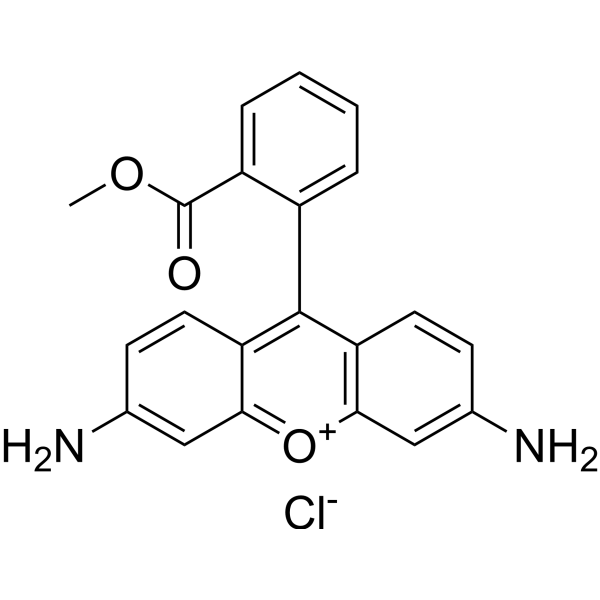
- HY-15534
-
JC-1
Maximum Cited Publications
212 Publications Verification
CBIC2
|
Fluorescent Dye
|
Others
|
|
JC-1 (CBIC2) is an ideal fluorescent probe widely used to detect mitochondrial membrane potential. JC-1 accumulates in mitochondria in a potential dependent manner and can be used to detect the membrane potential of cells, tissues or purified mitochondria. In normal mitochondria, JC-1 aggregates in the mitochondrial matrix to form a polymer, which emits strong red fluorescence (Ex=585 nm, Em=590 nm); When the mitochondrial membrane potential is low, JC-1 cannot aggregate in the matrix of mitochondria and produce green fluorescence (ex=510 nm, em= 527 nm) .
|
-
-
- HY-D0985A
-
|
Tetramethylrhodamine ethyl ester perchlorate
|
Fluorescent Dye
|
Others
|
|
Rhodamine dyes are membrane-permeable cationic fluorescent probes that specifically recognize mitochondrial membrane potentials, thereby attaching to mitochondria and producing bright fluorescence, and at certain concentrations, rhodamine dyes have low toxicity to cells, so they are commonly used to detect mitochondria in animal cells, plant cells, and microorganisms .
|
-
-
- HY-D0984A
-
|
T668
|
Fluorescent Dye
|
Others
|
|
Rhodamine dyes are membrane-permeable cationic fluorescent probes that specifically recognize mitochondrial membrane potentials, thereby attaching to mitochondria and producing bright fluorescence, and at certain concentrations, rhodamine dyes have low toxicity to cells, so they are commonly used to detect mitochondria in animal cells, plant cells, and microorganisms .
|
-
-
- HY-D0085
-
|
|
Fluorescent Dye
|
Cancer
|
|
DiSC3(5) is a fluorescent probe commonly used as a tracer dye to evaluate mitochondrial membrane potential. The excitation/emission wavelength of DiSC3(5) is up to 622/670 nm. DiSC3(5) can inhibit the respiratory system associated with mitochondrial NAD, and the IC50 value is 8 μM. DiSC3(5) in the presence of Na +/K +-ATPase inhibitor ouabain 2 can induce membrane hyperpolarization of Ehrlich ascites tumor cells .
|
-
-
- HY-D0816
-
|
RH-123; R-22420
|
Fluorescent Dye
|
Cardiovascular Disease
|
|
Rhodamine dyes are membrane-permeable cationic fluorescent probes that specifically recognize mitochondrial membrane potentials, thereby attaching to mitochondria and producing bright fluorescence, and at certain concentrations, rhodamine dyes have low toxicity to cells, so they are commonly used to detect mitochondria in animal cells, plant cells, and microorganisms .
|
-
-
- HY-125623
-
|
|
Fluorescent Dye
|
Others
|
|
MitoPerOx is a mitochondrial-targeted, lipid peroxidation-indicating fluorescent probe with BODIPY581/591 fluorophores. The triphenylphosphine cation (TPP+) of MitoPerOx can be selectively enriched in mitochondria (depending on membrane potential) and can be used to detect lipid peroxidation in the inner mitochondrial membrane. Under the action of lipid peroxides, the BODIPY581/591 fluorophores of MitoPerOx shift their emission wavelength from 590 nm (reduced state) to 520 nm (oxidized state), and ratiometric detection can be performed at an excitation wavelength of 488 nm. MitoPerOx can specifically monitor the peroxidation of mitochondrial phospholipids (especially cardiolipin) and is used in the study of oxidative stress-related diseases (such as aging, neurodegenerative diseases, and mitochondrial dysfunction)[1][2].
|
-
-
- HY-D0309
-
|
Basic Red 1
|
Environmental Pollutants
Fluorescent Dye
|
Cancer
|
|
Rhodamine dyes are membrane-permeable cationic fluorescent probes that specifically recognize mitochondrial membrane potentials, thereby attaching to mitochondria and producing bright fluorescence, and at certain concentrations, rhodamine dyes have low toxicity to cells, so they are commonly used to detect mitochondria in animal cells, plant cells, and microorganisms .
|
-
-
- HY-131442
-
|
Alkyne tyramide; Alk-Ph
|
Biochemical Assay Reagents
|
Others
|
|
Alkyne-phenol (Alk-Ph) is a clickable ascorbate peroxidase 2 (APEX2) probe. Alkyne-phenol substantially improves APEX-labeling efficiency in intact yeast cells, as it is more cell wall-permeant than APEX2 substrate biotin-phenol (BP). Alkyne-phenol also facilitates the identification of APEX-labeling sites, allowing the unambiguous assignment of membrane topology of mitochondrial proteins . Alkyne-phenol is a click chemistry reagent, it contains an Alkyne group and can undergo copper-catalyzed azide-alkyne cycloaddition (CuAAc) with molecules containing Azide groups.
|
-
-
- HY-121970
-
|
|
Phosphoglycerate Dehydrogenase (PHGDH)
Reactive Oxygen Species (ROS)
HIF/HIF Prolyl-Hydroxylase
|
Metabolic Disease
|
|
iGP-1 is a cell-permeable, selective mixed inhibitor of mitochondrial sn-glycerol-3-phosphate dehydrogenase (mGPDH), with IC50s of 6.3 μM and 13.6 μM for rat mGPDH activity and H2O2 production, respectively. iGP-1 specifically blocks the mitochondrial component of the glycerophosphate shuttle without affecting cytosolic GPDH. iGP-1 not only inhibits cell proliferation and glutaminolysis, and enhances glycolysis, but also significantly alters key cellular physiological processes such as apoptosis, ROS production, HIF-1α stability and mitochondrial membrane potential. iGP-1 remains active in neutrophil cultures under both normoxic and hypoxic conditions, and serves as an ideal probe for glycerol-3-phosphate metabolic mechanisms. iGP-1 has been applied to studies on prostate cancer and related metabolic pathways .
|
-

-
- HY-135009
-
|
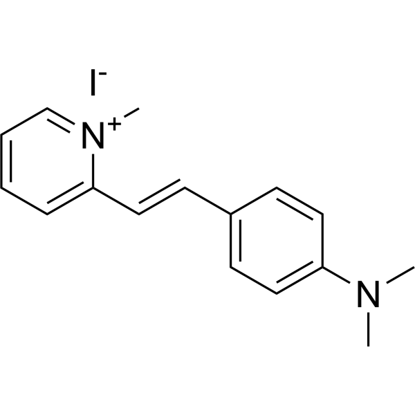
DASPI
|
G-quadruplex
|
Others
|
|
2-Di-1-ASP (DASPI; Compound 18a) is a mono-stryryl dye, and widely used as mitochondrial stain and groove-binding fluorescent probes for double-stranded DNA. 2-Di-1-ASP is selective for G-quadruplex (G4) and double-stranded DNA .
|
-
-
- HY-D0984
-
|
|
Fluorescent Dye
|
Inflammation/Immunology
|
|
Rhodamine dyes are membrane-permeable cationic fluorescent probes that specifically recognize mitochondrial membrane potentials, thereby attaching to mitochondria and producing bright fluorescence, and at certain concentrations, rhodamine dyes have low toxicity to cells, so they are commonly used to detect mitochondria in animal cells, plant cells, and microorganisms .
|
-
-
- HY-DY1003
-
|
|
Fluorescent Dye
|
Others
|
JC-1 (CBIC2) (solution) is an ideal fluorescent probe widely used to detect mitochondrial membrane potential. JC-1 accumulates in mitochondria in a potential dependent manner and can be used to detect the membrane potential of cells, tissues or purified mitochondria. In normal mitochondria, JC-1 aggregates in the mitochondrial matrix to form a polymer, which emits strong red fluorescence (Ex=585 nm, Em=590 nm) ; When the mitochondrial membrane potential is low, JC-1 cannot aggregate in the matrix of mitochondria and produce green fluorescence (ex=510 nm, em= 527 nm) .
Solvent and concentration: DMSO: 1.5 mM
|
-

-
- HY-DY1073
-
|
|
Fluorescent Dye
|
Others
|
MitoPerOx (solution) is a mitochondrial-targeted, lipid peroxidation-indicating fluorescent probe with BODIPY581/591 fluorophores. The triphenylphosphine cation (TPP+) of MitoPerOx can be selectively enriched in mitochondria (depending on membrane potential) and can be used to detect lipid peroxidation in the inner mitochondrial membrane. Under the action of lipid peroxides, the BODIPY581/591 fluorophores of MitoPerOx shift their emission wavelength from 590 nm (reduced state) to 520 nm (oxidized state) , and ratiometric detection can be performed at an excitation wavelength of 488 nm. MitoPerOx can specifically monitor the peroxidation of mitochondrial phospholipids (especially cardiolipin) and is used in the study of oxidative stress-related diseases (such as aging, neurodegenerative diseases, and mitochondrial dysfunction) .
Solvent and concentration: DMSO: 2 mM
|
-

-
- HY-DY1042
-
|
|
Fluorescent Dye
|
Others
|
Rhodamine dyes are membrane-permeable cationic fluorescent probes that specifically recognize mitochondrial membrane potentials, thereby attaching to mitochondria and producing bright fluorescence, and at certain concentrations, rhodamine dyes have low toxicity to cells, so they are commonly used to detect mitochondria in animal cells, plant cells, and microorganisms .
Solvent and concentration: DMSO: 10 mM
|
-

-
- HY-D2878
-
|
|
Fluorescent Dye
Ferroptosis
|
Others
|
|
MitoPeDPP is a mitochondrial-targeted fluorescent probe that is sensitive to LPO. MitoPeDPP is synthesized from diphenylpyrenephosphine. MitoPeDPP can be used to study the occurrence of mitochondrial LPO in RSL3-induced oligodendrocyte ferroptosis .
|
-

-
- HY-126220
-
|
|
Fluorescent Dye
|
Others
|
|
KMG-301AM is the acetoxy methyl esterified form of KMG-301. KMG-301AM successfully accumulates in mitochondria and then it is hydrolyzed to KMG-301. KMG-301 is an Mg 2+-selective fluorescent probe functional in mitochondria in intact cells. Since the mitochondrial membrane is impermeable to KMG-301, it is not released upon depolarization of the mitochondrial membrane potential. KMG-301 can indicate changes in mitochondrial Mg2+ concentration and shows Mg 2+ transport across the mitochondrial membrane in the early phases of a cellular model .
|
-
-
- HY-101876
-
|
|
Fluorescent Dye
|
Others
|
|
Rhodamine dyes are membrane-permeable cationic fluorescent probes that specifically recognize mitochondrial membrane potentials, thereby attaching to mitochondria and producing bright fluorescence, and at certain concentrations, rhodamine dyes have low toxicity to cells, so they are commonly used to detect mitochondria in animal cells, plant cells, and microorganisms .
|
-
-
- HY-DY1021
-
|
|
Fluorescent Dye
|
Others
|
DiSC3 (5) (solution) is a fluorescent probe commonly used as a tracer dye to evaluate mitochondrial membrane potential. The excitation/emission wavelength of DiSC3 (5) is up to 622/670 nm. DiSC3 (5) can inhibit the respiratory system associated with mitochondrial NAD, and the IC50 value is 8 μM. DiSC3 (5) in the presence of Na +/K +-ATPase inhibitor ouabain 2 can induce membrane hyperpolarization of Ehrlich ascites tumor cells .
Solvent and concentration: DMSO: 1 mM
The 1 mL volume is defined as the base specification. All larger sizes correspond to incremental volumes of this base.
|
-

-
- HY-D2346
-
|
|
Fluorescent Dye
|
Others
|
|
HBmito Crimson is a deep red fluorescent probe (λex: 658 nm, λem: 678 nm) for the inner mitochondrial membrane. HBmito Crimson is a cell membrane-permeable probe with high selectivity for the mitochondrial inner membrane, suitable for specific fluorescence staining of the inner mitochondrial membrane in living cells. HBmito Crimson has high photostability and brightness, suitable for long-term dynamic fluorescence imaging.
|
-

-
- HY-D1250
-
|
|
Fluorescent Dye
|
Cancer
|
|
Mito-TRFS, the first off-on probe, is used to image the mitochondrial thioredoxin reductase (TrxR2) in live cells .
|
-
-
- HY-D2312
-
|
|
Fluorescent Dye
Ferroptosis
|
Cancer
|
|
Mito-Rh-S is a ratiometric near-infrared (NIR) fluorescent probe that detects the fluctuation of mitochondrial HClO levels during ferroptosis in hepatocellular carcinoma (HCC) .
|
-

-
- HY-126220A
-
|
|
Fluorescent Dye
|
Others
|
|
KMG-301AM TFA is the acetoxy methyl esterified form of KMG-301. KMG-301AM TFA successfully accumulates in mitochondria and then it is hydrolyzed to KMG-301. KMG-301 is an Mg 2+-selective fluorescent probe functional in mitochondria in intact cells. Since the mitochondrial membrane is impermeable to KMG-301, it is not released upon depolarization of the mitochondrial membrane potential. KMG-301 can indicate changes in mitochondrial Mg2+ concentration and shows Mg 2+ transport across the mitochondrial membrane in the early phases of a cellular model .
|
-
-
- HY-DY1023
-
|
|
Fluorescent Dye
|
Others
|
Rhodamine dyes are membrane-permeable cationic fluorescent probes that specifically recognize mitochondrial membrane potentials, thereby attaching to mitochondria and producing bright fluorescence, and at certain concentrations, rhodamine dyes have low toxicity to cells, so they are commonly used to detect mitochondria in animal cells, plant cells, and microorganisms .
Solvent and concentration: DMSO: 10 mM
|
-

-
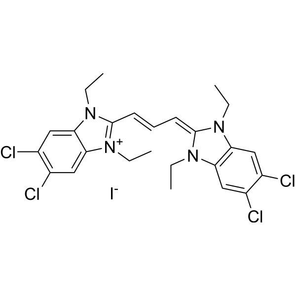
- HY-D1158
-
-
-
- HY-126474
-
|
|
Reactive Oxygen Species (ROS)
|
Inflammation/Immunology
|
|
MitoB bromide is an exomarker of mitochondrial hydrogen peroxide. MitoB bromide is a mitochondria-targeted ratiometric probe, and can be used to measure levels of one major ROS, hydrogen peroxide, within living animals .
|
-
-
- HY-DY1054
-
|
|
Fluorescent Dye
|
Cardiovascular Disease
|
Rhodamine dyes are membrane-permeable cationic fluorescent probes that specifically recognize mitochondrial membrane potentials, thereby attaching to mitochondria and producing bright fluorescence, and at certain concentrations, rhodamine dyes have low toxicity to cells, so they are commonly used to detect mitochondria in animal cells, plant cells, and microorganisms .
Solvent and concentration: DMSO: 5 mM
|
-

-
- HY-W020784
-
|
|
Biochemical Assay Reagents
|
Others
|
|
3-(N-Maleimidopropionyl)biocytin is a cysteine-specific labeling reagent and non-membrane-permeable probe. 3-(N-Maleimidopropionyl)biocytin covalently modifies the exposed cysteine residues of yeast Tim44, conjugating the biotin moiety to the polypeptide chain. 3-(N-Maleimidopropionyl)biocytin determines the topology of membrane proteins by modifying the exposed cysteine residues on the outer side of the inner mitochondrial membrane .
|
-
-
- HY-D2479
-
|
|
Fluorescent Dye
|
Metabolic Disease
|
|
DMANI is an intramolecular charge transfer (ICT)-based mitochondria-targeted ratiometric fluorescent probe. DMANI can monitor mitochondrial peroxynitrite (ONOO −) in living cells .
|
-

-
- HY-D0309R
-
|
Basic Red 1 (Standard)
|
Fluorescent Dye
Reference Standards
|
Cancer
|
|
Rhodamine 6G (Standard) is the analytical standard of Rhodamine 6G. This product is intended for research and analytical applications. Rhodamine dyes are membrane-permeable cationic fluorescent probes that specifically recognize mitochondrial membrane potentials, thereby attaching to mitochondria and producing bright fluorescence, and at certain concentrations, rhodamine dyes have low toxicity to cells, so they are commonly used to detect mitochondria in animal cells, plant cells, and microorganisms .
|
-

-
- HY-76573
-
|
|
Apoptosis
|
Cardiovascular Disease
Cancer
|
|
ML-10 is a small molecule apoptosis probe. Due to the presence of fluorine atoms, ML-10 can be radiolabeled with 18F isotopes and can be used for apoptosis positron emission tomography imaging studies. ML-10 is selectively taken up and accumulated in apoptotic cells, while being excluded from live or necrotic cells. In addition, the uptake of ML-10 is associated with apoptotic features such as caspase activation, Annexin-V binding, and disruption of mitochondrial membrane potential .
|
-

-
- HY-150175
-
|
|
Fluorescent Dye
|
Inflammation/Immunology
|
|
HKSOX-1 and its derivatives (HKSOX-1r and HKSOX-1m) are novel fluorescent probes designed for highly sensitive and selective detection of the superoxide anion radical (O2 •−) in cellular environments. These probes utilize an aryl trifluoromethanesulfonate group that undergoes O2 •−-mediated cleavage, releasing a free phenol and emitting fluorescence. They demonstrate excellent specificity and sensitivity across various pH ranges, withstand interference from strong oxidants and reductants typical in cellular contexts. HKSOX-1r, optimized for cellular retention, has been effectively employed in diverse assays including confocal imaging, flow cytometry, and zebrafish embryo studies, highlighting its utility in investigating O2 •− roles in inflammation, mitochondrial stress, and other physiological processes .
|
-

-
- HY-128536
-
|
|
Fluorescent Dye
|
Others
|
|
KMG-104AM is a Mg 2+ fluorescent probe. KMG-104AM is a membrane-permeable ester-modified derivative of KMG-104, which serves as a reporter and imaging agent. KMG-104AM can track the increase in intracellular free magnesium ion concentration induced by mitochondrial uncoupling. KMG-104AM enables visualization of the three-dimensional distribution of intracellular magnesium ion concentration. KMG-104AM is applicable to research related to the functions of intracellular magnesium ions .
|
-
-
- HY-123583
-
|
|
Fluorescent Dye
|
Others
|
|
MitoBADY is a mitochondria-selective Raman probe that successfully visualizes mitochondria in live HeLa cells at a concentration of 200-400 nM for 10 minutes. MitoBADY can be utilized for the study of mitochondrial dynamics .
|
-

-
- HY-D1752
-
|
D-22421
|
Fluorescent Dye
|
Others
|
|
JC-9 (D-22421) is a green-fluorescent probe used for ratiometric calculation of mitochondrial membrane potential.
|
-
-
- HY-N16300
-
|
|
Fluorescent Dye
|
Others
|
|
Mito-laurdan bromide, a derivative of Laurdan (HY-D0080), is a fluorescent probe. Mito-laurdan bromide contains a cationic triphenylphosphonium moiety, which accumulates at the inner mitochondrial membrane due to its negative membrane potential, connected via a 3 carbon linker .
|
-

-
- HY-W127775
-
-
-
- HY-158918
-
|
4-CF3-Triphenylphosphonium-DC
|
Biochemical Assay Reagents
|
Others
|
|
4-CF3-TPP-DC (4-CF3-Triphenylphosphonium-DC) is an inert mitochondrial targeting carrier, that delivers target drugs and probes to mitochondria without causing mitochondrial depolarization and cytotoxicity in cell C2C12 .
|
-

-
- HY-D1493
-
|
|
PKC
|
Others
|
|
FIM-1 is a fluorescent PKC (protein kinase C) probe that can be used for mitochondrial staining. FIM-1 inhibits PKC and acts as ATP-competitive catalytic site inhibitor .
|
-
-
- HY-126474S
-
-

-
- HY-D2279
-
|
|
Fluorescent Dye
|
Inflammation/Immunology
|
|
NFL-NH2 is a mitochondrial-targeted near-infrared ratiometric fluorescent probe. NFL-NH2 can rapidly detect NO levels associated with the inflammatory damage degree in rheumatoid arthritis (RA) mice models by ratiometric fluorescence imaging. The excitation wavelength and emission wavelength are 650 nm and 780 nm, respectively .
|
-
-
- HY-172342
-
|
|
Fluorescent Dye
|
Cardiovascular Disease
|
|
MitoA (bromide) is a mitochondria-targeted H2S probe, which can be used to assess relative changes in mitochondrial levels of H2S in vivo. MitoA (bromide) can accumulate inside mitochondria within tissues in vivo due to tryphenylphosphonium (TPP) cation. MitoA (bromide) can be studied in research on myocardial ischemia .
|
-

-
- HY-155062
-
|
|
Mitochondrial Metabolism
Reactive Oxygen Species (ROS)
β-catenin
Fluorescent Dye
PPAR
|
Cancer
|
|
IR-251 is a mitochondrion-targeting NIR fluorescent probe. IR-251 targets mitochondria via OATPs and causes mitochondrial damage in tumor cells. IR-251 IR-251 induced ROS overproduction by inhibiting PPARγ, and then inhibiting the β-catenin signaling pathway and downstream protein molecules related to the cell cycle and metastasis. IR-251 inhibits tumor proliferation and metastasis .
|
-
-
- HY-136675
-
|
|
Fluorescent Dye
|
Others
|
|
ASMI is a ratiometric, two-photon excited fluorescent probe, composed of a highly two-photon active and biocompatible merocyanine fluorophore and an acrylate moiety as a thiol reactive site. ASMI is able to selectively detect and monitor mitochondrial Cys with rapid responsiveness, imaging living cells and intact tissues with high contrast and brightness at a depth of 150 μm. The two-photon action cross section (Φσmax) of ASMI is 65.2 GM, corresponding to an excitation wavelength (λex) of 740 nm.
|
-

-
- HY-D3381
-
|
|
Fluorescent Dye
|
Others
|
|
MitoCLox is a mitochondria-targeted ratiometric fluorescent probe for measuring mitochondrial inner membrane lipid peroxidation .
|
-

-
- HY-P11637
-
|
|
Fluorescent Dye
|
Neurological Disease
|
|
Mitochondrial-targeted peptide BP19 is a fluorescent probe for the selective measurement of labile iron in the mitochondriairon. Mitochondrial-targeted peptide BP19 exhibits iron-selective sensing activity with mitochondrial accumulation, reduced fluorescence in iron-loaded cells, and fluorescence reinstatement upon iron chelation. Mitochondrial-targeted peptide BP19 evaluates mitochondrial labile iron levels in cultured fibroblasts with Friedreich's ataxia .
|
-

-
- HY-D2985
-
|
|
Mitochondrial Metabolism
Fluorescent Dye
|
Others
|
|
MI-BP-CC is a mitochondrial-targeting near-infrared fluorescent probe. MI-BP-CC can specifically localize in the mitochondria of living cells. MI-BP-CC visualizes viscosity with an emission wavelength of 722 nm for detecting mitochondrial viscosity .
|
-

-
- HY-D3240
-
|
|
Fluorescent Dye
|
Others
|
|
Photoactive NTR probe (Compound 1) is a covalent crosslinker and Fluorescent indicator targeting Nitroreductase. The Photoactive NTR probe undergoes a sequential activation process: it is first activated via nitroreductase-mediated nitro-to-amino conversion, and then forms a fluorescent product upon photoactivation. The Photoactive NTR probe can form covalent adducts with the side chains of cysteine, tyrosine, lysine and histidine in adjacent proteins to reduce fluorophore diffusion. The Photoactive NTR probe enables super-resolution (STORM) imaging of active mitochondrial nitroreductase microdomains in living cells .
|
-

-
- HY-D3166
-
|
|
Fluorescent Dye
|
Others
|
|
CEMT is a carboxylesterase (CE) substrate and a ratiometric two-photon fluorescent reporter probe. CEMT can be hydrolyzed by CE to generate HMT, which is used for mitochondrial pH sensing. After activation by CE, CEMT exhibits ratiometric fluorescence changes in response to pH variations. CEMT targets and covalently binds to mitochondria, and can avoid leakage during acidification, thus enabling in situ imaging .
|
-

-
- HY-156898
-
|
|
α-synuclein
Caspase
Reactive Oxygen Species (ROS)
|
Neurological Disease
|
|
NPT100-18A is an αSyn oligomerization inhibitor. NPT10018A rescues cleaved caspase-3 levels to control levels. NPT10018A attenuates mitochondrial oxidative stress probe levels in a compartment-specific manner and, at higher concentrations, increases ATP signals. NPT100-18A can be used for the study of Parkinson’s disease (PD) .
|
-

-
- HY-P11486
-
-

- HY-D3153
-
|
|
Apoptosis
Caspase
Microtubule/Tubulin
|
Inflammation/Immunology
|
|
PbQ is a tubulin inhibitor (with an IC50 of 5 μM against goat tubulin) and a fluorescent probe for cuprous ions Cu (I). PbQ can penetrate the membrane of peripheral blood mononuclear cells, form a stable 1:1 complex with Cu + ions, and exhibits low toxicity and good biocompatibility toward macrophage cell lines. In addition, PbQ promotes tubulin degradation and disrupts the microtubule network in lung epithelial cells without affecting actin. PbQ also possesses genotoxicity by forming DNA base adducts, and it can activate caspase-3 and apoptosis-related genes, induce loss of mitochondrial membrane potential, and trigger cell apoptosis. PbQ can be used in studies related to chronic obstructive pulmonary disease .
|
-

| Cat. No. |
Product Name |
Type |
-
- HY-15534
-
JC-1
Maximum Cited Publications
212 Publications Verification
CBIC2
|
Fluorescent Dye
|
|
JC-1 (CBIC2) is an ideal fluorescent probe widely used to detect mitochondrial membrane potential. JC-1 accumulates in mitochondria in a potential dependent manner and can be used to detect the membrane potential of cells, tissues or purified mitochondria. In normal mitochondria, JC-1 aggregates in the mitochondrial matrix to form a polymer, which emits strong red fluorescence (Ex=585 nm, Em=590 nm); When the mitochondrial membrane potential is low, JC-1 cannot aggregate in the matrix of mitochondria and produce green fluorescence (ex=510 nm, em= 527 nm) .
|
-
- HY-D0985A
-
|
Tetramethylrhodamine ethyl ester perchlorate
|
Fluorescent Dye
|
|
Rhodamine dyes are membrane-permeable cationic fluorescent probes that specifically recognize mitochondrial membrane potentials, thereby attaching to mitochondria and producing bright fluorescence, and at certain concentrations, rhodamine dyes have low toxicity to cells, so they are commonly used to detect mitochondria in animal cells, plant cells, and microorganisms .
|
-
- HY-D0984A
-
|
T668
|
Fluorescent Dye
|
|
Rhodamine dyes are membrane-permeable cationic fluorescent probes that specifically recognize mitochondrial membrane potentials, thereby attaching to mitochondria and producing bright fluorescence, and at certain concentrations, rhodamine dyes have low toxicity to cells, so they are commonly used to detect mitochondria in animal cells, plant cells, and microorganisms .
|
-
- HY-D0085
-
|
|
Fluorescent Dye
|
|
DiSC3(5) is a fluorescent probe commonly used as a tracer dye to evaluate mitochondrial membrane potential. The excitation/emission wavelength of DiSC3(5) is up to 622/670 nm. DiSC3(5) can inhibit the respiratory system associated with mitochondrial NAD, and the IC50 value is 8 μM. DiSC3(5) in the presence of Na +/K +-ATPase inhibitor ouabain 2 can induce membrane hyperpolarization of Ehrlich ascites tumor cells .
|
-
- HY-D0816
-
|
RH-123; R-22420
|
Fluorescent Dye
|
|
Rhodamine dyes are membrane-permeable cationic fluorescent probes that specifically recognize mitochondrial membrane potentials, thereby attaching to mitochondria and producing bright fluorescence, and at certain concentrations, rhodamine dyes have low toxicity to cells, so they are commonly used to detect mitochondria in animal cells, plant cells, and microorganisms .
|
-
- HY-125623
-
|
|
Fluorescent Dye
|
|
MitoPerOx is a mitochondrial-targeted, lipid peroxidation-indicating fluorescent probe with BODIPY581/591 fluorophores. The triphenylphosphine cation (TPP+) of MitoPerOx can be selectively enriched in mitochondria (depending on membrane potential) and can be used to detect lipid peroxidation in the inner mitochondrial membrane. Under the action of lipid peroxides, the BODIPY581/591 fluorophores of MitoPerOx shift their emission wavelength from 590 nm (reduced state) to 520 nm (oxidized state), and ratiometric detection can be performed at an excitation wavelength of 488 nm. MitoPerOx can specifically monitor the peroxidation of mitochondrial phospholipids (especially cardiolipin) and is used in the study of oxidative stress-related diseases (such as aging, neurodegenerative diseases, and mitochondrial dysfunction)[1][2].
|
-
- HY-D0309
-
|
Basic Red 1
|
Fluorescent Dye
|
|
Rhodamine dyes are membrane-permeable cationic fluorescent probes that specifically recognize mitochondrial membrane potentials, thereby attaching to mitochondria and producing bright fluorescence, and at certain concentrations, rhodamine dyes have low toxicity to cells, so they are commonly used to detect mitochondria in animal cells, plant cells, and microorganisms .
|
-
- HY-135009
-
|
DASPI
|
Fluorescent Dye
|
|
2-Di-1-ASP (DASPI; Compound 18a) is a mono-stryryl dye, and widely used as mitochondrial stain and groove-binding fluorescent probes for double-stranded DNA. 2-Di-1-ASP is selective for G-quadruplex (G4) and double-stranded DNA .
|
-
- HY-D0984
-
|
|
Fluorescent Dye
|
|
Rhodamine dyes are membrane-permeable cationic fluorescent probes that specifically recognize mitochondrial membrane potentials, thereby attaching to mitochondria and producing bright fluorescence, and at certain concentrations, rhodamine dyes have low toxicity to cells, so they are commonly used to detect mitochondria in animal cells, plant cells, and microorganisms .
|
-
- HY-DY1003
-
|
|
Fluorescent Dye
|
JC-1 (CBIC2) (solution) is an ideal fluorescent probe widely used to detect mitochondrial membrane potential. JC-1 accumulates in mitochondria in a potential dependent manner and can be used to detect the membrane potential of cells, tissues or purified mitochondria. In normal mitochondria, JC-1 aggregates in the mitochondrial matrix to form a polymer, which emits strong red fluorescence (Ex=585 nm, Em=590 nm) ; When the mitochondrial membrane potential is low, JC-1 cannot aggregate in the matrix of mitochondria and produce green fluorescence (ex=510 nm, em= 527 nm) .
Solvent and concentration: DMSO: 1.5 mM
|
-
- HY-DY1073
-
|
|
Fluorescent Dye
|
MitoPerOx (solution) is a mitochondrial-targeted, lipid peroxidation-indicating fluorescent probe with BODIPY581/591 fluorophores. The triphenylphosphine cation (TPP+) of MitoPerOx can be selectively enriched in mitochondria (depending on membrane potential) and can be used to detect lipid peroxidation in the inner mitochondrial membrane. Under the action of lipid peroxides, the BODIPY581/591 fluorophores of MitoPerOx shift their emission wavelength from 590 nm (reduced state) to 520 nm (oxidized state) , and ratiometric detection can be performed at an excitation wavelength of 488 nm. MitoPerOx can specifically monitor the peroxidation of mitochondrial phospholipids (especially cardiolipin) and is used in the study of oxidative stress-related diseases (such as aging, neurodegenerative diseases, and mitochondrial dysfunction) .
Solvent and concentration: DMSO: 2 mM
|
-
- HY-DY1042
-
|
|
Fluorescent Dye
|
Rhodamine dyes are membrane-permeable cationic fluorescent probes that specifically recognize mitochondrial membrane potentials, thereby attaching to mitochondria and producing bright fluorescence, and at certain concentrations, rhodamine dyes have low toxicity to cells, so they are commonly used to detect mitochondria in animal cells, plant cells, and microorganisms .
Solvent and concentration: DMSO: 10 mM
|
-
- HY-D2878
-
|
|
Fluorescent Dye
|
|
MitoPeDPP is a mitochondrial-targeted fluorescent probe that is sensitive to LPO. MitoPeDPP is synthesized from diphenylpyrenephosphine. MitoPeDPP can be used to study the occurrence of mitochondrial LPO in RSL3-induced oligodendrocyte ferroptosis .
|
-
- HY-101876
-
|
|
Fluorescent Dye
|
|
Rhodamine dyes are membrane-permeable cationic fluorescent probes that specifically recognize mitochondrial membrane potentials, thereby attaching to mitochondria and producing bright fluorescence, and at certain concentrations, rhodamine dyes have low toxicity to cells, so they are commonly used to detect mitochondria in animal cells, plant cells, and microorganisms .
|
-
- HY-DY1021
-
|
|
Fluorescent Dye
|
DiSC3 (5) (solution) is a fluorescent probe commonly used as a tracer dye to evaluate mitochondrial membrane potential. The excitation/emission wavelength of DiSC3 (5) is up to 622/670 nm. DiSC3 (5) can inhibit the respiratory system associated with mitochondrial NAD, and the IC50 value is 8 μM. DiSC3 (5) in the presence of Na +/K +-ATPase inhibitor ouabain 2 can induce membrane hyperpolarization of Ehrlich ascites tumor cells .
Solvent and concentration: DMSO: 1 mM
The 1 mL volume is defined as the base specification. All larger sizes correspond to incremental volumes of this base.
|
-
- HY-D2346
-
|
|
Fluorescent Dye
|
|
HBmito Crimson is a deep red fluorescent probe (λex: 658 nm, λem: 678 nm) for the inner mitochondrial membrane. HBmito Crimson is a cell membrane-permeable probe with high selectivity for the mitochondrial inner membrane, suitable for specific fluorescence staining of the inner mitochondrial membrane in living cells. HBmito Crimson has high photostability and brightness, suitable for long-term dynamic fluorescence imaging.
|
-
- HY-D1250
-
|
|
Fluorescent Dye
|
|
Mito-TRFS, the first off-on probe, is used to image the mitochondrial thioredoxin reductase (TrxR2) in live cells .
|
-
- HY-D2312
-
|
|
Fluorescent Dye
|
|
Mito-Rh-S is a ratiometric near-infrared (NIR) fluorescent probe that detects the fluctuation of mitochondrial HClO levels during ferroptosis in hepatocellular carcinoma (HCC) .
|
-
- HY-DY1023
-
|
|
Fluorescent Dye
|
Rhodamine dyes are membrane-permeable cationic fluorescent probes that specifically recognize mitochondrial membrane potentials, thereby attaching to mitochondria and producing bright fluorescence, and at certain concentrations, rhodamine dyes have low toxicity to cells, so they are commonly used to detect mitochondria in animal cells, plant cells, and microorganisms .
Solvent and concentration: DMSO: 10 mM
|
-
- HY-D1158
-
|
|
Fluorescent Dye
|
|
HKOCl-4m is a selective and mitochondria-targeting rhodol-based fluorescent probe for monitoring mitochondrial hypochlorous acid (HOCl) .
|
-
- HY-DY1054
-
|
|
Fluorescent Dye
|
Rhodamine dyes are membrane-permeable cationic fluorescent probes that specifically recognize mitochondrial membrane potentials, thereby attaching to mitochondria and producing bright fluorescence, and at certain concentrations, rhodamine dyes have low toxicity to cells, so they are commonly used to detect mitochondria in animal cells, plant cells, and microorganisms .
Solvent and concentration: DMSO: 5 mM
|
-
- HY-D2479
-
|
|
Fluorescent Dye
|
|
DMANI is an intramolecular charge transfer (ICT)-based mitochondria-targeted ratiometric fluorescent probe. DMANI can monitor mitochondrial peroxynitrite (ONOO −) in living cells .
|
-
- HY-D0309R
-
|
Basic Red 1 (Standard)
|
Fluorescent Dye
|
|
Rhodamine 6G (Standard) is the analytical standard of Rhodamine 6G. This product is intended for research and analytical applications. Rhodamine dyes are membrane-permeable cationic fluorescent probes that specifically recognize mitochondrial membrane potentials, thereby attaching to mitochondria and producing bright fluorescence, and at certain concentrations, rhodamine dyes have low toxicity to cells, so they are commonly used to detect mitochondria in animal cells, plant cells, and microorganisms .
|
-
- HY-128536
-
|
|
Fluorescent Dye
|
|
KMG-104AM is a Mg 2+ fluorescent probe. KMG-104AM is a membrane-permeable ester-modified derivative of KMG-104, which serves as a reporter and imaging agent. KMG-104AM can track the increase in intracellular free magnesium ion concentration induced by mitochondrial uncoupling. KMG-104AM enables visualization of the three-dimensional distribution of intracellular magnesium ion concentration. KMG-104AM is applicable to research related to the functions of intracellular magnesium ions .
|
-
- HY-D1752
-
|
D-22421
|
Fluorescent Dye
|
|
JC-9 (D-22421) is a green-fluorescent probe used for ratiometric calculation of mitochondrial membrane potential.
|
-
- HY-W127775
-
|
|
Fluorescent Dye
|
|
Demethyl Rhod-2 AM is a fluorescent mitochondrial probe (λex=552 nm, λem=581 nm).
|
-
- HY-D1493
-
|
|
Fluorescent Dye
|
|
FIM-1 is a fluorescent PKC (protein kinase C) probe that can be used for mitochondrial staining. FIM-1 inhibits PKC and acts as ATP-competitive catalytic site inhibitor .
|
-
- HY-D2279
-
|
|
Fluorescent Dye
|
|
NFL-NH2 is a mitochondrial-targeted near-infrared ratiometric fluorescent probe. NFL-NH2 can rapidly detect NO levels associated with the inflammatory damage degree in rheumatoid arthritis (RA) mice models by ratiometric fluorescence imaging. The excitation wavelength and emission wavelength are 650 nm and 780 nm, respectively .
|
-
- HY-D3381
-
|
|
Fluorescent Dye
|
|
MitoCLox is a mitochondria-targeted ratiometric fluorescent probe for measuring mitochondrial inner membrane lipid peroxidation .
|
-
- HY-D2985
-
|
|
Fluorescent Dye
|
|
MI-BP-CC is a mitochondrial-targeting near-infrared fluorescent probe. MI-BP-CC can specifically localize in the mitochondria of living cells. MI-BP-CC visualizes viscosity with an emission wavelength of 722 nm for detecting mitochondrial viscosity .
|
-
- HY-D3240
-
|
|
Fluorescent Dye
|
|
Photoactive NTR probe (Compound 1) is a covalent crosslinker and Fluorescent indicator targeting Nitroreductase. The Photoactive NTR probe undergoes a sequential activation process: it is first activated via nitroreductase-mediated nitro-to-amino conversion, and then forms a fluorescent product upon photoactivation. The Photoactive NTR probe can form covalent adducts with the side chains of cysteine, tyrosine, lysine and histidine in adjacent proteins to reduce fluorophore diffusion. The Photoactive NTR probe enables super-resolution (STORM) imaging of active mitochondrial nitroreductase microdomains in living cells .
|
-
- HY-D3166
-
|
|
Fluorescent Dye
|
|
CEMT is a carboxylesterase (CE) substrate and a ratiometric two-photon fluorescent reporter probe. CEMT can be hydrolyzed by CE to generate HMT, which is used for mitochondrial pH sensing. After activation by CE, CEMT exhibits ratiometric fluorescence changes in response to pH variations. CEMT targets and covalently binds to mitochondria, and can avoid leakage during acidification, thus enabling in situ imaging .
|
-
- HY-D3153
-
|
|
Fluorescent Dye
|
|
PbQ is a tubulin inhibitor (with an IC50 of 5 μM against goat tubulin) and a fluorescent probe for cuprous ions Cu (I). PbQ can penetrate the membrane of peripheral blood mononuclear cells, form a stable 1:1 complex with Cu + ions, and exhibits low toxicity and good biocompatibility toward macrophage cell lines. In addition, PbQ promotes tubulin degradation and disrupts the microtubule network in lung epithelial cells without affecting actin. PbQ also possesses genotoxicity by forming DNA base adducts, and it can activate caspase-3 and apoptosis-related genes, induce loss of mitochondrial membrane potential, and trigger cell apoptosis. PbQ can be used in studies related to chronic obstructive pulmonary disease .
|
| Cat. No. |
Product Name |
Type |
| Cat. No. |
Product Name |
Target |
Research Area |
-
- HY-P11637
-
|
|
Fluorescent Dye
|
Neurological Disease
|
|
Mitochondrial-targeted peptide BP19 is a fluorescent probe for the selective measurement of labile iron in the mitochondriairon. Mitochondrial-targeted peptide BP19 exhibits iron-selective sensing activity with mitochondrial accumulation, reduced fluorescence in iron-loaded cells, and fluorescence reinstatement upon iron chelation. Mitochondrial-targeted peptide BP19 evaluates mitochondrial labile iron levels in cultured fibroblasts with Friedreich's ataxia .
|
-
- HY-P11486
-
-
- HY-KD1029
-
|
|
|
Mito Red is a red-fluorescent fluorescent probe that specifically labels mitochondria in living mammalian cells.
|
| Cat. No. |
Product Name |
Category |
Target |
Chemical Structure |
-
- HY-N16300
-
|
|
Lipid
|
Fluorescent Dye
|
|
Mito-laurdan bromide, a derivative of Laurdan (HY-D0080), is a fluorescent probe. Mito-laurdan bromide contains a cationic triphenylphosphonium moiety, which accumulates at the inner mitochondrial membrane due to its negative membrane potential, connected via a 3 carbon linker .
|
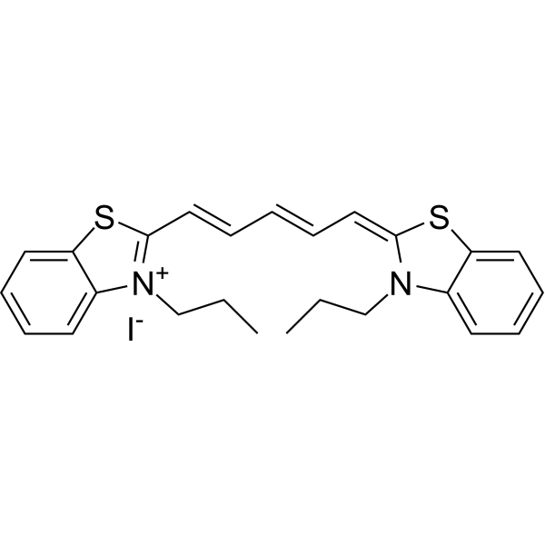
-
| Cat. No. |
Product Name |
Chemical Structure |
-
- HY-126474S
-
|
|
|
MitoB-d15 (bromide) is deuterium labeled MitoB (bromide). MitoB bromide is an exomarker of mitochondrial hydrogen peroxide. MitoB bromide is a mitochondria-targeted ratiometric probe, and can be used to measure levels of one major ROS, hydrogen peroxide, within living animals .
|
-
| Cat. No. |
Product Name |
|
Classification |
-
- HY-131442
-
|
Alkyne tyramide; Alk-Ph
|
|
Labeling and Fluorescence Imaging
Alkynes
|
|
Alkyne-phenol (Alk-Ph) is a clickable ascorbate peroxidase 2 (APEX2) probe. Alkyne-phenol substantially improves APEX-labeling efficiency in intact yeast cells, as it is more cell wall-permeant than APEX2 substrate biotin-phenol (BP). Alkyne-phenol also facilitates the identification of APEX-labeling sites, allowing the unambiguous assignment of membrane topology of mitochondrial proteins . Alkyne-phenol is a click chemistry reagent, it contains an Alkyne group and can undergo copper-catalyzed azide-alkyne cycloaddition (CuAAc) with molecules containing Azide groups.
|
Your information is safe with us. * Required Fields.
Inquiry Information
- Product Name:
- Cat. No.:
- Quantity:
- MCE Japan Authorized Agent:


