



JC-1 (solution)

Based on 1 Customer Validation
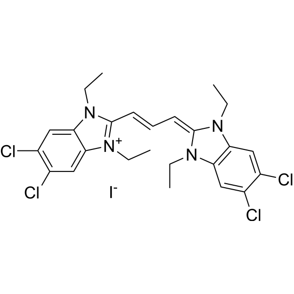
JC-1 (CBIC2) (solution) is an ideal fluorescent probe widely used to detect mitochondrial membrane potential. JC-1 accumulates in mitochondria in a potential dependent manner and can be used to detect the membrane potential of cells, tissues or purified mitochondria. In normal mitochondria, JC-1 aggregates in the mitochondrial matrix to form a polymer, which emits strong red fluorescence (Ex=585 nm, Em=590 nm) ; When the mitochondrial membrane potential is low, JC-1 cannot aggregate in the matrix of mitochondria and produce green fluorescence (ex=510 nm, em= 527 nm) .
Solvent and concentration: DMSO: 1.5 mM
For research use only. We do not sell to patients.
- CAS No.: 3520-43-2
- Formula: C25H27Cl4IN4
- Molecular Weight:652.22
-
Storage:
Please store the product under the recommended conditions in the Certificate of Analysis.


To place orders, for customer services and technical support, please contact: MedChemExpress USA
Tel: 609-228-6898 E-mail: [email protected] [email protected]


-
 Biological Activity
-
Chemical Information
- Protocols
- Purity & Documentation
- References
-
 Help & FAQs


-

HY-B0215
AcetylcysteineMucolytic Agent -
HY-15534
JC-1Mitochondrial Membrane Potential Probe -
HY-D0815
Propidium IodideFluorochrome -
HY-15924
Thiazolyl BlueCytotoxicity Detection Reagent -
HY-66019
FITCFluorochrome -
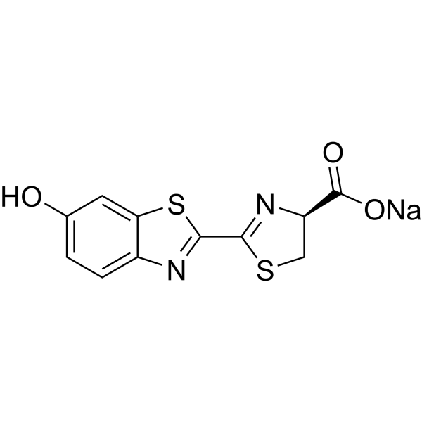
HY-12591
D-Luciferin sodiumSubstrate for Luciferase -
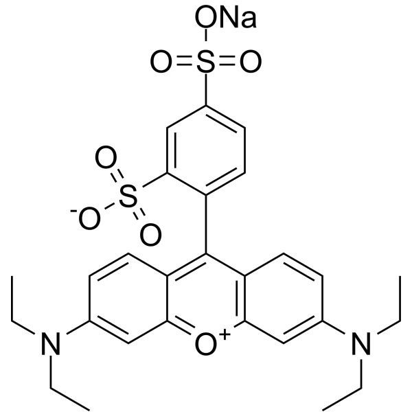
HY-D0974
Sulforhodamine B sodium saltFluorescent Dye -

HY-DY1006
Propidium Iodide (solution)Fluorochrome -
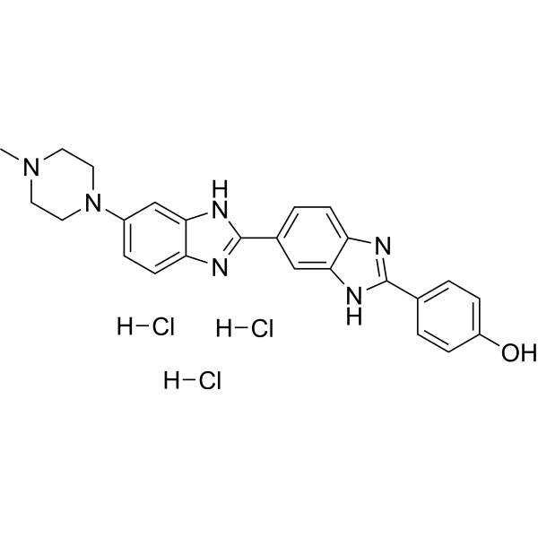
HY-15558A
Hoechst 33258 trihydrochlorideDNA Staining -
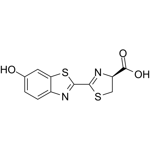
HY-12591A
D-LuciferinSubstrate for Luciferase -
HY-D0984A
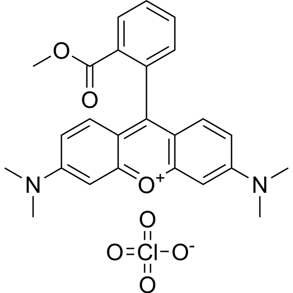
TMRM PerchlorateMitochondrial Membrane Potential Fluorescent Dye -
HY-D1048
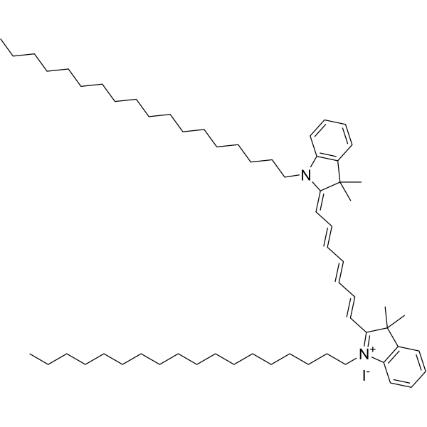
DiRMembrane Dye


Biological Activity
Description
Solvent and concentration: DMSO: 1.5 mM
In Vitro
Guidelines (Following is our recommended protocol. This protocol only provides a guideline, and should be modified according to your specific needs).
1. Preparation of JC-1 working solution:
1‰ DMSO + 89.9% ddH2O + 10% 10x PBS (HY-K3006). Prepare a 1.5-30 μM JC-1 working solution.
Note: 1) The JC-1 dye needs to be prepared immediately and used as soon as possible. It may precipitate if left for a period of time; try to prepare and use it under a light-proof condition.
2) At each step of the dilution process, ensure thorough mixing to achieve clarity. If necessary, ultrasonic assistance for dissolution at 37°C can be used.
3) Please do not directly dilute it with 1x PBS (HY-K3005) or serum-free medium to obtain the working solution; otherwise, it will lead to severe precipitation.
4) If the effect of JC-1 in entering the cells is not satisfactory, an appropriate amount of 20% Pluronic F127 solution can be added to the working solution, with a final concentration of 0.02-0.05%. Pluronic F127 can prevent JC-1 from aggregating in the buffer solution and help it enter the cells.
5) The storage solution of JC-1 is recommended to be aliquoted and stored at -20°C or -80°C in the dark.
2. JC-1 Staining:
2.1 Take the 6-well plate as an example for cell planking, and the density is 5×10⁵/mL. Incubate overnight in 5% CO2 incubator at 37°C.
Note: It is suggested that the cell density during apoptosis induction should not exceed 1×10⁶/mL, which can also be cultured to the appropriate density according to your own cell type.
2.2 Transfer 0.5 mL of the cell suspension into a sterile centrifuge tube.
2.3 Take 0.5 mL suspension into sterile centrifuge tube; 400 g centrifugation for 3-5 min; Discard the supernatant.
2.4 The cells were resuspended with 1mL JC-1 working solution and incubated in 5% CO2 incubator at 37°Cfor 15-30 min.
2.5 Centrifugation at room temperature for 5 min at 400 g; Suck of the supernatant.
2.6 The cells were resuspended with 2 mL cell culture medium or buffer, and then centrifuged at room temperature for 5 min at 400 g; Discard the supernatant and repeat twice.
2.7 Resuspend the cells with 1mL of fresh culture medium or buffer, and immediately conduct subsequent flow cytometry or fluorescence microscope observation.
2.8 Data analysis (flow cytometry) : mitochondria of healthy cells containing red JC-1 aggregates were detected by FL2 channel; Apoptotic or unhealthy cells containing green JC-1 monomer were detected by FL1 (FITC) channel.
Note: If used for enzyme labeling instrument, use 300 μL buffer resuspended cells; Then, transfer the stained cells to an opaque 96-well plate at a rate of 100 μL per well, and then conduct fluorescent enzyme label plate analysis.
MedChemExpress (MCE) has not independently confirmed the accuracy of these methods. They are for reference only.


Chemical Information
-
CAS No. 3520-43-2
-
Appearance Liquid
-
Molecular Weight 652.22
-
Formula C25H27Cl4IN4
-
Color Orange to red
-
SMILES
ClC1=C(Cl)C=C([N+](CC)=C(/C=C/C=C2N(C(C=C(Cl)C(Cl)=C3)=C3N\2CC)CC)N4CC)C4=C1.[I-]
-
Shipping
Room temperature in continental US; may vary elsewhere.
-
Storage
Please store the product under the recommended conditions in the Certificate of Analysis.


Purity & Documentation
-
Data Sheet (276 KB)
-
SDS (251 KB)
- English - EN (251 KB)
- Français - FR (251 KB)
- Deutsch - DE (251 KB)
- Norwegian - NO (251 KB)
- Español - ES (251 KB)
- Swedish - SV (251 KB)
- Italian - IT (251 KB)
- Korean - KR (251 KB)
- Portuguese - PT (251 KB)
-
Handling Instructions (2659 KB)
References
[1]. A Perelman, et al. JC-1: alternative excitation wavelengths facilitate mitochondrial membrane potential cytometry. Cell Death Dis. 2012 Nov 22;3:e430. [Content Brief]
[2]. Vera C. Keil, et al. Ratiometric high-resolution imaging of JC-1 fluorescence reveals the subcellular heterogeneity of astrocytic mitochondria. Pflügers Archiv - European Journal of Physiology. 2011,462(5): 693-708. [Content Brief]
[4]. Salvioli S, et al. JC-1, but not DiOC6(3) or rhodamine 123, is a reliable fluorescent probe to assess delta psi changes in intact cells: implications for studies on mitochondrial functionality during apoptosis. FEBS Lett. 1997 Jul 7;411(1):77-82. [Content Brief]

Calculators
Concentration (start) × Volume (start) = Concentration (final) × Volume (final)


JC-1 (solution) Related Classifications
HY-DY1003 Related Classifications
 Powered by Bioz