



Lometrexol
Based on 7 publication(s) in Google Scholar
Lometrexol (DDATHF), an antipurine antifolate, can inhibit the activity of glycinamide ribonucleotide formyltransferase (GARFT) but do not induce detectable levels of DNA strand breaks. Lometrexol can further inhibit de novo purine synthesis, causing abnormal cell proliferation and apoptosis, even cell cycle arrest. Lometrexol has anticancer activity. Lometrexol also is a potent human Serine hydroxymethyltransferase1/2 (hSHMT1/2) inhibitor.
商品は「研究用試薬」です。人や動物の医療用・臨床診断用・食品用の製品ではありません。
研究用途以外に使用した場合、当社は一切の責任を負いかねます。
- 純度 : 99.44%
- CAS 番号: 106400-81-1
- 分子式: C21H25N5O6
- 分子量:443.45
-
保管条件:Powder -20°C, 3 years , 4°C, 2 years ; In solvent -80°C, 6 months , -20°C, 1 month

To place orders, for customer services and technical support, please contact: MedChemExpress USA
Tel: 609-228-6898 E-mail: [email protected] [email protected]


-
 生物活性
生物活性
-
化学情報
-
溶剤 & 溶解度
- プロトコル
- 純度とドキュメンテーション
- 参考文献
-
 Help & FAQs


-
HY-10820
Pemetrexed -
HY-14519
MethotrexateDihydrofolate Reductase Inhibitor -
HY-10446
PralatrexateDihydrofolate Reductasean Inhibitor -
HY-108325
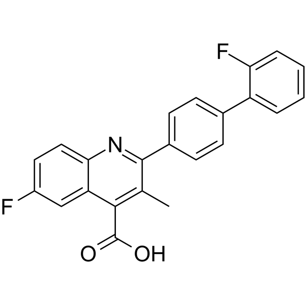
BrequinarDHODH Inhibitor -
HY-107981
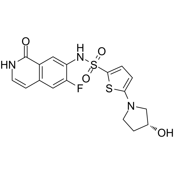
LSN 3213128AICARFT Inhibitor -
HY-B0510
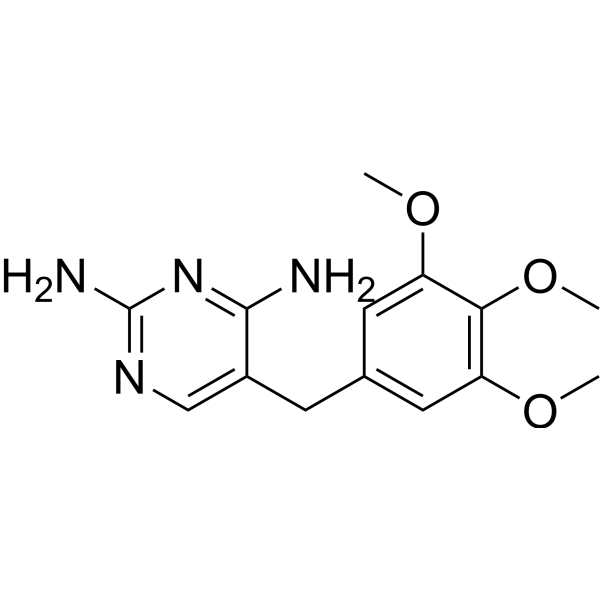
TrimethoprimBacteriostatic Antibiotic -
HY-13664
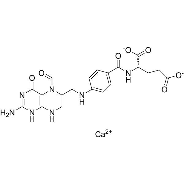
Folinic acid calciumBiological Folic Acid -
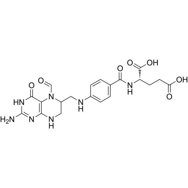
HY-17556
Folinic acidBiological Folic Acid -
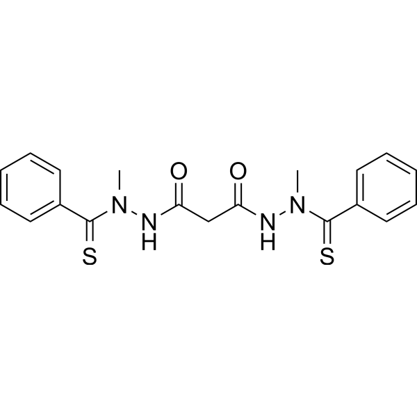
HY-12040
ElesclomolCuproptosis Inducer -
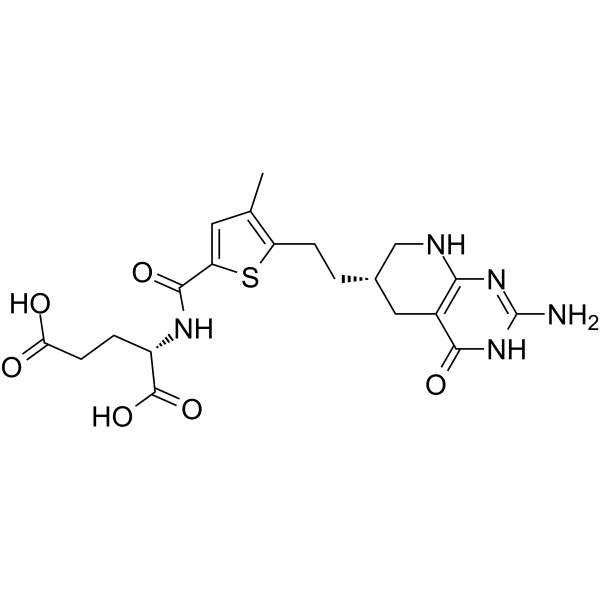
HY-14530
PelitrexolGARFT Inhibitor -

HY-B0510R
Trimethoprim (Standard)Bacteriostatic Antibiotic -
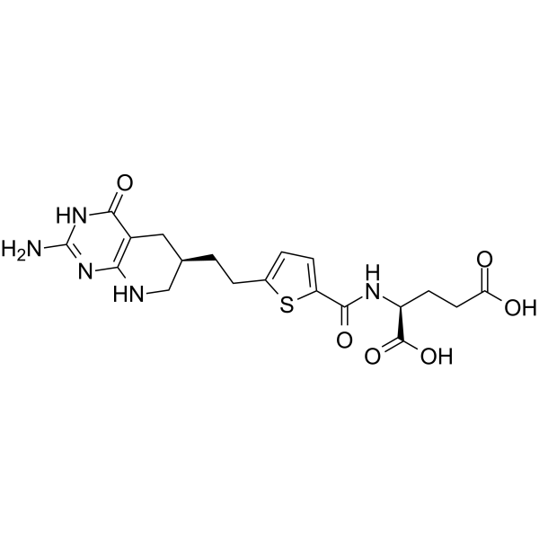
HY-10818
LY309887
MedChemExpress(MCE)の使用を引用している文献 Lometrexol
More Customer Validation & Images

-
Cell Proliferation/Viability Assay
-
Cell Proliferation/Viability Assay
Caspase アイソフォーム固有の製品をすべて表示
More

生物活性
製品説明
Cellular Effect
|
Cell Line

|
Type | Value | Description | References |
|---|---|---|---|---|
| CCRF-CEM | IC50 |
>100 μM
Compound: Lometrexol
|
Cytotoxicity in the absence of thymidine and presence of hypoxanthine against CCRF-CEM cell line
Cytotoxicity in the absence of thymidine and presence of hypoxanthine against CCRF-CEM cell line
|
[PMID: 10888335] |
| CCRF-CEM | IC50 |
>100 μM
Compound: Lometrexol
|
Cytotoxicity in the presence of thymidine and hypoxanthine against CCRF-CEM cell line
Cytotoxicity in the presence of thymidine and hypoxanthine against CCRF-CEM cell line
|
[PMID: 10888335] |
| CCRF-CEM | IC50 |
0.07 μM
Compound: Lometrexol
|
Cytotoxicity in the absence of hypoxanthine and presence of thymidine against CCRF-CEM cell line
Cytotoxicity in the absence of hypoxanthine and presence of thymidine against CCRF-CEM cell line
|
[PMID: 10888335] |
| CCRF-CEM | IC50 |
0.13 μM
Compound: Lometrexol
|
Cytotoxicity in the absence of thymidine and hypoxanthine against CCRF-CEM cell line
Cytotoxicity in the absence of thymidine and hypoxanthine against CCRF-CEM cell line
|
[PMID: 10888335] |
| CCRF-CEM | IC50 |
0.016 μM
Compound: DDATHF
|
Cytotoxicity against human lymphoblastic leukemic CCRF-CEM cell line was evaluated as the concentration required for 50% inhibition of the growth of the control value
Cytotoxicity against human lymphoblastic leukemic CCRF-CEM cell line was evaluated as the concentration required for 50% inhibition of the growth of the control value
|
[PMID: 1552503] |
| CCRF-CEM | IC50 |
>100 μM
Compound: lometrexol
|
Antiproliferative activity against human CCRF-CEM cell line in presence of hypoxanthine
Antiproliferative activity against human CCRF-CEM cell line in presence of hypoxanthine
|
[PMID: 16686541] |
| CCRF-CEM | IC50 |
0.2 μM
Compound: lometrexol
|
Antiproliferative activity against human CCRF-CEM cell line
Antiproliferative activity against human CCRF-CEM cell line
|
[PMID: 16686541] |
| CCRF-CEM | IC50 |
0.2 μM
Compound: lometrexol
|
Antiproliferative activity against human CCRF-CEM cell line in presence of thymidine
Antiproliferative activity against human CCRF-CEM cell line in presence of thymidine
|
[PMID: 16686541] |
| CCRF-CEM | IC50 |
25 μM
Compound: lometrexol
|
Antiproliferative activity against FPGS-deficient CCRF-CEM cell line
Antiproliferative activity against FPGS-deficient CCRF-CEM cell line
|
[PMID: 16686541] |
| CCRF-CEM | IC50 |
>10 μM
Compound: lometrexol, (6R)-5,10-DDATHF
|
Cytotoxicity against folyl-poly-glutamate synthetase-deficient human CCRF-CEM cells in absence of thymidine and hypoxanthine
Cytotoxicity against folyl-poly-glutamate synthetase-deficient human CCRF-CEM cells in absence of thymidine and hypoxanthine
|
[PMID: 18686942] |
| CCRF-CEM | IC50 |
>10 μM
Compound: lometrexol, (6R)-5,10-DDATHF
|
Cytotoxicity against human CCRF-CEM cells in absence of thymidine and presence of hypoxanthine
Cytotoxicity against human CCRF-CEM cells in absence of thymidine and presence of hypoxanthine
|
[PMID: 18686942] |
| CCRF-CEM | IC50 |
>100 μM
Compound: lometrexol, (6R)-5,10-DDATHF
|
Cytotoxicity against human CCRF-CEM cells expressing reduced folate carrier in absence of thymidine and hypoxanthine
Cytotoxicity against human CCRF-CEM cells expressing reduced folate carrier in absence of thymidine and hypoxanthine
|
[PMID: 18686942] |
| CCRF-CEM | IC50 |
0.2 μM
Compound: lometrexol, (6R)-5,10-DDATHF
|
Cytotoxicity against human CCRF-CEM cells in absence of thymidine and hypoxanthine
Cytotoxicity against human CCRF-CEM cells in absence of thymidine and hypoxanthine
|
[PMID: 18686942] |
| CCRF-CEM | IC50 |
0.2 μM
Compound: lometrexol, (6R)-5,10-DDATHF
|
Cytotoxicity against human CCRF-CEM cells in presence of thymidine and absence of hypoxanthine
Cytotoxicity against human CCRF-CEM cells in presence of thymidine and absence of hypoxanthine
|
[PMID: 18686942] |
| CCRF-CEM | IC50 |
0.007 μg/mL
Compound: 1 (DDATHF)
|
The compound was tested for its cytotoxicity against CCRF-CEM human leukemic cells using 72 hrs assay
The compound was tested for its cytotoxicity against CCRF-CEM human leukemic cells using 72 hrs assay
|
10.1016/S0960-894X(01)80214-6 |
| CCRF-CEM | IC50 |
0.007 μg/mL
Compound: DDATHF
|
Tested for the growth inhibition of CCRF-CEM, a human T-cell derived lymphoblastic leukemic cell line
Tested for the growth inhibition of CCRF-CEM, a human T-cell derived lymphoblastic leukemic cell line
|
10.1016/S0960-894X(01)80736-8 |
| CCRF-CEM | IC50 |
15.2 nM
Compound: LTX (2)
|
Tested in vitro for cellular cytotoxicity against human T-cell derived lymphoblastic leukemia (CCRF-CEM) cells
Tested in vitro for cellular cytotoxicity against human T-cell derived lymphoblastic leukemia (CCRF-CEM) cells
|
10.1016/S0960-894X(97)00041-3 |
| IGROV-1 | IC50 |
16 nM
Compound: lometrexol
|
Antiproliferative activity against human RFC and FRalpha expressing human IGROV1 cells in presence of folic acid
Antiproliferative activity against human RFC and FRalpha expressing human IGROV1 cells in presence of folic acid
|
[PMID: 18680275] |
| IGROV-1 | IC50 |
3.1 nM
Compound: lometrexol
|
Antiproliferative activity against human RFC and FRalpha expressing human IGROV1 cells
Antiproliferative activity against human RFC and FRalpha expressing human IGROV1 cells
|
[PMID: 18680275] |
| IGROV-1 | IC50 |
16 nM
Compound: LMTX, Lometrexol, 6-(R)DDATHF
|
Antiproliferative activity against human IGROV1 cells expressing human RFC, FRalpha and PCFT assessed as reduction of viable cells after 96 hrs in the presence of 200 nM folic acid
Antiproliferative activity against human IGROV1 cells expressing human RFC, FRalpha and PCFT assessed as reduction of viable cells after 96 hrs in the presence of 200 nM folic acid
|
[PMID: 21879757] |
| IGROV-1 | IC50 |
3.1 nM
Compound: LMTX, Lometrexol, 6-(R)DDATHF
|
Antiproliferative activity against human IGROV1 cells expressing human RFC, FRalpha and PCFT assessed as reduction of viable cells after 96 hrs
Antiproliferative activity against human IGROV1 cells expressing human RFC, FRalpha and PCFT assessed as reduction of viable cells after 96 hrs
|
[PMID: 21879757] |
| IGROV-1 | IC50 |
5.77 nM
Compound: LMTX
|
Inhibition of GARFTase in human IGROV1 cells assessed as reduction in [14C]glycine incorporation into [14C]formyl GAR incubated for 15 hrs in complete folate free RPMI medium in presence of 2 nM leucovorin
Inhibition of GARFTase in human IGROV1 cells assessed as reduction in [14C]glycine incorporation into [14C]formyl GAR incubated for 15 hrs in complete folate free RPMI medium in presence of 2 nM leucovorin
|
[PMID: 22243528] |
| KB | IC50 |
1.2 nM
Compound: lometrexol
|
Antiproliferative activity against human RFC and FRalpha expressing human KB cells
Antiproliferative activity against human RFC and FRalpha expressing human KB cells
|
[PMID: 18680275] |
| KB | IC50 |
14 nM
Compound: lometrexol
|
Inhibition of GARFTase in human KB cells assessed as inhibition of [14C]glycine incorporation into [14C]formylGAR in presence of azaserine
Inhibition of GARFTase in human KB cells assessed as inhibition of [14C]glycine incorporation into [14C]formylGAR in presence of azaserine
|
[PMID: 18680275] |
| KB | IC50 |
31 nM
Compound: lometrexol
|
Antiproliferative activity against human RFC and FRalpha expressing human KB cells in presence of folic acid
Antiproliferative activity against human RFC and FRalpha expressing human KB cells in presence of folic acid
|
[PMID: 18680275] |
| KB | IC50 |
14 nM
Compound: LMX
|
Inhibition of GARFTase in human KB cells assessed as inhibition of incorporation of [14C]glycine into [14C]formyl GAR after 30 mins in presence of azaserine
Inhibition of GARFTase in human KB cells assessed as inhibition of incorporation of [14C]glycine into [14C]formyl GAR after 30 mins in presence of azaserine
|
[PMID: 19371039] |
| KB | IC50 |
14 μM
Compound: LMTX, (R)-6-DDATHF
|
Inhibition of GARFtase in human KB cells assessed as [14C]glycine incorporation in to [14C]FGAR in folate free RPMI medium with 2 nM LCV by in-situassay
Inhibition of GARFtase in human KB cells assessed as [14C]glycine incorporation in to [14C]FGAR in folate free RPMI medium with 2 nM LCV by in-situassay
|
[PMID: 20085328] |
| KB | IC50 |
1.2 nM
Compound: LMTX, Lometrexol, 6-(R)DDATHF
|
Antiproliferative activity against human KB cells expressing human RFC, FRalpha and PCFT assessed as reduction of viable cells after 96 hrs
Antiproliferative activity against human KB cells expressing human RFC, FRalpha and PCFT assessed as reduction of viable cells after 96 hrs
|
[PMID: 21879757] |
| KB | IC50 |
31 nM
Compound: LMTX, Lometrexol, 6-(R)DDATHF
|
Antiproliferative activity against human KB cells expressing human RFC, FRalpha and PCFT assessed as reduction of viable cells after 96 hrs in the presence of 200 nM folic acid
Antiproliferative activity against human KB cells expressing human RFC, FRalpha and PCFT assessed as reduction of viable cells after 96 hrs in the presence of 200 nM folic acid
|
[PMID: 21879757] |
| KB | IC50 |
1.2 nM
Compound: LMTX, Lometrexol
|
Growth inhibition of human KB cells expressing human RFC/FRalpha/PCFT after 96 hrs by CellTiter-blue assay
Growth inhibition of human KB cells expressing human RFC/FRalpha/PCFT after 96 hrs by CellTiter-blue assay
|
[PMID: 24111942] |
| KB | IC50 |
31 nM
Compound: LMTX, Lometrexol
|
Growth inhibition of human KB cells expressing human RFC/FRalpha/PCFT after 96 hrs by CellTiter-blue assay in presence of folic acid
Growth inhibition of human KB cells expressing human RFC/FRalpha/PCFT after 96 hrs by CellTiter-blue assay in presence of folic acid
|
[PMID: 24111942] |
| MCF7 | IC50 |
18 μM
Compound: 1
|
Ability to inhibit growth of MCF-7 human breast adenocarcinoma in vitro
Ability to inhibit growth of MCF-7 human breast adenocarcinoma in vitro
|
[PMID: 8027993] |
| MOLT-4 | IC50 |
1.5 μM
Compound: 1
|
Ability to inhibit [3H]methotrexate transport into MOLT-4 cells in vitro
Ability to inhibit [3H]methotrexate transport into MOLT-4 cells in vitro
|
[PMID: 8027993] |
| R2 | IC50 |
>1000 nM
Compound: lometrexol
|
Antiproliferative activity against human RFC expressing Chinese hamster R2 cells
Antiproliferative activity against human RFC expressing Chinese hamster R2 cells
|
[PMID: 18680275] |
| R2 | IC50 |
29.7 nM
Compound: LMTX, Lometrexol, 6-(R)DDATHF
|
Antiproliferative activity against chinese hamster R2 cells expressing human PCFT assessed as inhibition of colony formation after 10 to 14 days
Antiproliferative activity against chinese hamster R2 cells expressing human PCFT assessed as inhibition of colony formation after 10 to 14 days
|
[PMID: 21879757] |
| R2 | IC50 |
38 nM
Compound: LMTX, Lometrexol, 6-(R)DDATHF
|
Antiproliferative activity against chinese hamster R2 cells expressing human PCFT assessed as reduction of viable cells after 96 hrs
Antiproliferative activity against chinese hamster R2 cells expressing human PCFT assessed as reduction of viable cells after 96 hrs
|
[PMID: 21879757] |
| R2 | IC50 |
>1000 nM
Compound: LMTX, Lometrexol, 6-(R)DDATHF
|
Antiproliferative activity against chinese hamster R2 cells assessed as reduction of viable cells after 96 hrs
Antiproliferative activity against chinese hamster R2 cells assessed as reduction of viable cells after 96 hrs
|
[PMID: 21879757] |
| R2 | IC50 |
38 nM
Compound: LMTX, Lometrexol
|
Growth inhibition of Chinese hamster R2 cells expressing human PCFT4 after 96 hrs by CellTiter-blue assay
Growth inhibition of Chinese hamster R2 cells expressing human PCFT4 after 96 hrs by CellTiter-blue assay
|
[PMID: 24111942] |
| R2 | IC50 |
38 nM
Compound: LMTX, lometrexol
|
Cytotoxicity against chinese hamster R2 cells expressing human PCFT4 after 96 hrs by CellTitre-Blue fluorescence assay
Cytotoxicity against chinese hamster R2 cells expressing human PCFT4 after 96 hrs by CellTitre-Blue fluorescence assay
|
[PMID: 24256410] |
| R2 | IC50 |
12 nM
Compound: LMTX; (6R)-5,10-dideazatetrahydrofolate
|
Cytotoxicity in RFC-null Chinese hamster R2 cells assessed as reduction in cell viability measured after 96 hrs by Cell-Titer Blue fluorescence analysis
Cytotoxicity in RFC-null Chinese hamster R2 cells assessed as reduction in cell viability measured after 96 hrs by Cell-Titer Blue fluorescence analysis
|
[PMID: 27458733] |
体外実験
Lometrexol (DDATHF) binds tightly to GART, resulting in a rapid and prolonged depletion of intracellular purine ribonucleotides[3].
Lometrexol (1-30 μM; 2-10 hours) induces rapid and complete growth inhibition in L1210 cells[3].
Lometrexol (1 μM; 2-24 hours) induces cell cycle arrest in murine leukemia L1210 cells[3].
MedChemExpress (MCE) has not independently confirmed the accuracy of these methods. They are for reference only.
-
Cell Line:Mouse leukemia L1210 cells
-
Concentration:1, 30 μM
-
Incubation Time:2, 4, 6, 8, 10 hours
-
Result:Induced rapid and complete growth inhibition.
-
Cell Line:L1210 cells
-
Concentration:1 μM
-
Incubation Time:2, 4, 8, 12, 24 hours
-
Result:Caused a rapid loss of the G2/M phase population of cells and an early S phase accumulation of cells by 8 hours. By 24 h, the S phase population appeared to be slowly shifting to higher DNA content, and hence, from mid-to-late S phase.
体内実験
Lometrexol (i.p.; 40 mg/kg; on gestation day 7.5) decreases glycinamide ribonucleotide formyl transferase (GARFT) activity and Changes of ATP, GTP, dATP and dGTP levels[1].
Lometrexol (i.p.; 40 mg/kg; on gestation day 7.5) induces abnormal proliferation and apoptosis exist in neural tube defects (NTDs)[1].
MedChemExpress (MCE) has not independently confirmed the accuracy of these methods. They are for reference only.
-
Animal Model:C57BL/6 mice (7-8 week, 18-20 g)[1]
-
Dosage:15, 30, 35, 40, 45 and 60 mg/kg
-
Administration:Intraperitoneal injection; on gestation day 7.5
-
Result:Increased the rate of embryonic resorption and growth retardation in a dose-dependent manner.
-
Animal Model:C57BL/6 mice (7-8 week, 18-20 g)[1]
-
Dosage:40 mg/kg
-
Administration:Intraperitoneal injection; on gestation day 7.5, for 0, 6, 24, 48 and 96 hours
-
Result:Inhibited glycinamide ribonucleotide formyl transferase (GARFT) activity and GARFT activity was maximally inhibited after at 6 hours.
Decreased the levels of ATP, GTP, dATP, and dGTP of NTDs embryonic brain tissue significantly at 6 hours.
-
Animal Model:C57BL/6 mice (7-8 week, 18-20 g)[1]
-
Dosage:40 mg/kg
-
Administration:Intraperitoneal injection; on gestation day 7.5, for 4 days
-
Result:Decreased the expression of proliferation-related genes (Pcna, Foxg1 and Ptch1) and increased the expression of apoptosis-related genes (Bax, Casp8 and Casp9) in NTD groups.
- Annexin V plus membrane-impermeant dye apoptosis staining
- BrdU Incorporation Assay
- Somatic Cell Culture
- CFSE Dye Dilution Proliferation Assay
- Dye-dilution cell tracking and proliferation staining
- CCK-8/WST-8 Cell Proliferation Assay
- Protocol for Cell Cycle
- Cell Counting-Based Growth Curve Assay
- Colony Formation (Clonogenic) Assay
- Cell Viability Determination by MTT Colorimetric Assay
- EdU Incorporation Assay (Click Chemistry-Based DNA Synthesis Measurement)
- Ki-67 Immunostaining Proliferation Assay
- Detection of Apoptosis
- TUNEL staining for apoptotic DNA fragmentation
- PCNA Immunodetection Proliferation Assay
- Flow cytometric DNA-content cell-cycle staining
- Protocol for Cell Counting and Cell Density Analysis
- Apoptosis Solutions
- MTT Cell Proliferation Assay


化学情報
-
CAS 番号 106400-81-1
-
性状 Solid
-
分子量 443.45
-
分子式 C21H25N5O6
-
Color White to light yellow
-
SMILES
O=C1C2=C(NC[C@H](CCC3=CC=C(C(N[C@@H](CCC(O)=O)C(O)=O)=O)C=C3)C2)N=C(N)N1
-
別名
DDATHF
-
輸送条件
Room temperature in continental US; may vary elsewhere.
-
保管条件
Powder -20°C 3 years 4°C 2 years In solvent -80°C 6 months -20°C 1 month


Publications (7)
-
Journal Impact Factor
-
Most Recent
-
Nature
2026 Jul;655(8125):1300-1308. PMID: 42457960 -
Nat Commun
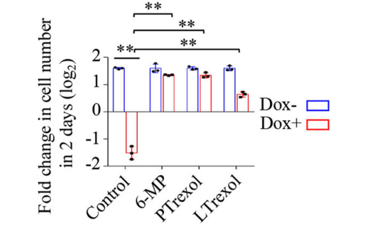
Identification of purine biosynthesis as an NADH-sensing pathway to mediate energy stress. [Abstract]2022 Nov 17;13(1):7031. PMID: 36396642
Lometrexol purchased from MedChemExpress. Usage Cited in: Nat Commun. 2022 Nov 17;13(1):7031. [Abstract]
Effects of purine biosynthesis inhibitors on proliferation of HeLaTet-on EcSTH cells (n = 3 biologically independent samples). Cells were pretreated with the indicated inhibitors for 2 h and counted after treatment with Dox (1 µg/mL) and inhibitors for 48 h. 6-Mercaptopurine (6-MP, 300 µM), pelitrexol (PTrexol, 10 µM), Lometrexol (LTrexol, 10 µM).
-
Sci Signal
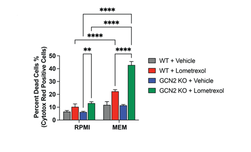
Coordination between the eIF2 kinase GCN2 and p53 signaling supports purine metabolism and the progression of prostate cancer. [Abstract]2024 Nov 26;17(864):eadp1375. PMID: 39591412
Lometrexol purchased from MedChemExpress. Usage Cited in: Sci Signal. 2024 Nov 26;17(864):eadp1375. [Abstract]
Measurements of cell death of 22Rv1 WT and GCN2 KO cells cultured in RPMI or MEM and treated with vehicle or Lometrexol (1 µM) for 6 days.
-
-
-
-


溶剤 & 溶解度
体外:
DMSO : 100 mg/mL (225.50 mM; Need ultrasonic; Hygroscopic DMSO has a significant impact on the solubility of product, please use newly opened DMSO)
Please refer to the solubility information to select the appropriate solvent. Once prepared, please aliquot and store the solution to prevent product inactivation from repeated freeze-thaw cycles.
Storage method and period of stock solution: -80°C, 6 months; -20°C, 1 month. When stored at -80°C, please use it within 6 months. When stored at -20°C, please use it within 1 month.
Please refer to the solubility information to select the appropriate solvent. Once prepared, please aliquot and store the solution to prevent product inactivation from repeated freeze-thaw cycles.
Storage method and period of stock solution: -80°C, 6 months; -20°C, 1 month. When stored at -80°C, please use it within 6 months. When stored at -20°C, please use it within 1 month.
濃度 (開始) × 体積 (開始) = 濃度 (終了) × 体積 (終了)
体内:
Select the appropriate dissolution method based on your experimental animal and administration route.
- For the following dissolution methods, please ensure to first prepare a clear stock solution using an In Vitro approach and then sequentially add co-solvents:
- To ensure reliable experimental results, the clarified stock solution can be appropriately stored based on storage conditions. As for the working solution for In Vivo experiments, it is recommended to prepare freshly and use it on the same day.
- The percentages shown for the solvents indicate their volumetric ratio in the final prepared solution. If precipitation or phase separation occurs during preparation, heat and/or sonication can be used to aid dissolution.
Add each solvent one by one: 10% DMSO 40% PEG300 5% Tween-80 45% Saline
Solubility: ≥ 2.5 mg/mL (5.64 mM); Clear solution
This protocol yields a clear solution of ≥ 2.5 mg/mL (saturation unknown).
Taking 1 mL working solution as an example, add 100 μL DMSO stock solution (25.0 mg/mL) to 400 μL PEG300, and mix evenly; then add 50 μL Tween-80 and mix evenly; then add 450 μL Saline to adjust the volume to 1 mL.
Preparation of Saline: Dissolve 0.9 g sodium chloride in ddH₂O and dilute to 100 mL to obtain a clear Saline solution.
Add each solvent one by one: 10% DMSO 90% (20% SBE-β-CD in Saline)
Solubility: ≥ 2.5 mg/mL (5.64 mM); Clear solution
This protocol yields a clear solution of ≥ 2.5 mg/mL (saturation unknown).
Taking 1 mL working solution as an example, add 100 μL DMSO stock solution (25.0 mg/mL) to 900 μL 20% SBE-β-CD in Saline, and mix evenly.
Preparation of 20% SBE-β-CD in Saline (4°C, storage for one week): 2 g SBE-β-CD powder is dissolved in 10 mL Saline, completely dissolve until clear.
For the following dissolution methods, please prepare the working solution directly:
It is recommended to prepare fresh solutions and use them promptly within a short period of time.
The percentages shown for the solvents indicate their volumetric ratio in the final prepared solution. If precipitation or phase separation occurs during preparation, heat and/or sonication can be used to aid dissolution.
In Vivo Dissolution Calculator
Please enter the basic information of animal experiments:
-
-
-
-
Recommended: Prepare an additional quantity of animals to account for potential losses during experiments.
Please enter your animal formula composition:
-
%DMSO +
Recommended: Keep the proportion of DMSO in working solution below 2% if your animal is weak.
-
%+
-
+%Tween-80 + +
-
%Saline +
The co-solvents required include: DMSO, . All of co-solvents are available by MedChemExpress (MCE). , Tween 80. All of co-solvents are available by MedChemExpress (MCE).
Working solution concentration: 0.22 mg/mL
Method for preparing stock solution: mg drug dissolved in μL DMSO. Stock solution concentration: mg/mL.
1. Take μL DMSO stock solution;
2. Add μL .
μL , mix evenly;
3. Then add μL Tween 80, mix evenly;
4. Then add μL
Please ensure that the stock solution in the first step is dissolved to a clear state, and add co-solvents in sequence. You can use ultrasonic heating (ultrasonic cleaner, recommended frequency 20-40 kHz), vortexing, etc. to assist dissolution.


純度とドキュメンテーション
-
データシート (278 KB)
-
SDS (420 KB)
- English - EN (420 KB)
- Français - FR (420 KB)
- Deutsch - DE (420 KB)
- Norwegian - NO (420 KB)
- Español - ES (420 KB)
- Swedish - SV (420 KB)
- Italian - IT (420 KB)
- Korean - KR (420 KB)
- Portuguese - PT (420 KB)
-
取扱説明書 (2659 KB)
参考文献
[1]. Xu L, et, al. The effect of inhibiting glycinamide ribonucleotide formyl transferase on the development of neural tube in mice. Nutr Metab (Lond). 2016 Aug 23;13(1):56. [Content Brief]
[2]. Scaletti E, et, al. Structural basis of inhibition of the human serine hydroxymethyltransferase SHMT2 by antifolate drugs. FEBS Lett. 2019 Jul;593(14):1863-1873. [Content Brief]
[3]. Bronder JL, et, al. Antifolates targeting purine synthesis allow entry of tumor cells into S phase regardless of p53 function. Cancer Res. 2002 Sep 15;62(18):5236-41. [Content Brief]


Complete Stock Solution Preparation Table
Please refer to the solubility information to select the appropriate solvent. Once prepared, please aliquot and store the solution to prevent product inactivation from repeated freeze-thaw cycles.
Storage method and period of stock solution: -80°C, 6 months; -20°C, 1 month. When stored at -80°C, please use it within 6 months. When stored at -20°C, please use it within 1 month.
| Optional Solvent | Concentration Solvent Mass | 1 mg | 5 mg | 10 mg | 25 mg |
|---|---|---|---|---|---|
| DMSO | 1 mM | 2.2550 mL | 11.2752 mL | 22.5505 mL | 56.3761 mL |
| 5 mM | 0.4510 mL | 2.2550 mL | 4.5101 mL | 11.2752 mL | |
| 10 mM | 0.2255 mL | 1.1275 mL | 2.2550 mL | 5.6376 mL | |
| 15 mM | 0.1503 mL | 0.7517 mL | 1.5034 mL | 3.7584 mL | |
| 20 mM | 0.1128 mL | 0.5638 mL | 1.1275 mL | 2.8188 mL | |
| 25 mM | 0.0902 mL | 0.4510 mL | 0.9020 mL | 2.2550 mL | |
| 30 mM | 0.0752 mL | 0.3758 mL | 0.7517 mL | 1.8792 mL | |
| 40 mM | 0.0564 mL | 0.2819 mL | 0.5638 mL | 1.4094 mL | |
| 50 mM | 0.0451 mL | 0.2255 mL | 0.4510 mL | 1.1275 mL | |
| 60 mM | 0.0376 mL | 0.1879 mL | 0.3758 mL | 0.9396 mL | |
| 80 mM | 0.0282 mL | 0.1409 mL | 0.2819 mL | 0.7047 mL | |
| 100 mM | 0.0226 mL | 0.1128 mL | 0.2255 mL | 0.5638 mL |


Lometrexol 関連分類
HY-14521 関連分類
 Powered by Bioz