



Pamidronic acid
Based on 1 publication(s) in Google Scholar
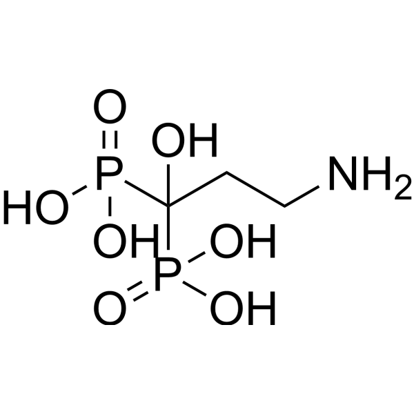
Pamidronic acid, the second-generation nitrogen-containing bisphosphonate, is an inhibitor of bone loss. Pamidronic acid significantly inhibits subchondral bone loss in early osteoarthritis by upregulating the expression of OPG in cartilage and subchondral bone, and inhibiting the expression of RANKL and MMP-9 in both tissues, as well as TLR-4 in cartilage, thereby alleviating cartilage degeneration. Additionally, Pamidronic acid can inhibit the signaling of Wnt and β-catenin, and is applicable for research on osteoporosis and osteosarcoma.
For research use only. We do not sell to patients.
- Purity : 99.05%
- CAS No.: 40391-99-9
- Formula: C3H11NO7P2
- Molecular Weight:235.07
-
Storage:
-20°C, protect from light
* In solvent : -80°C, 6 months; -20°C, 1 month (protect from light)

To place orders, for customer services and technical support, please contact: MedChemExpress USA
Tel: 609-228-6898 E-mail: [email protected] [email protected]


-
 Biological Activity
-
Chemical Information
-
Solvent & Solubility
- Protocols
- Purity & Documentation
- References
-
 Help & FAQs


-
HY-10218
EverolimusmTORC Inhibitor -
HY-14649
Retinoic acidRAR Agonist -
HY-17371
OxaliplatinDNA Synthesis Inhibitor -
HY-17394
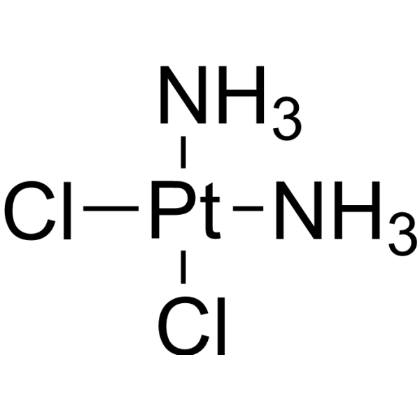
CisplatinDNA Alkylator -
HY-50878
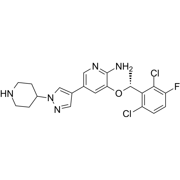
CrizotinibALK/c-Met/ROS1 Inhibitor -
HY-N0488
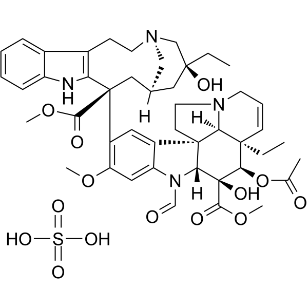
Vincristine sulfateAntineoplastic Agent -

HY-B0012R
Pamidronic acid (Standard)Bone Loss Inhibitors
Publications Citing Use of MedChemExpress (MCE) Pamidronic acid
More

Biological Activity
Description
Cellular Effect
|
Cell Line

|
Type | Value | Description | References |
|---|---|---|---|---|
| 13762 | IC50 |
33.6 μM
Compound: 38
|
Antiproliferative activity against rat 13762 cell line
Antiproliferative activity against rat 13762 cell line
|
[PMID: 16970405] |
| HepG2 | IC50 |
>200 μM
Compound: Pamidronate
|
Antimalarial activity against Plasmodium berghei NK65 infected in human HepG2 cells after 48 hrs by RT-PCR
Antimalarial activity against Plasmodium berghei NK65 infected in human HepG2 cells after 48 hrs by RT-PCR
|
[PMID: 20457823] |
| HFF | IC50 |
38 μM
Compound: 20
|
In vitro inhibitory concentration against the growth of Toxoplasma gondii in human foreskin fibroblast monolayer cells (HFF cells)
In vitro inhibitory concentration against the growth of Toxoplasma gondii in human foreskin fibroblast monolayer cells (HFF cells)
|
[PMID: 15857119] |
| MCF7 | IC50 |
141 μM
Compound: 38
|
Antiproliferative activity against human MCF7 cell line by MTT assay
Antiproliferative activity against human MCF7 cell line by MTT assay
|
[PMID: 16970405] |
| MCF7 | IC50 |
22.9 μM
Compound: Pamidronate
|
Cytotoxicity against human MCF7 cells assessed as cell growth inhibition measured after 24 to 48 hrs by SRB assay
Cytotoxicity against human MCF7 cells assessed as cell growth inhibition measured after 24 to 48 hrs by SRB assay
|
[PMID: 33132173] |
| NCI-H460 | IC50 |
71.8 μM
Compound: 38
|
Antiproliferative activity against human NCI-H460 cell line by MTT assay
Antiproliferative activity against human NCI-H460 cell line by MTT assay
|
[PMID: 16970405] |
| SF-268 | IC50 |
408 μM
Compound: 38
|
Antiproliferative activity against human SF-268 cell line by MTT assay
Antiproliferative activity against human SF-268 cell line by MTT assay
|
[PMID: 16970405] |
| T-cell | EC50 |
43 μM
Compound: 1
|
Effective concentration against human Gamma delta T cells
Effective concentration against human Gamma delta T cells
|
[PMID: 15828834] |
| T-cell | EC50 |
5.66 μM
Compound: pamidronate
|
Effective concentration to activate gammadelta T cells
Effective concentration to activate gammadelta T cells
|
[PMID: 12383012] |
| T-cell | IC50 |
62 μM
Compound: 34
|
Inhibitory activity, for stimulation of TNF-alpha release in gamma-delta T cells, using individual observed maximum TNF-alpha release
Inhibitory activity, for stimulation of TNF-alpha release in gamma-delta T cells, using individual observed maximum TNF-alpha release
|
[PMID: 14711309] |
| T-cell | IC50 |
941 μM
Compound: 34
|
Inhibitory activity, for stimulation of TNF-alpha release in gamma-delta T cells, using a constrained maximum TNF-alpha release of 2700 pg/mL
Inhibitory activity, for stimulation of TNF-alpha release in gamma-delta T cells, using a constrained maximum TNF-alpha release of 2700 pg/mL
|
[PMID: 14711309] |
| Vero | IC50 |
60 μM
Compound: 5
|
Inhibition of Trypanosoma cruzi Amastigotes was determined in Vero cells culture and fetal calf serum
Inhibition of Trypanosoma cruzi Amastigotes was determined in Vero cells culture and fetal calf serum
|
[PMID: 11300872] |
In Vitro
Pamidronic acid (0.001-1000 µM, 72 h) inhibits the cell viability of osteosarcoma cells POS, HMPOS and COS31, exhibits no cytotoxicity to fibroblasts[1].
MedChemExpress (MCE) has not independently confirmed the accuracy of these methods. They are for reference only. Further protocols information, click here.
-
Cell Line:POS, HMPOS and COS31
-
Concentration:0.001-1000 µM
-
Incubation Time:72 h
-
Result:Inhibited the cell viability.
In Vivo
MedChemExpress (MCE) has not independently confirmed the accuracy of these methods. They are for reference only.
-
Animal Model:Anterior cruciate ligament transection (ACLT)-induced osteoarthritis in rabbits[3]
-
Dosage:3 mg/kg
-
Administration:ear vein injection, once monthly for 14 weeks
-
Result:Increased bone volume fraction (BV/TV), trabecular thickness (TbTh) and trabecular number (TbN).
Reduced the erosion of cartilage surface, increase the content of chondrocytes and matrix.
Clinical Trial
| NCT Number | Sponsor | Condition | Start Date |
Phase
|
|---|---|---|---|---|
| NCT01329991 | Plexxikon| | 2011-05 | PHASE1 |


Chemical Information
-
CAS No. 40391-99-9
-
Appearance Solid
-
Molecular Weight 235.07
-
Formula C3H11NO7P2
-
Color White to off-white
-
SMILES
OC(P(O)(O)=O)(P(O)(O)=O)CCN
-
Shipping
Room temperature in continental US; may vary elsewhere.
-
Storage
-20°C, protect from light
* In solvent : -80°C, 6 months; -20°C, 1 month (protect from light)


Publications (1)
-
Journal Impact Factor
-
Most Recent
-
Cell Rep Methods
RECOVER identifies synergistic drug combinations in vitro through sequential model optimization. [Abstract]2023 Oct 23;3(10):100599. PMID: 37797618


Solvent & Solubility
In Vitro:
H2O : 5 mg/mL (21.27 mM; ultrasonic and warming and heat to 80°C)
DMSO : < 1 mg/mL (insoluble or slightly soluble)
Please refer to the solubility information to select the appropriate solvent. Once prepared, please aliquot and store the solution to prevent product inactivation from repeated freeze-thaw cycles.
Storage method and period of stock solution: -80°C, 6 months; -20°C, 1 month (protect from light). When stored at -80°C, please use it within 6 months. When stored at -20°C, please use it within 1 month.
* Note: If you choose water as the stock solution, please dilute it to the working solution, then filter and sterilize it with a 0.22 μm filter before use.
Please refer to the solubility information to select the appropriate solvent. Once prepared, please aliquot and store the solution to prevent product inactivation from repeated freeze-thaw cycles.
Storage method and period of stock solution: -80°C, 6 months; -20°C, 1 month (protect from light). When stored at -80°C, please use it within 6 months. When stored at -20°C, please use it within 1 month.
* Note: If you choose water as the stock solution, please dilute it to the working solution, then filter and sterilize it with a 0.22 μm filter before use.
Concentration (start) × Volume (start) = Concentration (final) × Volume (final)
In Vivo:
For the following dissolution methods, please prepare the working solution directly:
It is recommended to prepare fresh solutions and use them promptly within a short period of time.
The percentages shown for the solvents indicate their volumetric ratio in the final prepared solution. If precipitation or phase separation occurs during preparation, heat and/or sonication can be used to aid dissolution.
Add each solvent one by one: PBS
Solubility: 2 mg/mL (8.51 mM); Clear solution; Need ultrasonic

Protocols
-
RNA extraction experimental
By lysing cells, releasing RNA, and removing impurities such as proteins and DNA, high-purity RNA products are finally obtained. The commonly used traditional method is the guanidine isothiocyanate/phenol/chloroform method (Trizol), which is suitable for a variety of animal materials including animal tissues, microorganisms, cultured cells, etc., and most plant materials.
-
Mitochondrial membrane-potential fluorescent assay
Mitochondrial membrane potential fluorescent assays estimate ΔΨm in living cells using lipophilic cationic dyes such as TMRM, TMRE, rhodamine 123, and JC-1, which accumulate in mitochondria according to membrane polarization; loss of signal after FCCP or CCCP treatment is interpreted as mitochondrial depolarization. TMRM/TMRE and rhodamine 123 are commonly used for semi-quantitative live-cell microscopy or flow cytometry, while JC-1 can report a shift from red aggregate fluorescence to green monomer fluorescence during depolarization; interpretation requires controls because dye concentration, quenching mode, cell type, dye efflux, and mitochondrial mass can affect fluorescence independently of ΔΨm.
-
Mitochondrial membrane-potential and mitochondrial mass staining
Mitochondrial membrane potential staining measures the electrochemical polarization across the mitochondrial inner membrane in live cells using lipophilic cationic fluorescent probes; early rhodamine-based work showed that selective mitochondrial dye accumulation is lost when the mitochondrial transmembrane potential is dissipated. JC-1 reports mitochondrial polarization by shifting from green monomer fluorescence to red J-aggregate fluorescence as dye concentration increases within energized mitochondria; therefore, the red/green fluorescence ratio is used as a relative readout of mitochondrial membrane potential. TMRE or TMRM staining provides a single-channel relative readout because these cationic rhodamine esters accumulate in polarized mitochondria, and lower fluorescence indicates reduced mitochondrial polarization when acquisition and dye-loading conditions are controlled. Mitochondrial mass staining is commonly performed with MitoTracker Green FM or related MitoTracker dyes as


Purity & Documentation
-
Data Sheet (278 KB)
-
SDS (393 KB)
- English - EN (393 KB)
- Français - FR (393 KB)
- Deutsch - DE (393 KB)
- Norwegian - NO (393 KB)
- Español - ES (393 KB)
- Swedish - SV (393 KB)
- Italian - IT (393 KB)
- Korean - KR (393 KB)
- Portuguese - PT (393 KB)
-
Handling Instructions (2659 KB)
References
[1]. Ashton JA, et al. Investigation of the effect of pamidronate disodium on the in vitro viability of osteosarcoma cellsfrom dogs. Am J Vet Res. 2005 May;66(5):885-92. [Content Brief]
[2]. Xu Y, et al. Pamidronate Disodium Leads to Bone Necrosis via Suppression of Wnt/β-Catenin Signaling in Human Bone Marrow Mesenchymal Stem Cells In Vitro. J Oral Maxillofac Surg. 2017 Mar 23. [Content Brief]
[3]. Lv Y, et al. Effects of pamidronate disodium on the loss of osteoarthritic subchondral bone and the expression of cartilaginous and subchondral osteoprotegerin and RANKL in rabbits. BMC Musculoskelet Disord. 2014 Nov 6;15:371. [Content Brief]


Complete Stock Solution Preparation Table
Please refer to the solubility information to select the appropriate solvent. Once prepared, please aliquot and store the solution to prevent product inactivation from repeated freeze-thaw cycles.
Storage method and period of stock solution: -80°C, 6 months; -20°C, 1 month (protect from light). When stored at -80°C, please use it within 6 months. When stored at -20°C, please use it within 1 month.
| Optional Solvent | Concentration Solvent Mass | 1 mg | 5 mg | 10 mg | 25 mg |
|---|---|---|---|---|---|
| H2O | 1 mM | 4.2541 mL | 21.2703 mL | 42.5405 mL | 106.3513 mL |
| 5 mM | 0.8508 mL | 4.2541 mL | 8.5081 mL | 21.2703 mL | |
| 10 mM | 0.4254 mL | 2.1270 mL | 4.2541 mL | 10.6351 mL | |
| 15 mM | 0.2836 mL | 1.4180 mL | 2.8360 mL | 7.0901 mL | |
| 20 mM | 0.2127 mL | 1.0635 mL | 2.1270 mL | 5.3176 mL |
* Note: If you choose water as the stock solution, please dilute it to the working solution, then filter and sterilize it with a 0.22 μm filter before use.


Pamidronic acid Related Classifications
HY-B0012 Related Classifications
 Powered by Bioz