


Niraparib hydrochloride
Based on 87 publication(s) in Google Scholar
Niraparib hydrochloride (MK-4827 hydrochloride) is a highly potent and orally bioavailable PARP1 and PARP2 inhibitor with IC50s of 3.8 and 2.1 nM, respectively. Niraparib hydrochloride leads to inhibition of repair of DNA damage, activates apoptosis and shows anti-tumor activity.
For research use only. We do not sell to patients.
- Purity: 99.41%
- CAS No.: 1038915-64-8
- Formula: C19H21ClN4O
- Molecular Weight:356.85
-
Storage:
4°C, sealed storage, away from moisture
* In solvent : -80°C, 6 months; -20°C, 1 month (sealed storage, away from moisture)

To place orders, for customer services and technical support, please contact: MedChemExpress USA
Tel: 609-228-6898 E-mail: [email protected] [email protected]


-
 Biological Activity
-
Chemical Information
-
Solvent & Solubility
- Protocol
- Purity & Documentation
- References
-
 Help & FAQs
-


Anti-Infection Compound Library
HY-L002
-
Apoptosis Compound Library
HY-L003
-
Cell Cycle/DNA Damage Compound Library
HY-L004
-
Epigenetics Compound Library
HY-L005
-
FDA-Approved Drug Library
HY-L022
-
Anti-Cancer Compound Library
HY-L025
-
Anti-Aging Compound Library
HY-L034
-
Drug Repurposing Compound Library
HY-L035
-
Anti-COVID-19 Compound Library
HY-L052
-
Orally Active Compound Library
HY-L061
-
FDA Approved & Pharmacopeial Drug Library
HY-L066
-
Anti-Breast Cancer Compound Library
HY-L074
-
Anti-Pancreatic Cancer Compound Library
HY-L077
-
Targeted Therapy Drug Library
HY-L080
-
Rare Diseases Drug Library
HY-L102
-
Pediatric Drug Library
HY-L104
-
EMA-Approved Drug Library
HY-L116
-
FDA-Approved Anticancer Drug Library
HY-L122
-
Anti-Prostate Cancer Compound Library
HY-L124
-
Heterocyclic Compound Library
HY-L138
-
Cell Death Library
HY-L162
-
Anti-Ovarian Cancer Compound Library
HY-L173
-
Multi-Target Compound Library
HY-L176
-
Bioactive Compound Library Max
HY-L181
-
MCE Bioactive Compound Library
HY-L001V
-
Drug Repurposing Compound Library Plus
HY-L035P
-
FDA-Approved Drug Library Plus
HY-L022P
-
FDA-Approved Drug Library Mini
HY-L022M
-
Bioactive Compound Library
HY-L001
-
Non-Alcoholic Fatty Liver Disease (NAFLD) Compound Library
HY-L199
-
High-Throughput Bioactive Compound Library
HY-L205
-
Anti-Cancer Approved Drug Library
HY-L213


Publications Citing Use of MedChemExpress (MCE) Niraparib hydrochloride
More- Cancer Cell. 2020 Dec 14;38(6):844-856.e7. [Abstract]
- Cancer Discov. 2017 Sep;7(9):984-998. [Abstract]
- Nat Biomed Eng. 2024 Feb;8(2):165-176. [Abstract]
- Cancer Res. 2022 Jul 5;82(13):2361-2377. [Abstract]
- Drug Resist Updat. 2026 Jan:84:101319. [Abstract]
- Nat Commun. 2026 Feb 12;17(1):1214. [Abstract]
- Nat Commun. 2022 Nov 19;13(1):7107. [Abstract]
- Theranostics. 2020 Jul 25;10(21):9477-9494. [Abstract]
- Acta Pharm Sin B. 2025 Dec;15(12):6444-6460. [Abstract]
- J Clin Invest. 2026 Mar 17:e200260. [Abstract]
- J Exp Clin Cancer Res. 2025 Jun 16;44(1):175. [Abstract]
- J Clin Invest. 2019 Mar 1;129(3):1211-1228. [Abstract]
- Adv Sci (Weinh). 2025 Jul 16:e15585. [Abstract]
- Adv Sci (Weinh). 2022 Oct;9(30):e2201210. [Abstract]
- Sci Adv. Sci Adv. 2025 Apr 25;11(17):eadu0847. [Abstract]
- Cell Death Dis. 2026 Jun 3. [Abstract]
- Cell Death Dis. 2026 Jun 27;17(1):603. [Abstract]
- Cell Death Dis. 2020 Apr 6;11(4):219. [Abstract]
- Cancer Lett. 2022 Feb 1:526:180-192. [Abstract]
- Int J Biol Sci. 2020 Feb 21;16(8):1363-1375. [Abstract]
- Phytomedicine. 2026 Jun 11.
- EBioMedicine. 2020 Sep;59:102923. [Abstract]
- Mater Today Bio. 2020 Oct 22;8:100082. [Abstract]
- Clin Cancer Res. 2017 Feb 15;23(4):1001-1011. [Abstract]
- NPJ Precis Oncol. 2025 Aug 30;9(1):306. [Abstract]
- NPJ Precis Oncol. 2021 Jun 9;5(1):49. [Abstract]
- J Transl Med. 2025 Aug 5;23(1):860. [Abstract]
- Oncogene. 2022 Sep;41(37):4271-4281. [Abstract]
- Int J Biol Macromol. 2024 May 9:132275. [Abstract]
- Cell Rep. 2026 Jan 24;45(2):116910. [Abstract]
- J Med Chem. 2024 Feb 22;67(4):2349-2368. [Abstract]
- J Med Chem. 2023 Mar 23;66(6):4106-4130. [Abstract]
- Mol Cancer Ther. 2023 Apr 3;22(4):447-458. [Abstract]
- Mol Cancer Ther. 2022 Feb;21(2):245-256. [Abstract]
- JCI Insight. 2023 Nov 8;8(21):e165268. [Abstract]
- Biochem Pharmacol. 2025 May:235:116843. [Abstract]
- Int Immunopharmacol. 2025 Sep 23:162:115158. [Abstract]
- Int J Mol Sci. 2025 Mar 24;26(7):2921. [Abstract]
- J Mol Med (Berl). 2019 Aug;97(8):1183-1193. [Abstract]
- Am J Pathol. 2026 Jun 10:S0002-9440(26)00163-X. [Abstract]
- Int J Cancer. 2025 Jan 15;156(2):389-402. [Abstract]
- Am J Pathol. 2024 Nov;194(11):2007-2022. [Abstract]
- Mol Pharm. 2021 Dec 6;18(12):4371-4384. [Abstract]
- Sci Rep. 2021 Sep 27;11(1):19138. [Abstract]
- Cancers (Basel). 2024 Nov 5;16(22):3728. [Abstract]
- Cancers (Basel). 2023 May 11;15(10):2708. [Abstract]
- Appl Microbiol Biotechnol. 2019 Dec;103(23-24):9557-9568. [Abstract]
- Neurooncol Adv. 2024 Nov 19;6(1):vdae187. [Abstract]
- Bioengineering (Basel). 2025 Oct 19;12(10):1121. [Abstract]
- BMC Cancer. 2024 Dec 20;24(1):1562. [Abstract]
- BMC Cancer. 2022 Mar 23;22(1):312. [Abstract]
- Cancer Res Commun. 2024 May 20. [Abstract]
- Carcinogenesis. 2020 May 14;41(3):345-357. [Abstract]
- Front Oncol. 2021 Jul 9:11:681441. [Abstract]
- DNA Repair. 2019 Jan:73:64-70. [Abstract]
- Am J Cancer Res. 2024 Jan 15;14(1):378-389. [Abstract]
- Life Sci Alliance. 2021 Oct 5;4(12):e202101144. [Abstract]
- Nucl Med Biol. 2016 Dec;43(12):752-758. [Abstract]
- PeerJ. 2023 Nov 29:11:e16314. [Abstract]
- Urol Oncol. 2025 Sep 30:S1078-1439(25)00359-X. [Abstract]
- Cancer Chemother Pharmacol. 2017 Oct;80(4):861-867. [Abstract]
- Res Connect. 2026 May 28.
- bioRxiv. 2026 Mar 13.
- bioRxiv. 2025 July 08.
- Biomed Pharmacother. 2025 Jun 16:189:118273. [Abstract]
- bioRxiv. 2025 April 26.
- bioRxiv. 2025 January 14.
- Research Square Preprint. 2024 Nov 06.
- bioRxiv. 2024 Jul 10:2024.07.09.602803. [Abstract]
- J Oncol. 2022 Apr 5;2022:2800488. [Abstract]
- Elife. 2022 Apr 27;11:e72464. [Abstract]
- Patent. US20220048863A1.
- Research Square Preprint. 2022 Feb.
- Uppsala University. 2022 Feb.
- Patent. US20210299137A1.
- Elife. 2021 Jun 23:10:e69454. [Abstract]
- HAL. archives-ouvertes. 2020 Dec 4.
- Patent. US20200148645A1
- Patent. US20200129476A1
- Patent. US20200078369A1
- medRxiv. 2020 Jan.
- University of Belgrade. 2019 Aug.
- Patent. US20190092732A1.
- Patent. US20180362972A1.
- Patent. US20180134664A1.
- Patent. US9932310B2.
- Patent. US20170226063A1.
 Customer Validation & Images

-
WB
-
WB
-
Cell Proliferation/Viability Assay


Biological Activity
|
PARP-2 2.1 nM (IC50) |
PARP-1 3.8 nM (IC50) |
V-PARP 330 nM (IC50) |
TANK-1 570 nM (IC50) |
PARP-3 1300 nM (IC50) |
Niraparib inhibits PARP activity with EC50=4 nM and EC90=45 nM in a whole cell assay. Niraparib inhibits proliferation of cancer cells with mutant BRCA-1 and BRCA-2 with CC50 in the 10?100 nM range. Niraparib displays excellent PARP 1 and 2 inhibition with IC50=3.8 and 2.1 nM, respectively, and in a whole cell assay[1]. To validate that Niraparib inhibits PARP in these cell lines, A549 and H1299 cells are treated with 1 μM Niraparib for various times and measured PARP enzymatic activity using a chemiluminescent assay. The results show that Niraparib inhibits PARP within 15 minutes of treatment reaching about 85% inhibition in the A549 cells at 1 h and about 55% inhibition at 1 h for the H1299 cells[2].
MedChemExpress (MCE) has not independently confirmed the accuracy of these methods. They are for reference only.
MedChemExpress (MCE) has not independently confirmed the accuracy of these methods. They are for reference only.


Chemical Information
-
CAS No. 1038915-64-8
-
Appearance Solid
-
Molecular Weight 356.85
-
Formula C19H21ClN4O
-
Color White to light yellow
-
SMILES
NC(C1=CC=CC2=CN(C3=CC=C([C@H]4CNCCC4)C=C3)N=C21)=O.Cl
-
Synonyms
MK-4827 hydrochloride
-
Shipping
Room temperature in continental US; may vary elsewhere.
-
Storage
4°C, sealed storage, away from moisture
* In solvent : -80°C, 6 months; -20°C, 1 month (sealed storage, away from moisture)


Publications (87)
-
Journal Impact Factor
-
Most Recent
-
Cancer Cell
Elevated CXorf67 Expression in PFA Ependymomas Suppresses DNA Repair and Sensitizes to PARP Inhibitors. [Abstract]2020 Dec 14;38(6):844-856.e7. PMID: 33186520 -
Cancer Discov
Secondary Somatic Mutations Restoring RAD51C and RAD51D Associated with Acquired Resistance to the PARP Inhibitor Rucaparib in High-Grade Ovarian Carcinoma. [Abstract]2017 Sep;7(9):984-998. PMID: 28588062 -
Nat Biomed Eng
Functional annotation of variants of the BRCA2 gene via locally haploid human pluripotent stem cells. [Abstract]2024 Feb;8(2):165-176. PMID: 37488236 -
Cancer Res
2022 Jul 5;82(13):2361-2377. PMID: 35472077 -
Drug Resist Updat
ZBP1 antagonizes MRE11-mediated DNA end resection and confers synthetic lethality to PARP inhibition in ovarian cancer. [Abstract]2026 Jan:84:101319. PMID: 41192279 -
Nat Commun
Human iPSC-based Modeling of Pulmonary Fibrosis Reveals p300/CBP Inhibition Suppresses Alveolar Transitional Cell State. [Abstract]2026 Feb 12;17(1):1214. PMID: 41680175 -
Nat Commun
DNA mechanical flexibility controls DNA potential to activate cGAS-mediated immune surveillance. [Abstract]2022 Nov 19;13(1):7107. PMID: 36402783 -
Theranostics
Molecular signatures of BRCAness analysis identifies PARP inhibitor Niraparib as a novel targeted therapeutic strategy for soft tissue Sarcomas. [Abstract]2020 Jul 25;10(21):9477-9494. PMID: 32863940 -
Acta Pharm Sin B
The cytoskeletal protein smoothelin maintains homologous recombination repair by stabilizing RAD51 in an HUWE1-dependent manner in colorectal cancer. [Abstract]2025 Dec;15(12):6444-6460. PMID: 41477349 -
J Clin Invest
Targeting Wnt/β-Catenin and circadian regulator restores PRC2/EZH2 controlled chromatin bivalency and suppresses cell state diversity. [Abstract]2026 Mar 17:e200260. PMID: 41842971 -
J Exp Clin Cancer Res
2025 Jun 16;44(1):175. PMID: 40518539 -
J Clin Invest
PARP inhibition enhances tumor cell-intrinsic immunity in ERCC1-deficient non-small cell lung cancer. [Abstract]2019 Mar 1;129(3):1211-1228. PMID: 30589644 -
Adv Sci (Weinh)
Disruption of ARID1B Recruitment to the Nuclear Pore Complex as a New Anticancer Therapeutic Strategy. [Abstract]2025 Jul 16:e15585. PMID: 40671262 -
Adv Sci (Weinh)
Vanguard is a Glucose Deprivation-Responsive Long Non-Coding RNA Essential for Chromatin Remodeling-Reliant DNA Repair. [Abstract]2022 Oct;9(30):e2201210. PMID: 36047643 -
Sci Adv
Acute BRCAness induction and AR pathway blockage through CDK12/7/9 degradation enhances PARP inhibitor sensitivity in prostate cancer. [Abstract]Sci Adv. 2025 Apr 25;11(17):eadu0847. PMID: 40267193 -
Cell Death Dis
Cdk1-phosphorylated Nur77 accumulates at the centrosome during mitosis to regulate the Cep192-PLK1 signaling axis. [Abstract]2026 Jun 3. PMID: 42236697 -
Cell Death Dis
Targeting EZH2-driven cholesterol metabolic vulnerability through Napabucasin suppresses ovarian cancer metastasis. [Abstract]2026 Jun 27;17(1):603. PMID: 42373608 -
Cell Death Dis
Pan-cancer analysis reveals synergistic effects of CDK4/6i and PARPi combination treatment in RB-proficient and RB-deficient breast cancer cells. [Abstract]2020 Apr 6;11(4):219. PMID: 32249776 -
Cancer Lett
Inhibiting Src-mediated PARP1 tyrosine phosphorylation confers synthetic lethality to PARP1 inhibition in HCC. [Abstract]2022 Feb 1:526:180-192. PMID: 34762994 -
Int J Biol Sci
Quantitative determination of niraparib and olaparib tumor distribution by mass spectrometry imaging. [Abstract]2020 Feb 21;16(8):1363-1375. PMID: 32210725 -
-
EBioMedicine
Molecular correlates of sensitivity to PARP inhibition beyond homologous recombination deficiency in pre-clinical models of colorectal cancer point to wild-type TP53 activity. [Abstract]2020 Sep;59:102923. PMID: 32799124 -
Mater Today Bio
Low dose novel PARP-PI3K inhibition via nanoformulation improves colorectal cancer immunoradiotherapy. [Abstract]2020 Oct 22;8:100082. PMID: 33294836 -
Clin Cancer Res
Drug-Driven Synthetic Lethality: Bypassing Tumor Cell Genetics with a Combination of AsiDNA and PARP Inhibitors. [Abstract]2017 Feb 15;23(4):1001-1011. PMID: 27559053 -
NPJ Precis Oncol
Ex vivo 3D micro-tumour testing platform for predicting clinical response to platinum-based therapy in patients with high-grade serous ovarian cancer. [Abstract]2025 Aug 30;9(1):306. PMID: 40885787 -
NPJ Precis Oncol
PARP inhibitors promote stromal fibroblast activation by enhancing CCL5 autocrine signaling in ovarian cancer. [Abstract]2021 Jun 9;5(1):49. PMID: 34108603 -
J Transl Med
Ivabradine induces RAD51 degradation, potentiating PARP inhibitor efficacy in non-germline BRCA pathogenic variant triple-negative breast cancer. [Abstract]2025 Aug 5;23(1):860. PMID: 40764992 -
Oncogene
A genome-wide CRISPR-Cas9 knockout screen identifies novel PARP inhibitor resistance genes in prostate cancer. [Abstract]2022 Sep;41(37):4271-4281. PMID: 35933519 -
Int J Biol Macromol
Injectable bio-multifunctional hyaluronic acid-based hydrogels loaded with poly ADP-ribose polymerase inhibitors for ovarian cancer therapy. [Abstract]2024 May 9:132275. PMID: 38734345 -
Cell Rep
2026 Jan 24;45(2):116910. PMID: 41581148 -
J Med Chem
2024 Feb 22;67(4):2349-2368. PMID: 38299539 -
J Med Chem
Comparative Efficacy and Selectivity of Pharmacological Inhibitors of DYRK and CLK Protein Kinases. [Abstract]2023 Mar 23;66(6):4106-4130. PMID: 36876904 -
Mol Cancer Ther
Bepotastine sensitizes ovarian cancer to PARP inhibitors through suppressing NF-κB-triggered SASP in cancer-associated fibroblasts. [Abstract]2023 Apr 3;22(4):447-458. PMID: 36780236 -
Mol Cancer Ther
RP-3500: A Novel, Potent, and Selective ATR Inhibitor that is Effective in Preclinical Models as a Monotherapy and in Combination with PARP Inhibitors. [Abstract]2022 Feb;21(2):245-256. PMID: 34911817 -
JCI Insight
Mutant RB1 enhances therapeutic efficacy of PARPis in lung adenocarcinoma by triggering the cGAS/STING pathway. [Abstract]2023 Nov 8;8(21):e165268. PMID: 37937640 -
Biochem Pharmacol
Discovery of Bi-magnolignan as a novel BRD4 inhibitor inducing apoptosis and DNA damage for cancer therapy. [Abstract]2025 May:235:116843. PMID: 40024351 -
Int Immunopharmacol
Fluzoparib disrupts Golgi apparatus to inhibit O-GlcNAcylation and nuclear translocation of β-catenin to attenuate ovarian cancer invasion and metastasis. [Abstract]2025 Sep 23:162:115158. PMID: 40602265 -
Int J Mol Sci
Trabectedin Induces Synthetic Lethality via the p53-Dependent Apoptotic Pathway in Ovarian Cancer Cells Without BRCA Mutations When Used in Combination with Niraparib. [Abstract]2025 Mar 24;26(7):2921. PMID: 40243501 -
J Mol Med (Berl)
2019 Aug;97(8):1183-1193. PMID: 31201471 -
Am J Pathol
Low-Dose Radiotherapy Attenuates Pulmonary Granulomas Involving Ataxia-Telangiectasia Mutated-Dependent Modulation of the Interferon-β Response: A Host-Directed Therapeutic Strategy for Tuberculosis. [Abstract]2026 Jun 10:S0002-9440(26)00163-X. PMID: 42270072 -
Int J Cancer
Combined inhibition of RAD51 and CHK1 causes synergistic toxicity in cisplatin resistant cancer cells by triggering replication fork collapse. [Abstract]2025 Jan 15;156(2):389-402. PMID: 39239809 -
Am J Pathol
2024 Nov;194(11):2007-2022. PMID: 39168365 -
Mol Pharm
P-Glycoprotein (ABCB1/MDR1) Controls Brain Penetration and Intestinal Disposition of the PARP1/2 Inhibitor Niraparib. [Abstract]2021 Dec 6;18(12):4371-4384. PMID: 34730366 -
Sci Rep
2021 Sep 27;11(1):19138. PMID: 34580349 -
Cancers (Basel)
2024 Nov 5;16(22):3728. PMID: 39594684 -
Cancers (Basel)
[89Zr]-Atezolizumab-PET Imaging Reveals Longitudinal Alterations in PDL1 during Therapy in TNBC Preclinical Models. [Abstract]2023 May 11;15(10):2708. PMID: 37345044 -
Appl Microbiol Biotechnol
Autophagy suppression enhances DNA damage and cell death upon treatment with PARP inhibitor Niraparib in laryngeal squamous cell carcinoma. [Abstract]2019 Dec;103(23-24):9557-9568. PMID: 31686145
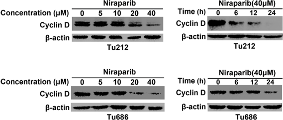
Niraparib hydrochloride purchased from MedChemExpress. Usage Cited in: Appl Microbiol Biotechnol. 2019 Dec;103(23-24):9557-9568. [Abstract]
Cyclin D is evaluated via western blot analysis in different cell lines with the treatment of Niraparib in different concentrations and times.
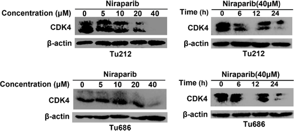
Niraparib hydrochloride purchased from MedChemExpress. Usage Cited in: Appl Microbiol Biotechnol. 2019 Dec;103(23-24):9557-9568. [Abstract]
CDK4 is evaluated via western blot analysis in different cell lines with the treatment of Niraparib in different concentrations and times.
-
Neurooncol Adv
Ion channel modulator DPI-201-106 significantly enhances antitumor activity of DNA damage response inhibitors in glioblastoma. [Abstract]2024 Nov 19;6(1):vdae187. PMID: 39659830 -
Bioengineering (Basel)
Precision Oncology for High-Grade Gliomas: A Tumor Organoid Model for Adjuvant Treatment Selection. [Abstract]2025 Oct 19;12(10):1121. PMID: 41155119 -
BMC Cancer
Regorafenib induces DNA damage and enhances PARP inhibitor efficacy in pancreatic ductal carcinoma. [Abstract]2024 Dec 20;24(1):1562. PMID: 39707244 -
BMC Cancer
PARP inhibitors chemopotentiate and synergize with cisplatin to inhibit bladder cancer cell survival and tumor growth. [Abstract]2022 Mar 23;22(1):312. PMID: 35321693 -
Cancer Res Commun
Nucleolar Localization of the RNA Helicase DDX21 Predicts Survival Outcomes in Gynecological Cancers. [Abstract]2024 May 20. PMID: 38767454 -
Carcinogenesis
2020 May 14;41(3):345-357. PMID: 31175354 -
Front Oncol
The Emerging Role of Poly (ADP-Ribose) Polymerase Inhibitors as Effective Therapeutic Agents in Renal Cell Carcinoma. [Abstract]2021 Jul 9:11:681441. PMID: 34307148 -
DNA Repair
Loss of the p12 subunit of DNA polymerase delta leads to a defect in HR and sensitization to PARP inhibitors. [Abstract]2019 Jan:73:64-70. PMID: 30470508 -
Am J Cancer Res
Novel dual action PARP and microtubule polymerization inhibitor AMXI-5001 powerfully inhibits growth of esophageal carcinoma both alone and in combination with radiotherapy. [Abstract]2024 Jan 15;14(1):378-389. PMID: 38323288 -
Life Sci Alliance
Suppression of isoprenylcysteine carboxylmethyltransferase compromises DNA damage repair. [Abstract]2021 Oct 5;4(12):e202101144. PMID: 34610973 -
Nucl Med Biol
Iodinated benzimidazole PARP radiotracer for evaluating PARP1/2 expression in vitro and in vivo. [Abstract]2016 Dec;43(12):752-758. PMID: 27689533 -
PeerJ
Niraparib restrains prostate cancer cell proliferation and metastasis and tumor growth in mice by regulating the lncRNA MEG3/miR-181-5p/GATA6 pathway. [Abstract]2023 Nov 29:11:e16314. PMID: 38047026 -
Urol Oncol
PTTG1-mediated reprogramming of asparagine metabolism enhances DNA damage repair and leads to compromised antitumor immunity in prostate cancer. [Abstract]2025 Sep 30:S1078-1439(25)00359-X. PMID: 41033896 -
Cancer Chemother Pharmacol
2017 Oct;80(4):861-867. PMID: 28756516
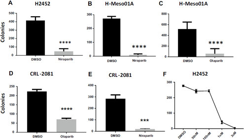
Niraparib hydrochloride purchased from MedChemExpress. Usage Cited in: Cancer Chemother Pharmacol. 2017 Oct;80(4):861-867. [Abstract]
PARP1 inhibition is lethal to MPM cells. Colony formation assays of clonal cell survival with continuous Niraparib or AZD2281.
-
-
-
-
Biomed Pharmacother
Synergistic suppression of cholangiocarcinoma cells via DNA damage response and cell cycle arrest by dual targeting PARP and ATM in DNA damage repair pathway. [Abstract]2025 Jun 16:189:118273. PMID: 40527032 -
-
-
-
bioRxiv
Acute BRCAness Induction and AR Signaling Blockage through CDK12/7/9 Degradation Enhances PARP Inhibitor Sensitivity in Prostate Cancer. [Abstract]2024 Jul 10:2024.07.09.602803. PMID: 39026842 -
J Oncol
The PARP1 Inhibitor Niraparib Represses DNA Damage Repair and Synergizes with Temozolomide for Antimyeloma Effects. [Abstract]2022 Apr 5;2022:2800488. PMID: 35422863 -
Elife
Development and characterization of new tools for detecting poly(ADP-ribose) in vitro and in vivo. [Abstract]2022 Apr 27;11:e72464. PMID: 35476036 -
-
-
-
-
Elife
Exosome component 1 cleaves single-stranded DNA and sensitizes human kidney renal clear cell carcinoma cells to poly(ADP-ribose) polymerase inhibitor. [Abstract]2021 Jun 23:10:e69454. PMID: 34159897 -
-
-
-
-
-
-
-
-
-
-


Solvent & Solubility
DMSO : 250 mg/mL (700.57 mM; Need ultrasonic; Hygroscopic DMSO has a significant impact on the solubility of product, please use newly opened DMSO)
H2O : 100 mg/mL (280.23 mM; Need ultrasonic)
Please refer to the solubility information to select the appropriate solvent. Once prepared, please aliquot and store the solution to prevent product inactivation from repeated freeze-thaw cycles.
Storage method and period of stock solution: -80°C, 6 months; -20°C, 1 month (sealed storage, away from moisture). When stored at -80°C, please use it within 6 months. When stored at -20°C, please use it within 1 month.
* Note: If you choose water as the stock solution, please dilute it to the working solution, then filter and sterilize it with a 0.22 μm filter before use.
Concentration (start) × Volume (start) = Concentration (final) × Volume (final)
Select the appropriate dissolution method based on your experimental animal and administration route.
- For the following dissolution methods, please ensure to first prepare a clear stock solution using an In Vitro approach and then sequentially add co-solvents:
- To ensure reliable experimental results, the clarified stock solution can be appropriately stored based on storage conditions. As for the working solution for In Vivo experiments, it is recommended to prepare freshly and use it on the same day.
- The percentages shown for the solvents indicate their volumetric ratio in the final prepared solution. If precipitation or phase separation occurs during preparation, heat and/or sonication can be used to aid dissolution.
Add each solvent one by one: 10% DMSO 40% PEG300 5% Tween-80 45% Saline
Solubility: ≥ 2.08 mg/mL (5.83 mM); Clear solution
This protocol yields a clear solution of ≥ 2.08 mg/mL (saturation unknown).
Taking 1 mL working solution as an example, add 100 μL DMSO stock solution (20.8 mg/mL) to 400 μL PEG300, and mix evenly; then add 50 μL Tween-80 and mix evenly; then add 450 μL Saline to adjust the volume to 1 mL.
Preparation of Saline: Dissolve 0.9 g sodium chloride in ddH₂O and dilute to 100 mL to obtain a clear Saline solution.
Add each solvent one by one: 10% DMSO 90% (20% SBE-β-CD in Saline)
Solubility: ≥ 2.08 mg/mL (5.83 mM); Clear solution
This protocol yields a clear solution of ≥ 2.08 mg/mL (saturation unknown).
Taking 1 mL working solution as an example, add 100 μL DMSO stock solution (20.8 mg/mL) to 900 μL 20% SBE-β-CD in Saline, and mix evenly.
Preparation of 20% SBE-β-CD in Saline (4°C, storage for one week): 2 g SBE-β-CD powder is dissolved in 10 mL Saline, completely dissolve until clear.
For the following dissolution methods, please prepare the working solution directly:
It is recommended to prepare fresh solutions and use them promptly within a short period of time.
The percentages shown for the solvents indicate their volumetric ratio in the final prepared solution. If precipitation or phase separation occurs during preparation, heat and/or sonication can be used to aid dissolution.
Add each solvent one by one: PBS
Solubility: 100 mg/mL (280.23 mM); Clear solution; Need ultrasonic
Please enter the basic information of animal experiments:
-
-
-
-
Recommended: Prepare an additional quantity of animals to account for potential losses during experiments.
Working solution concentration: 0.22 mg/mL
This product has good water solubility, please refer to the measured solubility data in water/PBS/Saline for details.

Protocol
Enzyme assay is conducted in buffer containing 25 mM Tris, pH 8.0, 1 mM DTT, 1 mM spermine, 50 mM KCl, 0.01% Nonidet P-40, and 1 mM MgCl2. PARP reaction contains 0.1 μCi [3H]NAD+ (200 000 DPM), 1.5 μM NAD+, 150 nM biotinylated NAD+, 1 μg/mL activated calf thymus, and 1−5 nM PARP-1. Autoreactions utilizing SPA bead-based detection are carried out in 50 μL volumes in white 96-well plates. Compounds (e.g., Niraparib) are prepared in 11-point serial dilution in 96-well plate, 5 μL/well in 5% DMSO/H2O (10× concentrated). Reactions are initiated by adding first 35 μL of PARP-1 enzyme in buffer and incubating for 5 min at room temperature and then 10 μL of NAD+ and DNA substrate mixture. After 3 h at room temperature, these reactions are terminated by the addition of 50 μL of streptavidin-SPA beads (2.5 mg/mL in 200 mM EDTA, pH 8). After 5 min, they are counted using a TopCount microplate scintillation counter. IC50 data is determined from inhibition curves at various substrate concentrations[1].
MedChemExpress (MCE) has not independently confirmed the accuracy of these methods. They are for reference only.
The inhibition of PARP is analyzed in A549 and H1299 cells using the HT Universal Chemiluminescent PARP Assay Kit. Briefly, cells are treated with DMSO or 1 μM niraparib for 15, 30, 60, or 120 minutes, trypsinized, and transferred to a pre-chilled tube. The cells are washed twice with ice cold PBS and resuspended in cold PARP extraction buffer. The cell suspensions are incubated on ice for 30 minutes with periodic vortexing to disrupt the cell membrane. The suspensions are centrifuged and the supernatant transferred to a pre-chilled tube on ice. The histone coated wells of the 96-well plate are rehydrated with 1X PARP buffer and incubated at room temperature for 30 minutes. The PARP buffer is removed and 20 μg of protein as determined by the Bio-Rad Protein Assay is added to each well followed by diluted PARP-HSA enzyme and 1X PARP buffer. The strip wells are then incubated at room temperature for 60 minutes, washed twice with PBS containing 0.1% Triton X-100, and then washed with PBS. Diluted Strep-HRP is added to the strip wells and incubated for 60 minutes at room temperature. The wells are washed again as before. Equal volumes of PeroxyGlow A and B are combined and added to the wells and chemiluminescent readings are obtained immediately using a plate-reader[2].
MedChemExpress (MCE) has not independently confirmed the accuracy of these methods. They are for reference only.
Mice[3]
Female nude mice (Ncr Nu/Nu) are randomly assigned to treatment groups consisting of 5 to 8 mice each when tumors grew to 6.0 mm in diameter at which time treatment with Niraparib is initiated. Niraparib is given at a dose of 25 mg/kg twice daily or 50 mg/kg once daily for either 21 days or is discontinued at 9 days from the time tumors reached 8 mm in diameter. Fractionated local tumor irradiation (XRT) is given when tumors reach 8.0 mm in diameter (7.7-8.2 mm). Radiation (2 Gy per fraction) is delivered to the tumor-bearing leg of mice once daily for 14 consecutive days or twice daily for 7 consecutive days using a small-animal irradiator consisting of two parallel-opposed 137Cs sources, at a dose rate of 5 Gy/min. During irradiation un-anesthetized mice are mechanically immobilized in a jig so that the tumor is centered within a 3.0 cm diameter radiation field and the animal’s body shielded from radiation exposure. On the day when both Niraparib and radiation are given, drug is administered 1 h before the first radiation dose of the day.
MedChemExpress (MCE) has not independently confirmed the accuracy of these methods. They are for reference only.


Purity & Documentation
-
Data Sheet (283 KB)
-
SDS (252 KB)
- English - EN (252 KB)
- Français - FR (252 KB)
- Deutsch - DE (252 KB)
- Norwegian - NO (252 KB)
- Español - ES (252 KB)
- Swedish - SV (252 KB)
- Italian - IT (252 KB)
- Korean - KR (252 KB)
- Portuguese - PT (252 KB)
-
Handling Instructions (2659 KB)
References
[1]. Jones P, et al. Discovery of 2-{4-[(3S)-piperidin-3-yl]phenyl}-2H-indazole-7-carboxamide (MK-4827): a novel oral poly(ADP-ribose)polymerase (PARP) inhibitor efficacious in BRCA-1 and -2 mutant tumors. J Med Chem. 2009 Nov 26;52(22):7170-85. [Content Brief]
[2]. Bridges KA, et al. Niraparib (MK-4827), a novel poly(ADP-Ribose) polymerase inhibitor, radiosensitizes human lung and breast cancer cells. Oncotarget. 2014 Jul 15;5(13):5076-86. [Content Brief]
[3]. Wang L, et al. MK-4827, a PARP-1/-2 inhibitor, strongly enhances response of human lung and breast cancer xenografts to radiation. Invest New Drugs. 2012 Dec;30(6):2113-20. [Content Brief]
[4]. Mirza MR, et al. Niraparib Maintenance Therapy in Platinum-Sensitive, Recurrent Ovarian Cancer. N Engl J Med. 2016 Dec 1;375(22):2154-2164. [Content Brief]


Complete Stock Solution Preparation Table
Please refer to the solubility information to select the appropriate solvent. Once prepared, please aliquot and store the solution to prevent product inactivation from repeated freeze-thaw cycles.
Storage method and period of stock solution: -80°C, 6 months; -20°C, 1 month (sealed storage, away from moisture). When stored at -80°C, please use it within 6 months. When stored at -20°C, please use it within 1 month.
| Optional Solvent | Concentration Solvent Mass | 1 mg | 5 mg | 10 mg | 25 mg |
|---|---|---|---|---|---|
| H2O / DMSO | 1 mM | 2.8023 mL | 14.0115 mL | 28.0230 mL | 70.0574 mL |
| 5 mM | 0.5605 mL | 2.8023 mL | 5.6046 mL | 14.0115 mL | |
| 10 mM | 0.2802 mL | 1.4011 mL | 2.8023 mL | 7.0057 mL | |
| 15 mM | 0.1868 mL | 0.9341 mL | 1.8682 mL | 4.6705 mL | |
| 20 mM | 0.1401 mL | 0.7006 mL | 1.4011 mL | 3.5029 mL | |
| 25 mM | 0.1121 mL | 0.5605 mL | 1.1209 mL | 2.8023 mL | |
| 30 mM | 0.0934 mL | 0.4670 mL | 0.9341 mL | 2.3352 mL | |
| 40 mM | 0.0701 mL | 0.3503 mL | 0.7006 mL | 1.7514 mL | |
| 50 mM | 0.0560 mL | 0.2802 mL | 0.5605 mL | 1.4011 mL | |
| 60 mM | 0.0467 mL | 0.2335 mL | 0.4670 mL | 1.1676 mL | |
| 80 mM | 0.0350 mL | 0.1751 mL | 0.3503 mL | 0.8757 mL | |
| 100 mM | 0.0280 mL | 0.1401 mL | 0.2802 mL | 0.7006 mL |
* Note: If you choose water as the stock solution, please dilute it to the working solution, then filter and sterilize it with a 0.22 μm filter before use.


 Powered by Bioz