



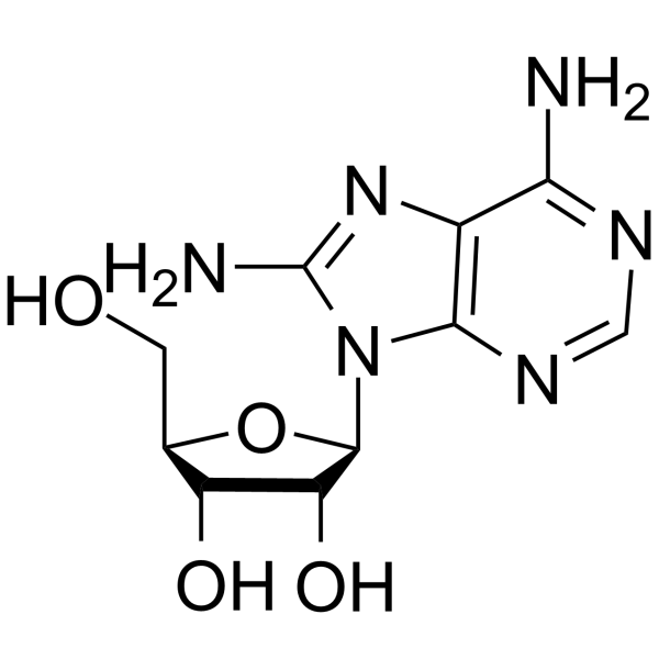
8-Aminoadenosine
Based on 1 publication(s) in Google Scholar
8-Aminoadenosine (8-NH2-Ado), a RNA-directed nucleoside analogue, reduces cellular ATP levels and inhibits mRNA synthesis. 8-Aminoadenosine blocks Akt/mTOR signaling and induces autophagy and apoptosis in a p53-independent manner. 8-Aminoadenosine has antitumor activity.
Nur für Forschungszwecke. Wir verkaufen nicht an Patienten.
- Reinheit : 99.05%
- CAS. Nr.: 3868-33-5
- Formel: C10H14N6O4
- Molecular Weight:282.26
-
Speicherung:Powder -20°C, 3 years , 4°C, 2 years ; In solvent -80°C, 6 months , -20°C, 1 month

To place orders, for customer services and technical support, please contact: MedChemExpress USA
Tel: 609-228-6898 E-mail: [email protected] [email protected]


-
 Biologische Aktivität
-
Chemical Information
-
Lösungsmittel & Löslichkeit
- Protokoll
- Reinheit & Dokumentation
- Verweise
-
 Help & FAQs


Publications Citing Use of MedChemExpress (MCE) 8-Aminoadenosine
MoreAlle DNA/RNA Synthesis Isoform-spezifische Produkte anzeigen
More

Biologische Aktivität
Beschreibung
|
|
mTOR |
Cellular Effect
|
Cell Line

|
Type | Value | Description | References |
|---|---|---|---|---|
| HCT-116 | GI50 |
0.05 μM
Compound: 4
|
Cytotoxicity against human HCT116 cells by SRB assay
Cytotoxicity against human HCT116 cells by SRB assay
|
[PMID: 19256508] |
| HCT-116 | GI50 |
0.05 μM
Compound: 2
|
Antiproliferative activity against human HCT116 cells
Antiproliferative activity against human HCT116 cells
|
[PMID: 21526763] |
In Vitro
8-Aminoadenosine (8-NH2-Ado; 0.1-10 μM; for 48 h) has IC50s of 1.5 μM and 8.88 μM in MM.1S and U266 cells, respectively[1].
8-Aminoadenosine (10 μM; for 24 h) induces significant apoptotic death of MCF-7 cells in p53-independent pathway. 8-Aminoadenosine causes PARP cleavage in MCF-7 cells[2].
8-Aminoadenosine (3 μM; 0.5-4 h) induces autophagy in the MM.1S cell line[1].
8-Aminoadenosine (3 μM; 2-16 h) causes a greater drop in ATP levels in the MM.1S cells[1].
8-Aminoadenosine (3 μM; 5 h) causes a 50% reduction in glucose consumption in MM.1S cells[1].
8-Aminoadenosine (3 μM; 5 h) indicates a time-dependent decrease in GLUT1 expression at 5 h, whereas at 24 h there was a down-regulation of both transporters (GLUT1 and GLUT4) in MM.1S cells[1].
8-Aminoadenosine inhibits cell proliferation, activated cell death, and does not activate transcription of the p53 target gene p21 or increase protein levels of either p53 or p21[1].
The toxic effects of 8-Aminoadenosine require adenosine kinase activity to convert 8-Aminoadenosine to 8-NH2-ATP in adenosine kinase-deficient cells[1].
MedChemExpress (MCE) has not independently confirmed the accuracy of these methods. They are for reference only.
-
Cell Line:MM.1S and U266 cells
-
Concentration:0.1, 0.3, 1, 3, 10 μM
-
Incubation Time:For 48 hours
-
Result:Had IC50s of 1.5 μM and 8.88 μM in MM.1S and U266 cells, respectively.
-
Cell Line:MCF-7 cells
-
Concentration:10 μM
-
Incubation Time:For 24 hours
-
Result:Induced significant apoptotic death.
Apoptosis was not inhibited by knockdown of functional p53.
-
Cell Line:MM.1S cell line
-
Concentration:3 μM
-
Incubation Time:0.5, 1, 1.5, 2, 2.5, 3, 3.5, 4 hours
-
Result:Induced the formation of LC3-II protein.
Caused the appearance of a population with a high AVO content with 1 μM for 24 hours.


Chemical Information
-
CAS. Nr. 3868-33-5
-
Appearance Solid
-
Molecular Weight 282.26
-
Formel C10H14N6O4
-
Color White to off-white
-
SMILES
O[C@H]1[C@@H](O)[C@H](N2C(N=CN=C3N)=C3N=C2N)O[C@@H]1CO
-
Synonyms
8-NH2-Ado
-
Structure Classification
-
Initial Source
Penicillium expansum
-
Versand
Room temperature in continental US; may vary elsewhere.
-
Speicherung
Powder -20°C 3 years 4°C 2 years In solvent -80°C 6 months -20°C 1 month


Publications (1)
-
Journal Impact Factor
-
Most Recent


Lösungsmittel & Löslichkeit
In Vitro:
DMSO : 83.33 mg/mL (295.22 mM; Need ultrasonic; Hygroscopic DMSO has a significant impact on the solubility of product, please use newly opened DMSO)
Please refer to the solubility information to select the appropriate solvent. Once prepared, please aliquot and store the solution to prevent product inactivation from repeated freeze-thaw cycles.
Storage method and period of stock solution: -80°C, 6 months; -20°C, 1 month. When stored at -80°C, please use it within 6 months. When stored at -20°C, please use it within 1 month.
Please refer to the solubility information to select the appropriate solvent. Once prepared, please aliquot and store the solution to prevent product inactivation from repeated freeze-thaw cycles.
Storage method and period of stock solution: -80°C, 6 months; -20°C, 1 month. When stored at -80°C, please use it within 6 months. When stored at -20°C, please use it within 1 month.
Konzentration (Stammlösung) × Volumen (Stammlösung) = Konzentration (Ziellösung) × Volumen (Ziellösung)


Reinheit & Dokumentation
-
Data Sheet (296 KB)
-
SDS (420 KB)
- English - EN (420 KB)
- Français - FR (420 KB)
- Deutsch - DE (420 KB)
- Norwegian - NO (420 KB)
- Español - ES (420 KB)
- Swedish - SV (420 KB)
- Italian - IT (420 KB)
- Korean - KR (420 KB)
- Portuguese - PT (420 KB)
-
Handling Instructions (2659 KB)
Verweise
[1]. Mala Shanmugam, et al. Targeting glucose consumption and autophagy in myeloma with the novel nucleoside analogue 8-aminoadenosine. J Biol Chem. 2009 Sep 25;284(39):26816-30. [Content Brief]
[2]. Alla Polotskaia, et al. 8-Amino-adenosine activates p53-independent cell death of metastatic breast cancers. Mol Cancer Ther. 2012 Nov;11(11):2495-504. [Content Brief]
[3]. Jennifer Ann Frey, et al. 8-Amino-adenosine inhibits multiple mechanisms of transcription. Mol Cancer Ther. 2010 Jan;9(1):236-45. [Content Brief]


Complete Stock Solution Preparation Table
Please refer to the solubility information to select the appropriate solvent. Once prepared, please aliquot and store the solution to prevent product inactivation from repeated freeze-thaw cycles.
Storage method and period of stock solution: -80°C, 6 months; -20°C, 1 month. When stored at -80°C, please use it within 6 months. When stored at -20°C, please use it within 1 month.
| Optional Solvent | Concentration Solvent Mass | 1 mg | 5 mg | 10 mg | 25 mg |
|---|---|---|---|---|---|
| DMSO | 1 mM | 3.5428 mL | 17.7142 mL | 35.4283 mL | 88.5708 mL |
| 5 mM | 0.7086 mL | 3.5428 mL | 7.0857 mL | 17.7142 mL | |
| 10 mM | 0.3543 mL | 1.7714 mL | 3.5428 mL | 8.8571 mL | |
| 15 mM | 0.2362 mL | 1.1809 mL | 2.3619 mL | 5.9047 mL | |
| 20 mM | 0.1771 mL | 0.8857 mL | 1.7714 mL | 4.4285 mL | |
| 25 mM | 0.1417 mL | 0.7086 mL | 1.4171 mL | 3.5428 mL | |
| 30 mM | 0.1181 mL | 0.5905 mL | 1.1809 mL | 2.9524 mL | |
| 40 mM | 0.0886 mL | 0.4429 mL | 0.8857 mL | 2.2143 mL | |
| 50 mM | 0.0709 mL | 0.3543 mL | 0.7086 mL | 1.7714 mL | |
| 60 mM | 0.0590 mL | 0.2952 mL | 0.5905 mL | 1.4762 mL | |
| 80 mM | 0.0443 mL | 0.2214 mL | 0.4429 mL | 1.1071 mL | |
| 100 mM | 0.0354 mL | 0.1771 mL | 0.3543 mL | 0.8857 mL |


8-Aminoadenosine Related Classifications
HY-125927 Related Classifications
 Powered by Bioz