From 11:00 pm to 12:00 pm EST ( 8:00 pm to 9:00 pm PST ) on January 6th, the website will be under maintenance. We are sorry for the inconvenience. Please arrange your schedule properly.
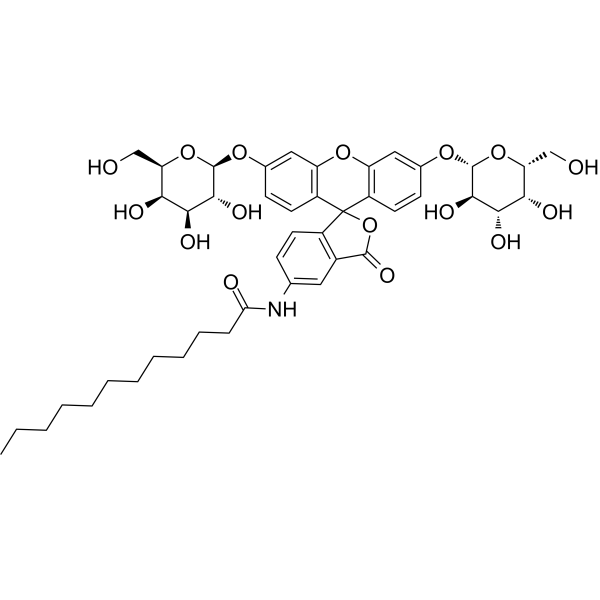
C12FDG (5-Dodecanoylaminofluorescein di-β-D-Galactopyranoside) is a lipophilic green fluorescent substrate for β-galactosidase detection. C12-FDG is more sensitive than Fluorescein di(β-D-galactopyranoside) (HY-101895) for beta-galactosidase activity determinations in animal cells (Ex/Em = 488/523 nm) .
X-GAL (BCIG) is a widely used chromogenic β-galactosidase substrate. X-GAL is a colorless compound until cleaved by β-galactosidase, at which point X-GAL turns to an insoluble and detectable blue compound, making X-GAL particularly useful in techniques such as blue-white screening for cloning in bacteria. X-GAL can also be used for detection of β-galactosidase activity .
1-Oleoyl lysophosphatidic acid (1-Oleoyl-sn-glycero-3-phosphate) is an abundant lysophosphatidic acid (LPA) species with high biological activity due to its strong affinity for the LPA receptors. 1-Oleoyl lysophosphatidic acid is commonly used in most laboratories as a reagent for LPA receptor activation . 1-Oleoyl lysophosphatidic acid increases SRE-driven β-galactosidase activity .
β-Galactosidase, E. coli (EC 3.2.1.23; GAL) is a glycoside hydrolase that hydrolyzes the β-glycosidic bonds formed between galactose and its organic moieties. β-Galactosidase, E. coli can hydrolyze lactose to form glucose and galactose, and enter glycolysis; it can also catalyze the transgalactosylation of lactose into allolactose; allolactose can be cracked into monosaccharides .
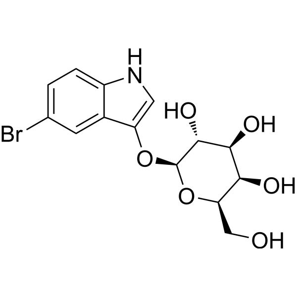
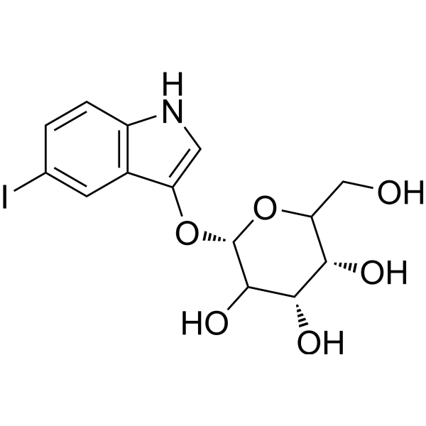
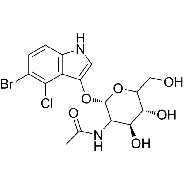
5-Bromo-3-indolyl β-D-galactopyranoside (Bluo-Gal) is a chromogenic substrate for β-galactosidase. 5-Bromo-3-indolyl β-D-galactopyranoside is hydrolyzed by the enzyme to generate a 5-bromoindole intermediate, which is further oxidized to form an insoluble blue precipitate. 5-Bromo-3-indolyl β-D-galactopyranoside can specifically recognize bacterial β-galactosidases (such as the product of the Escherichia coli lacZ gene) and reacts at pH 7.4, making it suitable for light and electron microscopic observations. 5-Bromo-3-indolyl β-D-galactopyranoside can be used in histochemical detection of reporter gene expression in transgenic organisms, such as the localization analysis of β-galactosidase activity in mouse embryos or muscle tissues .
Chlorophenol red-β-D-galactopyranoside (CPRG) is an efficient and sensitive chromogenic substrate for β-galactosidase (HY-P2869), widely used in colorimetric assays. Chlorophenol red-β-D-galactopyranoside itself appears pale yellow. When it is specifically hydrolyzed by β-galactosidase, it releases chlorophenol red. The released chlorophenol red turns purple-red under alkaline or neutral pH conditions. This color change from yellow to red can be quantitatively detected at wavelengths of 540-572 nm using visible spectrophotometry .
X-GAL (BCIG) (solution) is a widely used chromogenic β-galactosidase substrate. X-GAL is a colorless compound until cleaved by β-galactosidase, at which point X-GAL turns to an insoluble and detectable blue compound, making X-GAL particularly useful in techniques such as blue-white screening for cloning in bacteria. X-GAL can also be used for detection of β-galactosidase activity . Solvent and Concentration: DMF: 20 mg/mL
SSK1, a senescence-specific killing compound, is a β-galactosidase-targeted proagent attenuates inflammation. SSK1 is activated by lysosomal β-galactosidase and selectively killed senescent cells through the activation of p38 MAPK and induction of apoptosis .
RVG-Cys (RVG29-Cys) is a peptide derived from rabies virus glycoprotein (RVG29) with Cys attached to facilitate subsequent conjugation. RVG-Cys enhances the specific targeted delivery of proteins in brain tissue and neurons .
Beta-galactose dehydrogenase is a selective catalyst for β-galactose. Under pH 8.6 conditions, beta-galactose dehydrogenase catalyzes the oxidation of β-galactose, produced by the hydrolysis of lactose by β-galactosidase, with nicotinamide adenine dinucleotide (NAD) to produce reduced nicotinamide adenine dinucleotide (NADH). Beta-galactose dehydrogenase specifically mediates this oxidation reaction for the quantitative detection of the substrate, used in the analysis of lactose concentration in samples such as breast milk .
Gypenoside L is a saponin that can be found in Gynostemma pentaphyllum. Gypenoside L increases the SA-β-galactosidase activity, promotes the production of senescence-associated secretory cytokines. Gypenoside L also can activate p38 and ERK MAPK pathways and NF-κB pathway to induce senescence. Gypenoside L exhibits anti-tumor and anti-inflammatory activities .
DDAO is a promising near-infrared (NIR) red fluorescent probewith tunable excitation wavelength (600-650nm) and longemission wavelength(λem=656nm). DDAO can de desiged for detection of the activities of different enzymes such asβ-galactosidase,sulfatase, proteinphosphatase2A,carboxylesterase 2, humanalbumin andesterases .
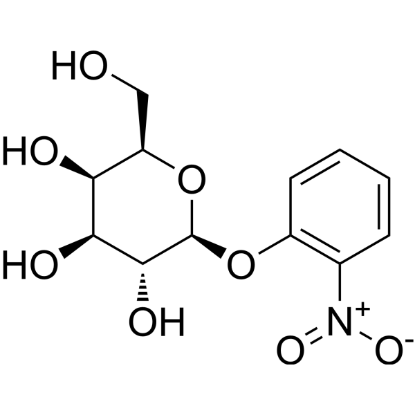
4-Aminophenyl-β-D-galactopyranoside, 98% is a highly efficient substrate for β-galactosidase. It is specifically hydrolyzed by this enzyme to release galactose and electroactive p-aminophenol. 4-Aminophenyl-β-D-galactopyranoside, 98% is widely used in colorimetric and electrochemical assays for detecting β-galactosidase activity and determining enzyme kinetics, such as in biosensing fields including cellular senescence, pathogen and contaminant detection. In addition, since β-galactosidase is often overexpressed in primary ovarian cancer, 4-Aminophenyl-β-D-galactopyranoside, 98% can also be applied to related research on primary ovarian cancer .
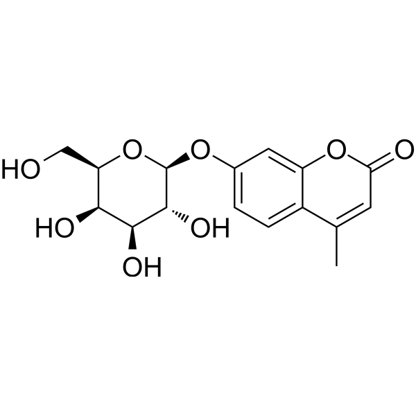
4-Methylumbelliferyl β-D-galactopyranoside is a fluorescent substrate for β-galactosidase which, when cleaved, produces a water-soluble blue fluorescent coumarin fluorophore that can be detected using a fluoroenzymeter or fluorometer .
C12FDG (5-Dodecanoylaminofluorescein di-β-D-Galactopyranoside) (solution) is a lipophilic green fluorescent substrate for β-galactosidase detection. C12-FDG is more sensitive than Fluorescein di (β-D-galactopyranoside) (HY-101895) for beta-galactosidase activity determinations in animal cells (Ex/Em = 488/523 nm) . Solvent and concentration: DMSO: 10 mM
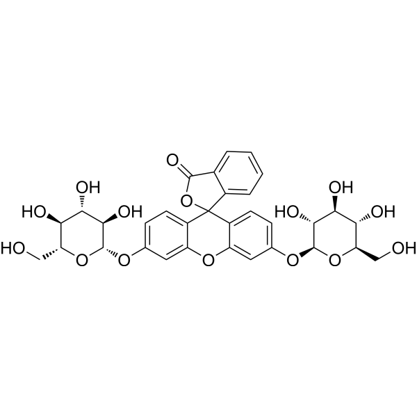
Rose-β-D-Gal is a chromogenic substrate, is also a β-galactosidase substrate. Rose-β-D-Gal creates a pink/magenta color after the reaction and has been used for detection of β-gal activity .
Resorufin-β-D-galactopyranoside is a commonly used substrate in various biochemical assays to measure the activity of β-galactosidase, an important enzyme involved in lactose metabolism and regulation of gene expression. Resorufin-β-D-galactopyranoside has unique chemical properties and can be hydrolyzed by β-galactosidase to form a red fluorescent product called resorufin. This makes it a useful tool for detecting and quantifying β-galactosidase activity in biological samples such as bacteria or mammalian cells.
5-Bromo-4-chloro-3-indolyl β-D-glucopyranoside, a chromogenic substrate for the detection of β-galactosidase activity. It is commonly used in molecular biology techniques such as gene expression analysis and reporter gene analysis. When β-galactosidase cleaves X-Gluc, a blue precipitate is produced, which can be observed by microscopy or other detection methods. X-Gluc has high sensitivity and specificity for the detection of β-galactosidase activity, making it a widely used tool in molecular biology research.
BMVC is a potent G-quadruplex (G4) stabilizer and a selective telomerase inhibitor with an IC50 of ~0.2 μM. BMVC inhibits Taq DNA polymerase with an IC50 of ~2.5 μM. BMVC increases the melting temperature of G4 structure of telomere and accelerates telomere length shortening. Anticancer activities .
NIR-βgal-2 is a β-galactosidase-activated near-infrared fluorescent probe with superior sensitivity. NIR-βgal-2 can be used for visualizing β-galactosidase in breast cancer .
β-Galactosidase, Sweet almond is a glycoside hydrolase that hydrolyzes the β-glycosidic bonds formed between galactose and its organic moieties. β-Galactosidase, Sweet almond can hydrolyze lactose to form glucose and galactose, and enter glycolysis; it can also catalyze the transgalactosylation of lactose into allolactose; allolactose can be cracked into monosaccharides .
β-galactosidase mRNA encodes β-galactosidase, a protein product of the bacterial LacZ gene. β-galactosidase catalyzes the conversion of β-galactosides into monosaccharides which could be used as a common marker to assess transfection efficiency.
6-O-β-D-Galactopyranosyl-D-galactose, a disaccharide, is a part of the polysaccharide main chain with β-(1→6)-glycoside bonds with a side chain bonded to the main one by the β-(1→3) bond. 6-O-β-D-Galactopyranosyl-D-galactose can be isolated from enzyme-hydrolyzed peach gum .
Tryptophanase is a zymogen-converting enzyme and inducible enzyme that can convert its inactive precursor form into an active enzyme without additional polypeptide synthesis. In Escherichia coli K12, tryptophanase functions as an inducible enzyme, and its induction kinetics are similar to those of β-galactosidase. Tryptophanase catalyzes the conversion of L-tryptophan to indole .
PFB-FDG is a non-fluorescent galactosidase substrate that can be hydrolysed to green fluorescent PFB-F (Ex=485 nm, Em=535 nm). PFB-FDG can be used for the determination of β-galactosidase activity .
β-galactosidase mRNA (5moU) encodes β-galactosidase, a protein product of the bacterial LacZ gene. β-galactosidase catalyzes the conversion of β-galactosides into monosaccharides which could be used as a common marker to assess transfection efficiency. The incorporation of 5moU can reduce the immunogenicity of the resulting mRNA.
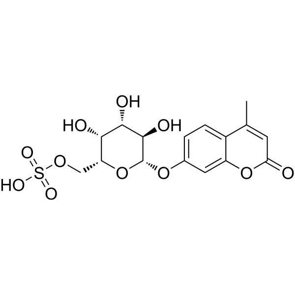
4-Methylumbelliferyl-β-D-galactopyranoside 6-sulfate is a fluorescent dye. 4-Methylumbelliferyl-β-D-galactopyranoside 6-sulfate undergoes desulphation by galactose-6-sulphate sulphatase to form 4-methylumbelliferyl-β-D-galactopyranoside, which is cleaved by β-galactosidase to release fluorescent 4-methylumbelliferone. 4-Methylumbelliferyl-β-D-galactopyranoside 6-sulfate interacts with N-acetylgalactosamine-6-sulfate sulfatase (GALNS) via hydrogen bonds, electrostatic interactions, and steric interactions. 4-Methylumbelliferyl-β-D-galactopyranoside 6-sulfate serves as a substrate in assays measuring galactose-6-sulphate sulphatase and GALNS activity. 4-Methylumbelliferyl-β-D-galactopyranoside 6-sulfate can be used for the research of Morquio disease type A (mucopolysaccharidosis IV A) .
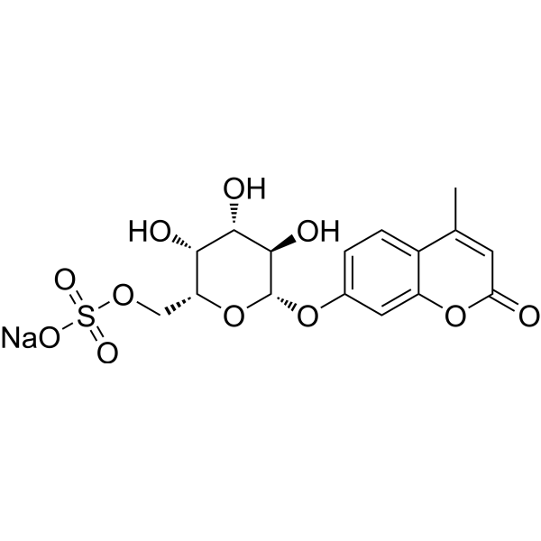
4-Methylumbelliferyl-β-D-galactopyranoside 6-sulfate sodium is a fluorescent dye. 4-Methylumbelliferyl-β-D-galactopyranoside 6-sulfate sodium undergoes desulphation by galactose-6-sulphate sulphatase to form 4-methylumbelliferyl-β-D-galactopyranoside, which is cleaved by β-galactosidase to release fluorescent 4-methylumbelliferone. 4-Methylumbelliferyl-β-D-galactopyranoside 6-sulfate sodium interacts with N-acetylgalactosamine-6-sulfate sulfatase (GALNS) via hydrogen bonds, electrostatic interactions, and steric interactions. 4-Methylumbelliferyl-β-D-galactopyranoside 6-sulfate sodium serves as a substrate in assays measuring galactose-6-sulphate sulphatase and GALNS activity. 4-Methylumbelliferyl-β-D-galactopyranoside 6-sulfate sodium can be used for the research of Morquio disease type A (mucopolysaccharidosis IV A) .
(2R)-Glycerol-O-β-D-galactopyranoside (3-O-β-D-Galactopyranosyl-sn-glycerol) is a good substrate for all three components of the lac operon, i.e. β-galactosidase, the lactose transporter and thiogalactoside transacetylase .
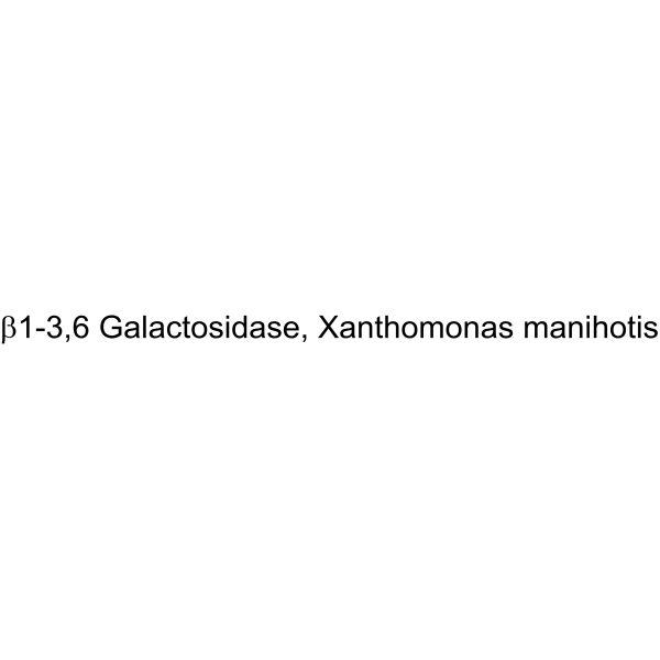
β1-3,6 Galactosidase, Xanthomonas manihotis is an exoglycosidase that catalyzes the hydrolysis of terminal β(1-3)- and β(1-6)-linked galactose residues .
Chlorophenol red-β-D-galactopyranoside (CPRG) sodium is an efficient and sensitive chromogenic substrate for β-galactosidase (HY-P2869), widely used in colorimetric assays. Chlorophenol red-β-D-galactopyranoside sodium itself appears pale yellow. When it is specifically hydrolyzed by β-galactosidase, it releases chlorophenol red. The released chlorophenol red turns purple-red under alkaline or neutral pH conditions. This color change from yellow to red can be quantitatively detected at wavelengths of 540-572 nm using visible spectrophotometry .
β-Galactosidase Mutein, E. coli is a hydrolase enzyme that catalyzes the hydrolysis of β-galactosides into monosaccharides. Substrates of different β-galactosidases include ganglioside GM1, lactosylceramides, lactose, and various glycoproteins.
β-Galactosidase 42A, Bifidobacterium longum (EC 3.2.1.23) is a hydrolase enzyme that catalyzes the hydrolysis of β-galactosides into monosaccharides. Substrates of different β-galactosidases include ganglioside GM1, lactosylceramides, lactose, and various glycoproteins.
β-Galactosidase 42A, Thermotoga maritima (EC 3.2.1.23) is a hydrolase enzyme that catalyzes the hydrolysis of β-galactosides into monosaccharides. Substrates of different β-galactosidases include ganglioside GM1, lactosylceramides, lactose, and various glycoproteins.
β-Galactosidase 2A, Bacteroides thetaiotaomicron (EC 3.2.1.23) is a hydrolase enzyme that catalyzes the hydrolysis of β-galactosides into monosaccharides. Substrates of different β-galactosidases include ganglioside GM1, lactosylceramides, lactose, and various glycoproteins.
β-Galactosidase 1A, Sulfolobus solfataricus (EC 3.2.1.23) is a hydrolase enzyme that catalyzes the hydrolysis of β-galactosides into monosaccharides. Substrates of different β-galactosidases include ganglioside GM1, lactosylceramides, lactose, and various glycoproteins.
β-Galactosidase 42A, Caldicellulosiruptor saccharolyticus (EC 3.2.1.23) is a hydrolase enzyme that catalyzes the hydrolysis of β-galactosides into monosaccharides. Substrates of different β-galactosidases include ganglioside GM1, lactosylceramides, lactose, and various glycoproteins.
β-Galactosidase 2B, Bacteroides thetaiotaomicron (EC 3.2.1.23) is a hydrolase enzyme that catalyzes the hydrolysis of β-galactosides into monosaccharides. Substrates of different β-galactosidases include ganglioside GM1, lactosylceramides, lactose, and various glycoproteins.
DDAO phosphate diammonium is a fluorescent phosphatase substrate. DDAO phosphate diammonium has tunable excitation wavelength (600-650nm) and long emission wavelength (λem=656nm). DDAO phosphate diammonium can be used to detect the activity of different enzymes such as β-galactosidase, sulfatase, protein phosphatase 2A, carboxylesterase 2, human albumin and esterase.
β-galactosidase mRNA (N1-Methylpseudo-UTP) encodes β-galactosidase, a protein product of the bacterial LacZ gene. β-galactosidase catalyzes the conversion of β-galactosides into monosaccharides which could be used as a common marker to assess transfection efficiency. The incorporation of N1-Methyl-pseudo-UTP can reduce the immunogenicity of the resulting mRNA.
Xentry is a cell-penetrating peptide (CCP) consisting of only 7 amino acids of hepatitis B virus: LCLRPVG. Xentry-linked anti-B-raf antibodies and siRNAs demonstrates the capability to kill B-raf-dependent melanoma cells. Xentry alone or conjugated to β-galactosidase leads to its delivery to most tissues in mice, except circulating blood cells. Xentry can be used for the delivery of large molecules (antibodies, siRNA, enzymes) .
NIR-BG2 is a near-infrared fluorescent probe targeting senescence-associated β-galactosidase (SA-β-Gal). NIR-BG2 is activated by SA-β-Gal and undergoes hydrolysis to release electrophilic quinone methide that covalently binds to surrounding proteins for in situ labeling, with a 16-fold enhancement of fluorescence signal at 709 nm . NIR-BG2 is promising for research of vivo imaging of cellular senescence .
Purple-β-D-Gal is a chromogenic β-galactosidase substrate. Intracellular enzymatic hydrolysis of Purple-β-D-Gal generates free indoxyl molecules, which undergo in situ oxidation and subsequent dimerization to produce chromogenic, water-insoluble, indigo precipitates. Purple-β-D-Gal can be used for the detection of β-galactosidase activity .
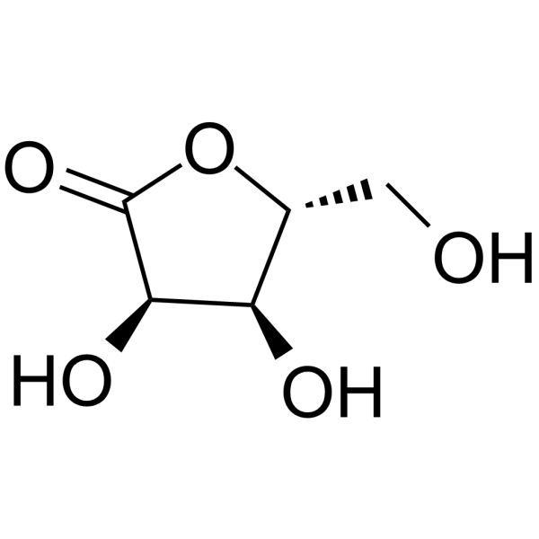
D-Ribonolactone (Standard) is the analytical standard of D-Ribonolactone. This product is intended for research and analytical applications. D-Ribonolactone is sugar lactone and an inhibitor of β-galactosidase of Escherichia coli with a Ki of 26 mM .
β-Galactosidase, Kluyveromyces lactis is a hydrolase enzyme that catalyzes the hydrolysis of β-galactosides into monosaccharides. SubstRates of different β-galactosidases include ganglioside GM1, lactosylceramides, lactose, and various glycoproteins.
Beta-gal-nonoate is a β-galactosidase dependent nitric oxide (NO) donor that releases NO once activated by β-galactosidase. β-Gal-NONOate has bactericidal activity and can be used as a bactericide .
6-Bromo-2-naphthyl-β-D-galactopyranoside is a chromogenic substrate commonly used to measure β-galactosidase enzyme activity in food, enzyme substrates, and culture media. Upon hydrolysis by β-galactosidase, it generates a yellow precipitate indicating the enzyme's presence.
Afegostat hydrochloride (D-Isofagomine hydrochloride) is a potent β-galactosidase inhibitor with activity ameliorating GM1-gangliosidosis and Morquio B disease-associated mutations. Afegostat hydrochloride is able to induce the maturation of mutant β-galactosidase in fibroblasts from patients with GM1-gangliosidosis. Afegostat hydrochloride also promotes the reduction of keratin sulfate and oligosaccharide load in patient cells .
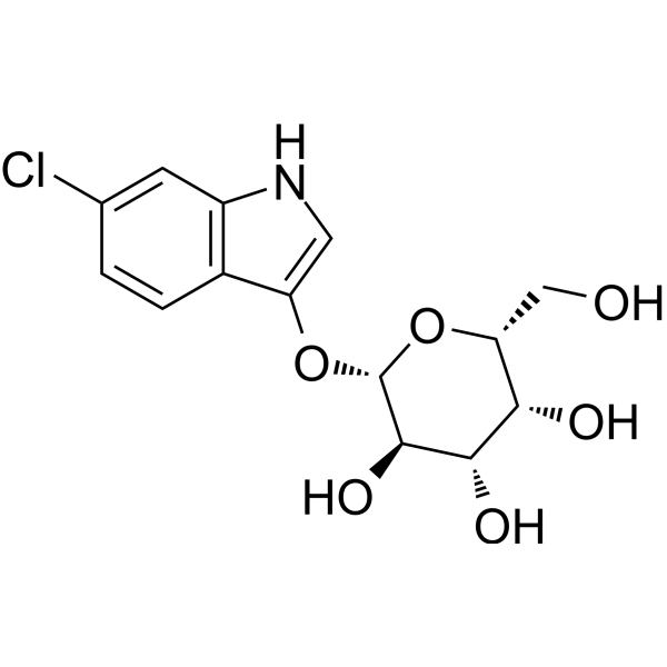
6-Fluoro-3-indolyl-β-D-galactopyranoside is a chromogenic enzyme substrate commonly used in microbiology to detect the expression of β-galactosidase in bacterial colonies.
2-Chloro-4-nitrophenyl-β-D-galactopyranoside is a chromogenic substrate used for testing enzyme activity. It is commonly used in molecular biology research to detect and measure the activity of β-galactosidase (biosynth: EC03318).
5-Bromo-6-chloro-3-indolyl-N-acetyl-β-D-glucosaminide, 98% is a chromogenic substrate used to measure β-galactosidase activity in food, enzyme substrates, and culture media.
5-Bromo-6-chloro-3-indolyl β-D-Galactopyranoside (contains ca. 10% Ethyl Acetate) (5-Bromo-6-chloro-3-indolyl-beta-D-galactoside) is a chromogenic substrate of β-galactosidase (β-gal) .
5-Bromo-4-chloro-1H-indol-3-yl nonanoate is a fluorescent substrate used to detect enzymes. The substrate reacts with various enzymes to produce a fluorescent product, which has been shown to be active against β-galactosidase, α-chymotrypsin, and β-glucuronidase.
KSA02 is a two-dimensional intelligent fluorescent probe. KSA02 not only detects the activity of the aging-related β-galactosidase (SA-β-gal), but also can simultaneously sense the pH value changes of the lysosomal microenvironment where SA-β-gal is located. KSA02 can distinguish between aging and cancer, track the aging process, and evaluate the efficacy of anti-aging agents. KSA02 can be used for the study of aging biology .
X-GalNAc is a chromogenic substrate for for N-acetyl-β-galactosidase, used to determine the presence or absence of a cloned DNA insert in bacteria growing on agar plates .
DCM-gal-CF is a fluorescent probe containing a DCM fluorophore scaffold and a D-galactose recognition moiety. DCM-gal-CF itself shows weak fluorescence, and its fluorescence is enhanced after cleavage by β-galactosidase. DCM-gal-CF can be used to detect β-galactosidase activity .
HMRef-βGal is a fluorescent probe and a substrate responsive to β-galactosidase(β-galactosidase) (Ex/Em=498 nm/505-600 nm). After being cleaved by β-galactosidase, HMRef-βGal triggers significant fluorescence enhancement via intramolecular spirocyclic function regulation. HMRef-βGal generates bright fluorescence in cancer cells with elevated β-galactosidase activity, enabling visualization of tiny peritoneal metastases in mouse models. HMRef-βGal exhibits low in vitro cytotoxicity and low acute in vivo toxicity in mice. HMRef-βGal can be used for preclinical fluorescence-guided diagnosis and cytoreductive surgery of peritoneal metastases, including compatibility with real-time naked-eye detection and endoscopic imaging, as well as for studies related to peritoneal metastases of ovarian cancer .
β-Galactosidase-biotin labeled, Escherichia coli is a hydrolase enzyme that catalyzes the hydrolysis of β-galactosides into monosaccharides. SubstRates of different β-galactosidases include ganglioside GM1, lactosylceramides, lactose, and various glycoproteins.
N-Octyl-α-4-epivalienamine is an orally active and CNS-penetrant molecular chaperone that induces high expression of the deficient β-galactosidase activity. N-Octyl-α-4-epivalienamine ameliorates symptoms and increase survival rate in a mouse model of GM1-gangliosidosis. N-Octyl-α-4-epivalienamine can be used for neurogenetic disease research .
o-Nitrophenyl β-D-galactopyranoside-6-phosphate cyclohexylammonium is a chromogenic substrate with activity for β-galactosidase detection. The use of o-Nitrophenyl β-D-galactopyranoside-6-phosphate cyclohexylammonium can help researchers quickly identify and quantify β-galactosidase activity.
CH-123 is an orally active lipid-lowering agent. CH-123 inhibits the elevated activities of β-glucuronidase, β-galactosidase, N-acetyl-β-glucosaminidase and acid phosphatase in aortic smooth muscle cells and hepatocytes. CH-123 reduces serum total lipid and cholesterol levels, as well as intracellular cholesterol content in aortic smooth muscle cells. CH-123 significantly inhibits lysosomal enzyme activity. CH-123 can be used in the research of atherosclerosis .
5-Amino-1-(pyridin-3-yl)-1H-imidazole-4-carboxamide is a riboswitch ligand. 5-Amino-1-(pyridin-3-yl)-1H-imidazole-4-carboxamide effectively activates riboswitches during in vitro transcription and induces β-galactosidase expression .
β 1-4,6-Galactosidase, Jack bean (EC 3.2.1.23) is a hydrolase enzyme that catalyzes the hydrolysis of β-galactosides into monosaccharides. Substrates of different β-galactosidases include ganglioside GM1, lactosylceramides, lactose, and various glycoproteins.
Millewanin H is a Flavonoids product that can be isolated from the herbs of Millettia pachycarpa . Millewanin H has antiestrogenic activity and inhibit 17β-estradiol-induced-β-galactosidase activity . Millewanin H showes α-glucosidase inhibition .
Gal-dMor-Gem is a selective senescent cell scavenger, Apoptosis inducer, and a prodrug of Gemcitabine (HY-17026). Gal-dMor-Gem releases Gemcitabine upon activation by Esterases and β-gal. Gal-dMor-Gem reduces SA-β-gal, preferentially induces apoptosis in senescent cells, regulates apoptosis-related proteins, accumulates in senescent tissues, and ameliorates senescence-related organ phenotypes. Gal-dMor-Gem is applicable to research on chemotherapy-induced senescence .
C12FDG (5-Dodecanoylaminofluorescein di-β-D-Galactopyranoside) is a lipophilic green fluorescent substrate for β-galactosidase detection. C12-FDG is more sensitive than Fluorescein di(β-D-galactopyranoside) (HY-101895) for beta-galactosidase activity determinations in animal cells (Ex/Em = 488/523 nm) .
X-GAL (BCIG) is a widely used chromogenic β-galactosidase substrate. X-GAL is a colorless compound until cleaved by β-galactosidase, at which point X-GAL turns to an insoluble and detectable blue compound, making X-GAL particularly useful in techniques such as blue-white screening for cloning in bacteria. X-GAL can also be used for detection of β-galactosidase activity .
Chlorophenol red-β-D-galactopyranoside (CPRG) is an efficient and sensitive chromogenic substrate for β-galactosidase (HY-P2869), widely used in colorimetric assays. Chlorophenol red-β-D-galactopyranoside itself appears pale yellow. When it is specifically hydrolyzed by β-galactosidase, it releases chlorophenol red. The released chlorophenol red turns purple-red under alkaline or neutral pH conditions. This color change from yellow to red can be quantitatively detected at wavelengths of 540-572 nm using visible spectrophotometry .
X-GAL (BCIG) (solution) is a widely used chromogenic β-galactosidase substrate. X-GAL is a colorless compound until cleaved by β-galactosidase, at which point X-GAL turns to an insoluble and detectable blue compound, making X-GAL particularly useful in techniques such as blue-white screening for cloning in bacteria. X-GAL can also be used for detection of β-galactosidase activity . Solvent and Concentration: DMF: 20 mg/mL
DDAO is a promising near-infrared (NIR) red fluorescent probewith tunable excitation wavelength (600-650nm) and longemission wavelength(λem=656nm). DDAO can de desiged for detection of the activities of different enzymes such asβ-galactosidase,sulfatase, proteinphosphatase2A,carboxylesterase 2, humanalbumin andesterases .
4-Methylumbelliferyl β-D-galactopyranoside is a fluorescent substrate for β-galactosidase which, when cleaved, produces a water-soluble blue fluorescent coumarin fluorophore that can be detected using a fluoroenzymeter or fluorometer .
C12FDG (5-Dodecanoylaminofluorescein di-β-D-Galactopyranoside) (solution) is a lipophilic green fluorescent substrate for β-galactosidase detection. C12-FDG is more sensitive than Fluorescein di (β-D-galactopyranoside) (HY-101895) for beta-galactosidase activity determinations in animal cells (Ex/Em = 488/523 nm) . Solvent and concentration: DMSO: 10 mM
Rose-β-D-Gal is a chromogenic substrate, is also a β-galactosidase substrate. Rose-β-D-Gal creates a pink/magenta color after the reaction and has been used for detection of β-gal activity .
NIR-βgal-2 is a β-galactosidase-activated near-infrared fluorescent probe with superior sensitivity. NIR-βgal-2 can be used for visualizing β-galactosidase in breast cancer .
PFB-FDG is a non-fluorescent galactosidase substrate that can be hydrolysed to green fluorescent PFB-F (Ex=485 nm, Em=535 nm). PFB-FDG can be used for the determination of β-galactosidase activity .
4-Methylumbelliferyl-β-D-galactopyranoside 6-sulfate is a fluorescent dye. 4-Methylumbelliferyl-β-D-galactopyranoside 6-sulfate undergoes desulphation by galactose-6-sulphate sulphatase to form 4-methylumbelliferyl-β-D-galactopyranoside, which is cleaved by β-galactosidase to release fluorescent 4-methylumbelliferone. 4-Methylumbelliferyl-β-D-galactopyranoside 6-sulfate interacts with N-acetylgalactosamine-6-sulfate sulfatase (GALNS) via hydrogen bonds, electrostatic interactions, and steric interactions. 4-Methylumbelliferyl-β-D-galactopyranoside 6-sulfate serves as a substrate in assays measuring galactose-6-sulphate sulphatase and GALNS activity. 4-Methylumbelliferyl-β-D-galactopyranoside 6-sulfate can be used for the research of Morquio disease type A (mucopolysaccharidosis IV A) .
4-Methylumbelliferyl-β-D-galactopyranoside 6-sulfate sodium is a fluorescent dye. 4-Methylumbelliferyl-β-D-galactopyranoside 6-sulfate sodium undergoes desulphation by galactose-6-sulphate sulphatase to form 4-methylumbelliferyl-β-D-galactopyranoside, which is cleaved by β-galactosidase to release fluorescent 4-methylumbelliferone. 4-Methylumbelliferyl-β-D-galactopyranoside 6-sulfate sodium interacts with N-acetylgalactosamine-6-sulfate sulfatase (GALNS) via hydrogen bonds, electrostatic interactions, and steric interactions. 4-Methylumbelliferyl-β-D-galactopyranoside 6-sulfate sodium serves as a substrate in assays measuring galactose-6-sulphate sulphatase and GALNS activity. 4-Methylumbelliferyl-β-D-galactopyranoside 6-sulfate sodium can be used for the research of Morquio disease type A (mucopolysaccharidosis IV A) .
DDAO phosphate diammonium is a fluorescent phosphatase substrate. DDAO phosphate diammonium has tunable excitation wavelength (600-650nm) and long emission wavelength (λem=656nm). DDAO phosphate diammonium can be used to detect the activity of different enzymes such as β-galactosidase, sulfatase, protein phosphatase 2A, carboxylesterase 2, human albumin and esterase.
Purple-β-D-Gal is a chromogenic β-galactosidase substrate. Intracellular enzymatic hydrolysis of Purple-β-D-Gal generates free indoxyl molecules, which undergo in situ oxidation and subsequent dimerization to produce chromogenic, water-insoluble, indigo precipitates. Purple-β-D-Gal can be used for the detection of β-galactosidase activity .
KSA02 is a two-dimensional intelligent fluorescent probe. KSA02 not only detects the activity of the aging-related β-galactosidase (SA-β-gal), but also can simultaneously sense the pH value changes of the lysosomal microenvironment where SA-β-gal is located. KSA02 can distinguish between aging and cancer, track the aging process, and evaluate the efficacy of anti-aging agents. KSA02 can be used for the study of aging biology .
X-GalNAc is a chromogenic substrate for for N-acetyl-β-galactosidase, used to determine the presence or absence of a cloned DNA insert in bacteria growing on agar plates .
DCM-gal-CF is a fluorescent probe containing a DCM fluorophore scaffold and a D-galactose recognition moiety. DCM-gal-CF itself shows weak fluorescence, and its fluorescence is enhanced after cleavage by β-galactosidase. DCM-gal-CF can be used to detect β-galactosidase activity .
HMRef-βGal is a fluorescent probe and a substrate responsive to β-galactosidase(β-galactosidase) (Ex/Em=498 nm/505-600 nm). After being cleaved by β-galactosidase, HMRef-βGal triggers significant fluorescence enhancement via intramolecular spirocyclic function regulation. HMRef-βGal generates bright fluorescence in cancer cells with elevated β-galactosidase activity, enabling visualization of tiny peritoneal metastases in mouse models. HMRef-βGal exhibits low in vitro cytotoxicity and low acute in vivo toxicity in mice. HMRef-βGal can be used for preclinical fluorescence-guided diagnosis and cytoreductive surgery of peritoneal metastases, including compatibility with real-time naked-eye detection and endoscopic imaging, as well as for studies related to peritoneal metastases of ovarian cancer .
4-Aminophenyl-β-D-galactopyranoside, 98% is a highly efficient substrate for β-galactosidase. It is specifically hydrolyzed by this enzyme to release galactose and electroactive p-aminophenol. 4-Aminophenyl-β-D-galactopyranoside, 98% is widely used in colorimetric and electrochemical assays for detecting β-galactosidase activity and determining enzyme kinetics, such as in biosensing fields including cellular senescence, pathogen and contaminant detection. In addition, since β-galactosidase is often overexpressed in primary ovarian cancer, 4-Aminophenyl-β-D-galactopyranoside, 98% can also be applied to related research on primary ovarian cancer .
Resorufin-β-D-galactopyranoside is a commonly used substrate in various biochemical assays to measure the activity of β-galactosidase, an important enzyme involved in lactose metabolism and regulation of gene expression. Resorufin-β-D-galactopyranoside has unique chemical properties and can be hydrolyzed by β-galactosidase to form a red fluorescent product called resorufin. This makes it a useful tool for detecting and quantifying β-galactosidase activity in biological samples such as bacteria or mammalian cells.
5-Bromo-4-chloro-3-indolyl β-D-glucopyranoside, a chromogenic substrate for the detection of β-galactosidase activity. It is commonly used in molecular biology techniques such as gene expression analysis and reporter gene analysis. When β-galactosidase cleaves X-Gluc, a blue precipitate is produced, which can be observed by microscopy or other detection methods. X-Gluc has high sensitivity and specificity for the detection of β-galactosidase activity, making it a widely used tool in molecular biology research.
Chlorophenol red-β-D-galactopyranoside (CPRG) sodium is an efficient and sensitive chromogenic substrate for β-galactosidase (HY-P2869), widely used in colorimetric assays. Chlorophenol red-β-D-galactopyranoside sodium itself appears pale yellow. When it is specifically hydrolyzed by β-galactosidase, it releases chlorophenol red. The released chlorophenol red turns purple-red under alkaline or neutral pH conditions. This color change from yellow to red can be quantitatively detected at wavelengths of 540-572 nm using visible spectrophotometry .
6-Bromo-2-naphthyl-β-D-galactopyranoside is a chromogenic substrate commonly used to measure β-galactosidase enzyme activity in food, enzyme substrates, and culture media. Upon hydrolysis by β-galactosidase, it generates a yellow precipitate indicating the enzyme's presence.
6-Fluoro-3-indolyl-β-D-galactopyranoside is a chromogenic enzyme substrate commonly used in microbiology to detect the expression of β-galactosidase in bacterial colonies.
2-Chloro-4-nitrophenyl-β-D-galactopyranoside is a chromogenic substrate used for testing enzyme activity. It is commonly used in molecular biology research to detect and measure the activity of β-galactosidase (biosynth: EC03318).
5-Bromo-6-chloro-3-indolyl-N-acetyl-β-D-glucosaminide, 98% is a chromogenic substrate used to measure β-galactosidase activity in food, enzyme substrates, and culture media.
5-Bromo-4-chloro-1H-indol-3-yl nonanoate is a fluorescent substrate used to detect enzymes. The substrate reacts with various enzymes to produce a fluorescent product, which has been shown to be active against β-galactosidase, α-chymotrypsin, and β-glucuronidase.
o-Nitrophenyl β-D-galactopyranoside-6-phosphate cyclohexylammonium is a chromogenic substrate with activity for β-galactosidase detection. The use of o-Nitrophenyl β-D-galactopyranoside-6-phosphate cyclohexylammonium can help researchers quickly identify and quantify β-galactosidase activity.
RVG-Cys (RVG29-Cys) is a peptide derived from rabies virus glycoprotein (RVG29) with Cys attached to facilitate subsequent conjugation. RVG-Cys enhances the specific targeted delivery of proteins in brain tissue and neurons .
Xentry is a cell-penetrating peptide (CCP) consisting of only 7 amino acids of hepatitis B virus: LCLRPVG. Xentry-linked anti-B-raf antibodies and siRNAs demonstrates the capability to kill B-raf-dependent melanoma cells. Xentry alone or conjugated to β-galactosidase leads to its delivery to most tissues in mice, except circulating blood cells. Xentry can be used for the delivery of large molecules (antibodies, siRNA, enzymes) .
Gypenoside L is a saponin that can be found in Gynostemma pentaphyllum. Gypenoside L increases the SA-β-galactosidase activity, promotes the production of senescence-associated secretory cytokines. Gypenoside L also can activate p38 and ERK MAPK pathways and NF-κB pathway to induce senescence. Gypenoside L exhibits anti-tumor and anti-inflammatory activities .
6-O-β-D-Galactopyranosyl-D-galactose, a disaccharide, is a part of the polysaccharide main chain with β-(1→6)-glycoside bonds with a side chain bonded to the main one by the β-(1→3) bond. 6-O-β-D-Galactopyranosyl-D-galactose can be isolated from enzyme-hydrolyzed peach gum .
NIR-BG2 is a near-infrared fluorescent probe targeting senescence-associated β-galactosidase (SA-β-Gal). NIR-BG2 is activated by SA-β-Gal and undergoes hydrolysis to release electrophilic quinone methide that covalently binds to surrounding proteins for in situ labeling, with a 16-fold enhancement of fluorescence signal at 709 nm . NIR-BG2 is promising for research of vivo imaging of cellular senescence .
D-Ribonolactone (Standard) is the analytical standard of D-Ribonolactone. This product is intended for research and analytical applications. D-Ribonolactone is sugar lactone and an inhibitor of β-galactosidase of Escherichia coli with a Ki of 26 mM .
Millewanin H is a Flavonoids product that can be isolated from the herbs of Millettia pachycarpa . Millewanin H has antiestrogenic activity and inhibit 17β-estradiol-induced-β-galactosidase activity . Millewanin H showes α-glucosidase inhibition .
Beta-galactosidase Protein, with catalytic activity, hydrolyzes terminal non-reducing beta-D-galactose residues in beta-D-galactosides. Its enzymatic function involves breaking these specific bonds, facilitating the cleavage of beta-D-galactosides and contributing to the degradation of molecules containing these terminal galactose residues. Beta-galactosidase Protein, Saccharolobus solfataricus is the recombinant Beta-galactosidase protein, expressed by E. coli , with tag free.
Beta-galactosidase Protein, with catalytic activity, hydrolyzes terminal non-reducing beta-D-galactose residues in beta-D-galactosides. Its enzymatic function involves breaking these specific bonds, facilitating the cleavage of beta-D-galactosides and contributing to the degradation of molecules containing these terminal galactose residues. Beta-galactosidase Protein, Saccharolobus solfataricus (FLAG, His) is the recombinant Beta-galactosidase protein, expressed by E. coli , with N-6*His, N-Flag labeled tag.
Beta-galactosidase/GLB1 protein is a hydrolase with catalytic activity and binding sites of Mg2+, Mn2+ and Na+. Beta-galactosidase/GLB1 protein can be used for research in genetics, molecular biology, and other life sciences. Beta-galactosidase/GLB1 Protein, E.coli is the recombinant E. coli-derived Beta-galactosidase/GLB1 protein, expressed by E. coli , with tag free.
The beta-galactosidase/GLB1 protein cleaves beta-linked terminal galactose residues in gangliosides and glycoproteins, contributing to elastogenesis and connective tissue development. Beta-galactosidase/GLB1 Protein, Human (HEK293, His) is the recombinant human-derived Beta-galactosidase/GLB1 protein, expressed by HEK293 , with C-6*His labeled tag.
β-galactosidase mRNA encodes β-galactosidase, a protein product of the bacterial LacZ gene. β-galactosidase catalyzes the conversion of β-galactosides into monosaccharides which could be used as a common marker to assess transfection efficiency.
β-galactosidase mRNA (5moU) encodes β-galactosidase, a protein product of the bacterial LacZ gene. β-galactosidase catalyzes the conversion of β-galactosides into monosaccharides which could be used as a common marker to assess transfection efficiency. The incorporation of 5moU can reduce the immunogenicity of the resulting mRNA.
β-galactosidase mRNA (N1-Methylpseudo-UTP) encodes β-galactosidase, a protein product of the bacterial LacZ gene. β-galactosidase catalyzes the conversion of β-galactosides into monosaccharides which could be used as a common marker to assess transfection efficiency. The incorporation of N1-Methyl-pseudo-UTP can reduce the immunogenicity of the resulting mRNA.
Western blot analysis of extracts from THP-1(lane 2(20μg), Jurkat (lane 3(20μg) and NIH3T3(lane 4(20μg) using FOXO1A (HY-P80132) Rabbit mAb. Proteins were transferred
to a PVDF membrane and blocked with 5% non-fat milk in TBST for 2 hour at room temperature. The primary antibody (1/1000) and Loading control antibody (Beta Actin, HY-P80438, 1/10000) was
used in 5% non-fat milk in TBST at 4°C overnight. Goat Anti-Mouse/Rabbit IgG-HRP Secondary Antibody (1/10000) was used for 1 hour at room temperature.
Western blot analysis of extracts from THP-1(lane 2(20μg), Jurkat (lane 3(20μg) and NIH3T3(lane 4(20μg) using FOXO1A (HY-P80132) Rabbit mAb. Proteins were transferred
to a PVDF membrane and blocked with 5% non-fat milk in TBST for 2 hour at room temperature. The primary antibody (1/1000) and Loading control antibody (Beta Actin, HY-P80438, 1/10000) was
MedchemExpress Validation 03
Western blot analysis of extracts from THP-1(lane 2(20μg), Jurkat (lane 3(20μg) and NIH3T3(lane 4(20μg) using FOXO1A (HY-P80132) Rabbit mAb. Proteins were transferred
MedchemExpress Validation 04
Western blot analysis of extracts from THP-1(lane 2(20μg), Jurkat (lane 3(20μg) and NIH3T3(lane 4(20μg) using FOXO1A (HY-P80132) Rabbit mAb. Proteins were transferred
to a PVDF membrane and blocked with 5% non-fat milk in TBST for 2 hour at room temperature. The primary antibody (1/1000) and Loading control antibody (Beta Actin, HY-P80438, 1/10000) was
used in 5% non-fat milk in TBST at 4°C overnight. Goat Anti-Mouse/Rabbit IgG-HRP Secondary Antibody (1/10000) was used for 1 hour at room temperature.
MedchemExpress Validation
Western blot analysis of extracts from THP-1(lane 2(20μg), Jurkat (lane 3(20μg) and NIH3T3(lane 4(20μg) using FOXO1A (HY-P80132) Rabbit mAb. Proteins were transferred
to a PVDF membrane and blocked with 5% non-fat milk in TBST for 2 hour at room temperature. The primary antibody (1/1000) and Loading control antibody (Beta Actin, HY-P80438, 1/10000) was
used in 5% non-fat milk in TBST at 4°C overnight. Goat Anti-Mouse/Rabbit IgG-HRP Secondary Antibody (1/10000) was used for 1 hour at room temperature.
MedchemExpress Validation
Western blot analysis of extracts from THP-1(lane 2(20μg), Jurkat (lane 3(20μg) and NIH3T3(lane 4(20μg) using FOXO1A (HY-P80132) Rabbit mAb. Proteins were transferred
to a PVDF membrane and blocked with 5% non-fat milk in TBST for 2 hour at room temperature. The primary antibody (1/1000) and Loading control antibody (Beta Actin, HY-P80438, 1/10000) was
used in 5% non-fat milk in TBST at 4°C overnight. Goat Anti-Mouse/Rabbit IgG-HRP Secondary Antibody (1/10000) was used for 1 hour at room temperature.
MedChemExpress values your privacy and your trust is important to us. We use cookies to enhance your website experience. Some cookies are necessary to run the website.
Privacy and Cookie Policy