



X-GAL (solution)

Based on 1 Customer Validation
X-GAL (BCIG) (solution) is a widely used chromogenic β-galactosidase substrate. X-GAL is a colorless compound until cleaved by β-galactosidase, at which point X-GAL turns to an insoluble and detectable blue compound, making X-GAL particularly useful in techniques such as blue-white screening for cloning in bacteria. X-GAL can also be used for detection of β-galactosidase activity.
Solvent and Concentration: DMF: 20 mg/mL
For research use only. We do not sell to patients.
- CAS No.: 7240-90-6
- Formula: C14H15BrClNO6
- Molecular Weight:408.63
-
Storage:
Please store the product under the recommended conditions in the Certificate of Analysis.


To place orders, for customer services and technical support, please contact: MedChemExpress USA
Tel: 609-228-6898 E-mail: [email protected] [email protected]


-
 Biological Activity
-
Chemical Information
- Purity & Documentation
- References
-
 Help & FAQs


-
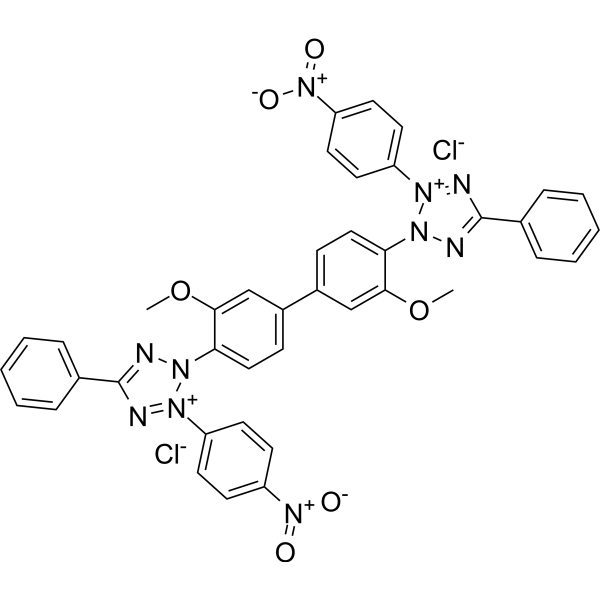
HY-15925
Nitro blue tetrazolium chlorideDehydrogenases Substrate -
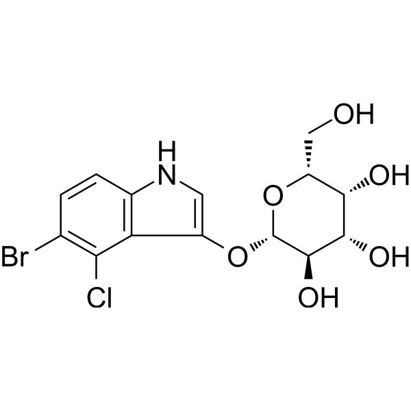
HY-15934
X-GALβ-Galactosidase Substrate -
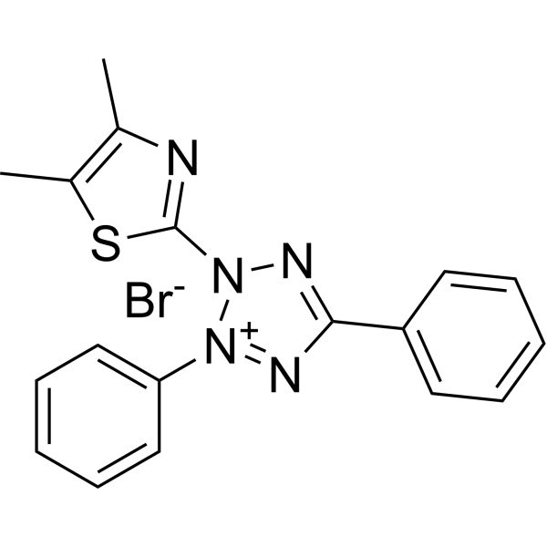
HY-15924
Thiazolyl BlueCytotoxicity Detection Reagent -
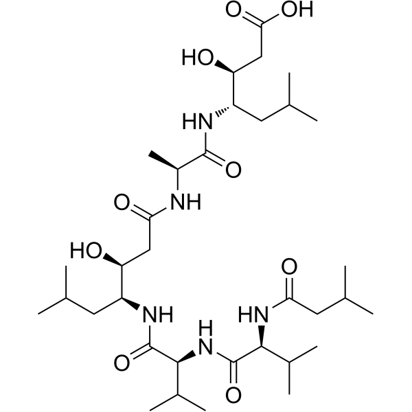
HY-P0018
PepstatinAspartic Protease Inhibitor -
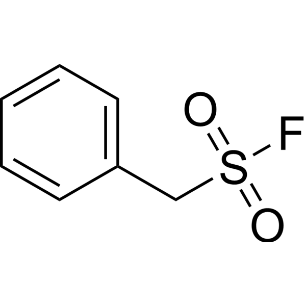
HY-B0496
PMSFCathepsin Inhibitor -
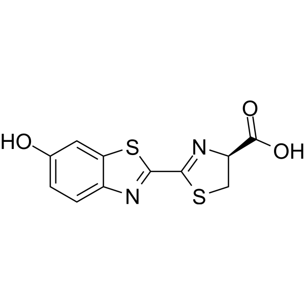
HY-12591A
D-LuciferinSubstrate for Luciferase -
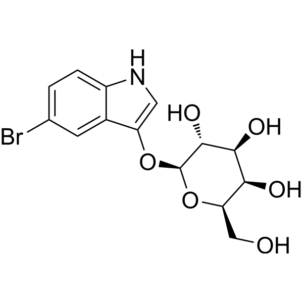
HY-137276
5-Bromo-3-indolyl β-D-galactopyranosideβ-galactosidase Substrate -
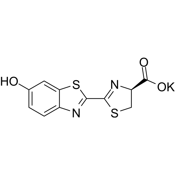
HY-12591B
D-Luciferin potassiumSubstrate for Luciferase -
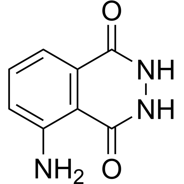
HY-15922
LuminolChemiluminescent Substance -
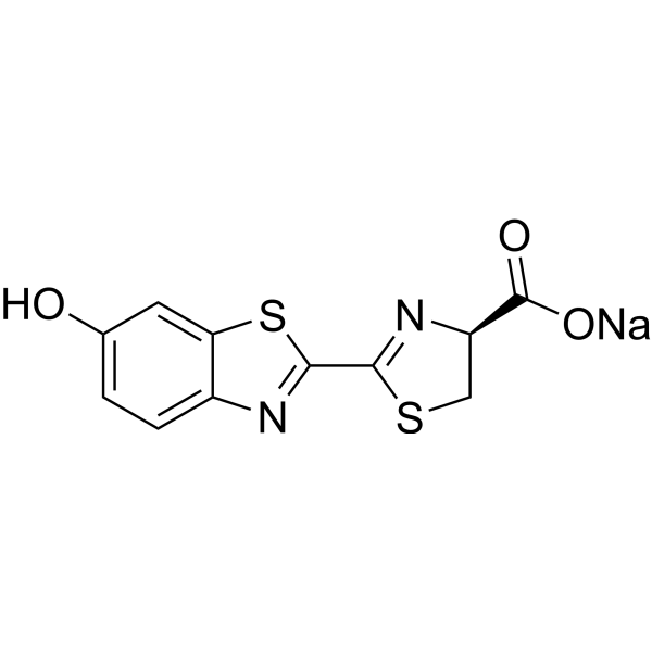
HY-12591
D-Luciferin sodiumSubstrate for Luciferase -
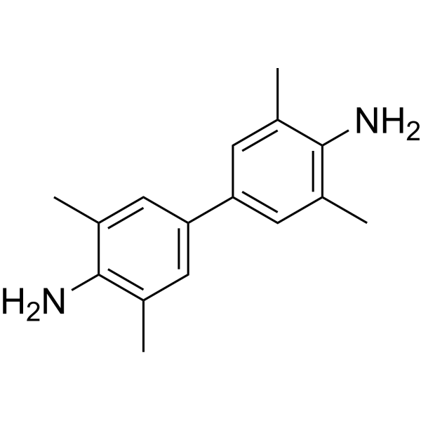
HY-15930
TMBChromogenic Agent -
HY-N0186
Indole-3-butyric acidEndogenous Metabolite


Biological Activity
Description
Solvent and Concentration: DMF: 20 mg/mL
In Vitro
Guide (The following is our recommended protocol. This protocol is only a guide and should be modified according to your specific needs).
1. Solution preparation[1]
1.1 Stock solution
Storage: Store at -20°C or -80°C in dark after aliquoting. Avoid repeated freezing and thawing.
1.2 Preparation of working solution
Dilute X-gal with 1 mL agar medium, and add 1 μL of IPTG (HY-15921) (24 mg/mL) and 1 μL of AMP (HY-A0181) (100 mg/mL) (optimized according to the experiment). The corresponding stock solution can be diluted according to the actual situation. Note that if the solvent is DMSO, the cytotoxicity of DMSO must be considered, and a solvent control should be prepared; if the solvent is pure water, the working solution needs to be filtered and sterilized before adding cells.
Note: The working solution should be prepared and used immediately. Keep it away from light.
2. Blue/white colony screening
2.1 After the DNA fragment is inserted into the pUC series vector (or other vectors with lacZ, Amp genes) and then transformed into lacZ-deficient cells, apply the above-mentioned X-gal, IPTG, and AMP plate culture medium.
2.2 Incubate at 37°C until blue-white colonies appear on the agar surface.
2.3 Cultivate the blue-white colonies further and identify them by double enzyme digestion.
2.4 Aspirate the dye working solution, wash with PBS 2-3 times, and observe under a microscope.
3. Cellular level detection[2]
3.1 When cells are cultured to 80% confluence in culture dishes, add 50 μmol/L H2O2 to the medium and incubate for 12 h.
3.2 Harvest cells with trypsin/EDTA, wash three times with PBS, and fix with 4% formaldehyde for 10 min.
3.3 Prepare cells without H2O2 treatment in the same way as the control group.
3.4 Perform X-gal staining at 37 ℃.
4. Tissue-level detection[2]
4.1 Excise intestinal tissues and cut them longitudinally. Rinse off intestinal contents with phosphate buffered saline, then place the intestinal tissues on glass slides with the inner surface facing upward.
4.2 Dilute a single blue colony containing β-gal (Escherichia coli DH5α carrying pUC18) in 100 μL PBS, and then add 10 μL of the dilution to the tissues.
4.3 After incubating for 1 hour, wash the tissues with PBS solution. For the control group, prepare tissues without blue colonies using the same method.
4.4 Fix all tissues with 4% formaldehyde for 10 minutes, and further stain with X-gal at 37 ℃.
MedChemExpress (MCE) has not independently confirmed the accuracy of these methods. They are for reference only. .


Chemical Information
-
CAS No. 7240-90-6
-
Appearance Liquid (Density: 1.882±0.06 g/cm3)
-
Molecular Weight 408.63
-
Formula C14H15BrClNO6
-
Color Colorless to light yellow
-
SMILES
O[C@H]([C@@H](O)[C@H]1O)[C@@H](O[C@@H]1CO)OC2=CNC3=C2C(Cl)=C(Br)C=C3
-
Shipping
Room temperature in continental US; may vary elsewhere.
-
Storage
Please store the product under the recommended conditions in the Certificate of Analysis.


Purity & Documentation
-
Data Sheet (276 KB)
-
SDS (251 KB)
- English - EN (251 KB)
- Français - FR (251 KB)
- Deutsch - DE (251 KB)
- Norwegian - NO (251 KB)
- Español - ES (251 KB)
- Swedish - SV (251 KB)
- Italian - IT (251 KB)
- Korean - KR (251 KB)
- Portuguese - PT (251 KB)
-
Handling Instructions (2659 KB)
References
[1]. Sanchez-Ramos J, et al. The X-gal caution in neural transplantation studies. Cell Transplant. 2000 Sep-Oct;9(5):657-67. [Content Brief]
[2]. Li S, et al. In-situ SERS readout strategy to improve the reliability of beta-galactosidase activity assay based on X-gal staining in shortening incubation times. Talanta. 2021 Nov 1;234:122689. [Content Brief]

Calculators
Concentration (start) × Volume (start) = Concentration (final) × Volume (final)


X-GAL (solution) Related Classifications
HY-DY2002 Related Classifications
 Powered by Bioz