



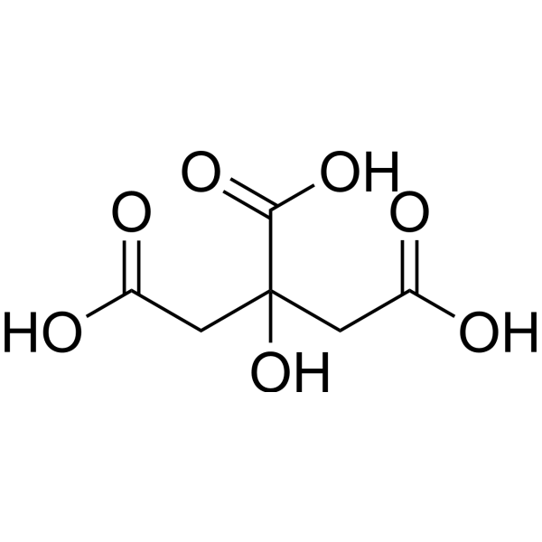
Citric acid
Based on 44 publication(s) in Google Scholar
Citric acid is a natural preservative and food tartness enhancer. Citric acid induces apoptosis and cell cycle arrest at G2/M phase and S phase in HaCaT cells. Citric acid cause oxidative damage of the liver by means of the decrease of antioxidative enzyme activities. Citric acid is also an acidulant, emulsifier, sequestrant and buffering agent widely used across many industries.
Nos produits utilisent uniquement pour la recherche. Nous ne vendons pas aux patients.
- Pureté : 99.98%
- CAS No.: 77-92-9
- Formule: C6H8O7
- Masse moléculaire:192.12
-
Stockage:
Store at room temperature 3 years.
In solvent -80°C, 2 years , -20°C, 1 year

To place orders, for customer services and technical support, please contact: MedChemExpress USA
Tel: 609-228-6898 E-mail: [email protected] [email protected]


-
 Activité biologique
-
Chemical Information
-
Solvant et solubilité
- Protocole
- Pureté et documentation
- Références
-
 Help & FAQs


-
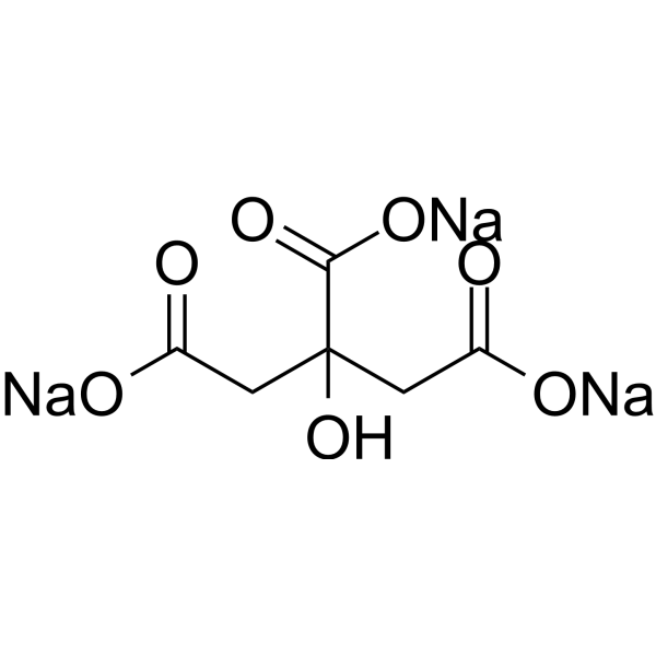
HY-B2201
Citric acid trisodiumOrganic Acid -
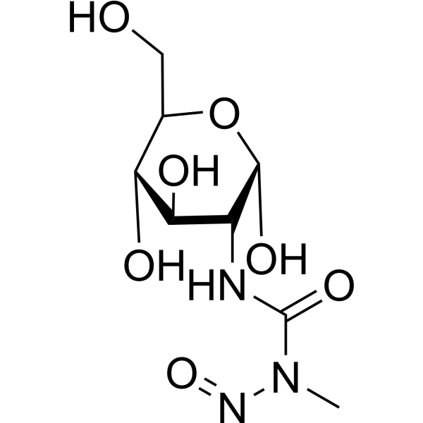
HY-13753
StreptozotocinDiabetes Modeling Agent -
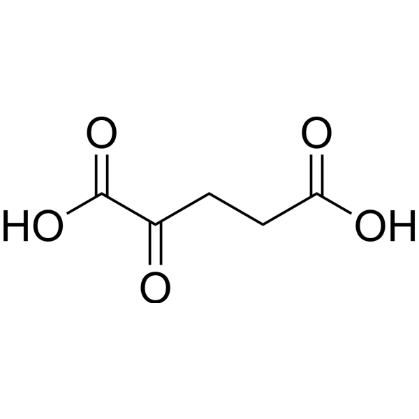
HY-W013636
2-Ketoglutaric acidKrebs Cycle Intermediate -
HY-B1610
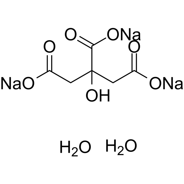
Sodium citrate dihydrateAnticancer/Antibacterial Agent -
HY-B0389
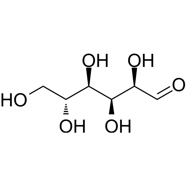
D-GlucoseMonosaccharide -
HY-N0420
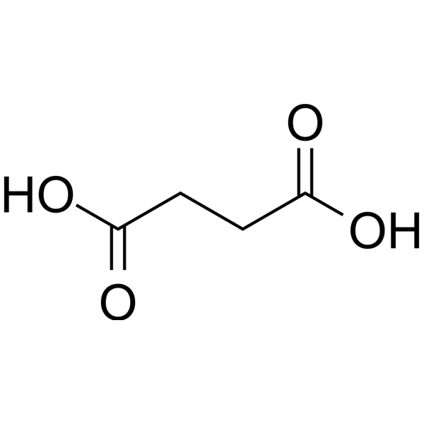
Succinic acidFood Additive, Platform chemical -
HY-Y1311
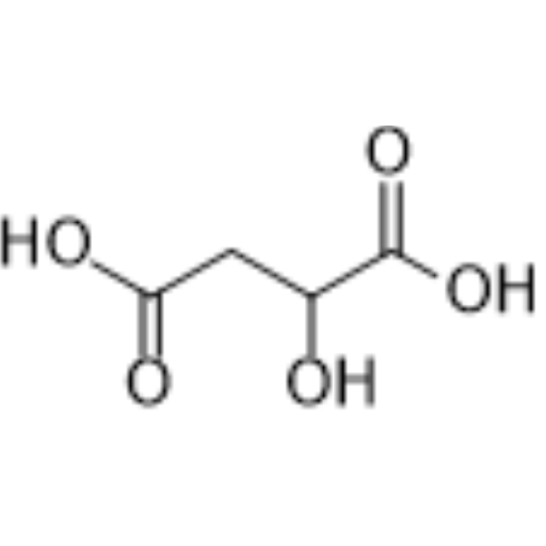
Malic acidDicarboxylic Acid -
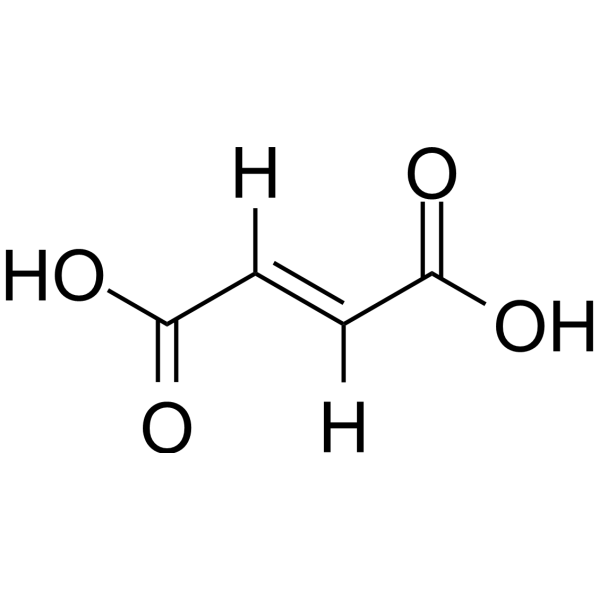
HY-W015883
Fumaric acidAnti-inflammatory agent -

HY-N1428R
Citric acid (standard)Standard of Citric acid -
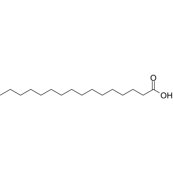
HY-N0830
Palmitic acidEndogenous Metabolite -
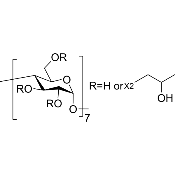
HY-101103
HP-β-CDCo-solvent -
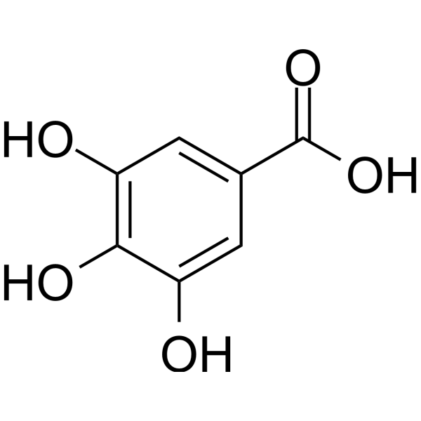
HY-N0523
Gallic acidBioactive Polyhydroxyphenolic Compound
Publications Citing Use of MedChemExpress (MCE) Citric acid
More- Nature. 2025 Jul;643(8070):192-200. [Abstract]
- Cell. 2026 Jun 11;189(12):3553-3570.e30. [Abstract]
- Mol Cancer. 2025 Oct 2;24(1):238. [Abstract]
- Nat Cancer. 2026 Sep;7(9):1387-1405. [Abstract]
- Autophagy. 2026 Jul 24:1-20.
- J Exp Clin Cancer Res. 2025 Jul 8;44(1):195. [Abstract]
- Adv Sci (Weinh). 2025 Nov 6:e07384. [Abstract]
- Adv Sci (Weinh). 2025 Nov 7:e18323. [Abstract]
- Adv Sci (Weinh). 2025 Jul;12(28):e2505359. [Abstract]
- Cell Rep Med. 2026 Feb 17;7(2):102621. [Abstract]
- Int J Biol Sci. 2024 Aug 6;20(11):4222-4237. [Abstract]
- Phytomedicine. 2025 Jun:141:156702. [Abstract]
- Food Chem. 2025 Oct 15:489:144992. [Abstract]
- Food Chem. 2023 Mar 30;405(Pt A):134807. [Abstract]
- Diabetes. 2026 Jun 1;75(6):1007-1023. [Abstract]
- Food Res Int. 2026 Feb 6.
- Regen Biomater. 2024 Aug 22:11:rbae098. [Abstract]
- Free Radic Biol Med. 2026 Mar 16:246:334-349. [Abstract]
- Front Immunol. 2024 Oct 1:15:1397727. [Abstract]
- Virulence. 2026 Dec 31;17(1):2709258.
- J Agric Food Chem. 2026 Apr 29;74(16):13213-13229. [Abstract]
- Biochem Pharmacol. 2026 Jun 25;251(Pt 2):118188. [Abstract]
- Life Sci. 2024 Jun 15:347:122682. [Abstract]
- Drug Des Devel Ther. 2024 Feb 1:18:259-275. [Abstract]
- Int J Mol Sci. 2026 Apr 8;27(8):3357. [Abstract]
- mBio. 2025 Apr 9;16(4):e0012725. [Abstract]
- BMC Complement Med Ther. 2026 Apr 2;26(1):177. [Abstract]
- Front Mol Biosci. 2022 Jul 22:9:952608. [Abstract]
- Biol Trace Elem Res. 2025 Jun 13. [Abstract]
- Biol Trace Elem Res. 2024 Feb;202(2):701-712. [Abstract]
- Insect Biochem Mol Biol. 2023 Jun:157:103958. [Abstract]
- Bone. 2025 Apr:193:117411. [Abstract]
- PLoS Negl Trop Dis. 2025 Dec 11;19(12):e0013815. [Abstract]
- J Sep Sci. 2025 Nov;48(11):e70312. [Abstract]
- J Biochem Mol Toxicol. 2025 Oct;39(10):e70484. [Abstract]
- Tissue Cell. 2026 Jul 6;104(Pt 1):103757.
- New J Chem. 03 Aug 2022.
- bioRxiv. 2026 Jun 2.
- Res Sq. 2025 Aug 08.
- bioRxiv. 2025 Jul 24:2025.07.24.666650. [Abstract]
- Biomed Pharmacother. 2023 Oct:166:115333. [Abstract]
- Oxid Med Cell Longev. 2022 Apr 25;2022:3846217. [Abstract]
- Bioengineered. 2021 Dec;12(2):11610-11621. [Abstract]
- Research Square Preprint. 2021 Jul.
 Customer Validation & Images

-
WB
-
Cell Proliferation/Viability Assay
-
Bio/Physico-chemical Assay
-
Cell Imaging/Staining
-
Cell Imaging/Staining
Voir tous les produits spécifiques à Isoform Endogenous Metabolite
More

Activité biologique
Description
In Vitro
Citric acid (0-12.5 mM; 24 h) shows antiproliferative activity in a dose dependent manner[3].
Citric acid (12.5 mM; 72 h) induces apoptosis and cell cycle arrest at G2/M phase and S phase in a dosedependent manner[3].
Citric acid (12.5 mM; 48 h) increases the expression of FAS, BAX, BID, AIF, EndoG, cytochrome c, PARP, GADD153, GRP78 and caspase-3, -8, -9, and decreases of BCL-2 and BCL-Xl[3].
MedChemExpress (MCE) has not independently confirmed the accuracy of these methods. They are for reference only.
-
Cell Line:HaCaT cells
-
Concentration:0, 2.5, 5, 7.5, 10, 12.5 mM
-
Incubation Time:24 h
-
Result:Inhibited the cell viability in a dose dependent manner.
-
Cell Line:HaCaT cells
-
Concentration:12.5 mM
-
Incubation Time:0, 12, 24, 48, 72 h
-
Result:Induced apoptosis and cell cycle arrest at G2/M phase and S phase in a dose-dependent manner.
-
Cell Line:HaCaT cells
-
Concentration:12.5 mM
-
Incubation Time:12, 24, 48 h
-
Result:Increased the expression of FAS, BAX, BID, AIF, EndoG, cytochrome c, PARP, GADD153, GRP78 and caspase-3, -8, -9, and decreased of BCL-2 and BCL-Xl.
In Vivo
Citric acid (120, 240, and 480 mg/kg; i.p.) induces apoptosis by increases caspase-3 activity in a dose-dependent manner in mouse hepatocytes[1].
Citric acid (120, 240, and 480 mg/kg; i.p.; weekly for 3 weeks) causes renal toxicity in mice[2].
MedChemExpress (MCE) has not independently confirmed the accuracy of these methods. They are for reference only.
-
Animal Model:20 g male Kunming mice[2]
-
Dosage:120, 240, 480 mg/kg
-
Administration:i.p.; weekly for 3 weeks
-
Result:T-SOD and GSH-Px activities in the treated groups decreased with increasing doses of citric acid, NOS activity tended to increase, and H2O2 and MDA contents gradually decreased.
- EdU Incorporation Assay (Click Chemistry-Based DNA Synthesis Measurement)
- Detection of Apoptosis
- TUNEL staining for apoptotic DNA fragmentation
- Flow cytometric DNA-content cell-cycle staining
- Annexin V plus membrane-impermeant dye apoptosis staining
- BrdU Incorporation Assay
- ROS/oxidative-stress fluorescent staining
- Apoptosis Solutions
- Protocol for Cell Cycle
Essai clinique
| NCT Number | Sponsor | Condition | Start Date |
Phase

|
|---|---|---|---|---|
| NCT01329991 | 2011-05 | PHASE1 |


Chemical Information
-
CAS No. 77-92-9
-
Appearance Solid
-
Masse moléculaire 192.12
-
Formule C6H8O7
-
Color White to off-white
-
SMILES
OC(CC(CC(O)=O)(C(O)=O)O)=O
-
Structure Classification
-
Initial Source
-
Livraison
Room temperature in continental US; may vary elsewhere.
-
Stockage
Store at room temperature 3 years
In solvent -80°C 2 years -20°C 1 year


Publications (44)
-
Journal Impact Factor
-
Most Recent
-
Nature
2025 Jul;643(8070):192-200. PMID: 39695227 -
Cell
2026 Jun 11;189(12):3553-3570.e30. PMID: 42049018 -
Mol Cancer
Hsa_circ_0038737 promotes PARPi resistance in castration-resistant prostate cancer via IGF2BP3-mediated DNPH1 mRNA stabilization. [Abstract]2025 Oct 2;24(1):238. PMID: 41039575 -
Nat Cancer
Galanin impairs tumor immunity in glioblastoma by promoting infiltration and ferroptosis resistance of myeloid-derived suppressor cells. [Abstract]2026 Sep;7(9):1387-1405. PMID: 42642633 -
-
J Exp Clin Cancer Res
Genomic profiling of a collection of patient-derived xenografts and cell lines identified ixabepilone as an active drug against chemo-resistant osteosarcoma. [Abstract]2025 Jul 8;44(1):195. PMID: 40624718 -
Adv Sci (Weinh)
Iron Overload Mediates the Differential Cell Fate of Astrocytes from Neurons and Its Regulatory Mechanisms in Ischemic Stroke. [Abstract]2025 Nov 6:e07384. PMID: 41195589 -
Adv Sci (Weinh)
Fe-S Protein FDX1 Triggers Tumor-Intrinsic Innate Immunity via Mitochondrial Nucleic Acids Release to Orchestrate Ferroptosis in CCRCC. [Abstract]2025 Nov 7:e18323. PMID: 41199656
Citric acid purchased from MedChemExpress. Usage Cited in: Adv Sci (Weinh). 2025 Nov 7:e18323. [Abstract]
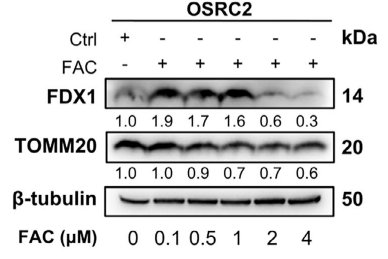
Immunoblot analysis of FDX1 and mitochondrial marker TOMM20 in OSRC2 cells treated for 24 h with either FAC (Ammonium iron(III) citrate) 0–4 μM.
-
Adv Sci (Weinh)
PHOSPHO1 Suppresses Ferroptosis in Retinal Pigment Epithelial Cells by Reducing the Levels of Phosphatidylethanolamine Molecular Species. [Abstract]2025 Jul;12(28):e2505359. PMID: 40396905
Citric acid purchased from MedChemExpress. Usage Cited in: Adv Sci (Weinh). 2025 Jul;12(28):e2505359. [Abstract]
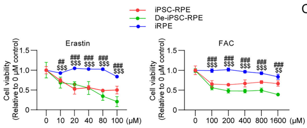
Ferroptosis was induced by erastin or FAC (Ammonium iron(III) citrate; 0-1600 μM) with different doses, and cell viability was analyzed by the CCK-8 after cells were treated for 2 days.
Citric acid purchased from MedChemExpress. Usage Cited in: Adv Sci (Weinh). 2025 Jul;12(28):e2505359. [Abstract]
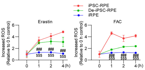
Intracellular ROS was assessed using the DCFH-DA probe after being treated with 30 µM erastin or 500 µM FAC (Ammonium iron(III) citrate) for 4 h.
Citric acid purchased from MedChemExpress. Usage Cited in: Adv Sci (Weinh). 2025 Jul;12(28):e2505359. [Abstract]
Intracellular Fe2+ levels under 30 µM erastin or 500 µM FAC (Ammonium iron(III) citrate) treatment for 4 h were detected with the RhoNox-1(1μM; 1 h) fluorescent probe.
Citric acid purchased from MedChemExpress. Usage Cited in: Adv Sci (Weinh). 2025 Jul;12(28):e2505359. [Abstract]
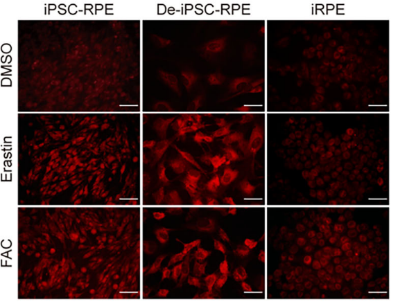
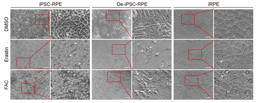
Representative images of ferroptotic cells among iPSC-RPE cells, De-iPSC-RPE cells, and iRPE cells treated with 30 µM erastin or 500 µM FAC (Ammonium iron(III) citrate) for 2 days.
-
Cell Rep Med
Bacterial vesicles from intratumoral L. salivarius enhance PD-1 blockade via FPR1-mediated macrophage polarization in gastric cancer. [Abstract]2026 Feb 17;7(2):102621. PMID: 41707647 -
Int J Biol Sci
IL-6 signaling accelerates iron overload by upregulating DMT1 in endothelial cells to promote aortic dissection. [Abstract]2024 Aug 6;20(11):4222-4237. PMID: 39247821 -
Phytomedicine
Tanshinone IIA promotes ferroptosis in cutaneous melanoma via STAT1-mediated upregulation of PTGS2 expression. [Abstract]2025 Jun:141:156702. PMID: 40222167 -
Food Chem
Flavonoid-mediated metabolic underpinning quality variation in red bud-sport pear mutants. [Abstract]2025 Oct 15:489:144992. PMID: 40466530 -
Food Chem
Discovery of novel ascorbic acid derivatives and other metabolites in fruit of Rosa roxburghii Tratt through untargeted metabolomics and feature-based molecular networking. [Abstract]2023 Mar 30;405(Pt A):134807. PMID: 36370576 -
Diabetes
Cooperative Action of Cathepsin K Inhibitor and hUMSC-EVs in Attenuating Ferroptosis Sensitivity for Superior Diabetic Wound Healing. [Abstract]2026 Jun 1;75(6):1007-1023. PMID: 41979994 -
-
Regen Biomater
Polyzwitterion-grafted decellularized bovine intercostal arteries as new substitutes of small-diameter arteries for vascular regeneration. [Abstract]2024 Aug 22:11:rbae098. PMID: 39224131 -
Free Radic Biol Med
Inhibiting Arachidonate-5-lipoxygenase expression ameliorates osteoarthritis progression by suppressing ferroptosis via the JAK2/STAT3 signaling pathway. [Abstract]2026 Mar 16:246:334-349. PMID: 41581577 -
Front Immunol
2024 Oct 1:15:1397727. PMID: 39430747 -
-
J Agric Food Chem
Polystyrene Microplastics Induced Hepatocytes Pyroptosis, Apoptosis and Ferroptosis via GSDMD-N-Mediated Mitochondrial Damage. [Abstract]2026 Apr 29;74(16):13213-13229. PMID: 41980172 -
Biochem Pharmacol
5-Methoxyseselin inhibits neuronal ferroptosis and β-amyloid production in female APP/PS1 transgenic mice. [Abstract]2026 Jun 25;251(Pt 2):118188. PMID: 42349617 -
Life Sci
Homogentisic acid metabolism inhibits papillary thyroid carcinoma proliferation through ROS and p21-induced cell cycle arrest. [Abstract]2024 Jun 15:347:122682. PMID: 38702025 -
Drug Des Devel Ther
Systematic Pharmacology and Experimental Validation to Reveal the Alleviation of Astragalus membranaceus Regulating Ferroptosis in Osteoarthritis. [Abstract]2024 Feb 1:18:259-275. PMID: 38318502 -
Int J Mol Sci
SLC25A28 Ameliorates Hyperoxic Lung Injury by Improving Mitochondrial Oxidative Phosphorylation in Alveolar Epithelial Cells. [Abstract]2026 Apr 8;27(8):3357. PMID: 42074001 -
mBio
The long noncoding RNA APR attenuates PPRV infection-induced accumulation of intracellular iron to inhibit membrane lipid peroxidation and viral replication. [Abstract]2025 Apr 9;16(4):e0012725. PMID: 40126010 -
BMC Complement Med Ther
Hepatotoxicity prediction for traditional Chinese medicine: a two-step in silico framework integrating network and machine learning approaches. [Abstract]2026 Apr 2;26(1):177. PMID: 41923057 -
Front Mol Biosci
NPC1 Deficiency Contributes to Autophagy-Dependent Ferritinophagy in HEI-OC1 Auditory Cells. [Abstract]2022 Jul 22:9:952608. PMID: 35936782 -
Biol Trace Elem Res
SIRT1 Regulates Iron Metabolism to Attenuate β-Cell Dedifferentiation by Inhibiting FoxO1 Acetylation. [Abstract]2025 Jun 13. PMID: 40512369 -
Biol Trace Elem Res
Antagonizing Effects of Chromium Against Iron-Decreased Glucose Uptake by Regulating ROS-Mediated PI3K/Akt/GLUT4 Signaling Pathway in C2C12. [Abstract]2024 Feb;202(2):701-712. PMID: 37156991 -
Insect Biochem Mol Biol
Development of an efficient insecticide substrate and inhibitor screening system of insect P450s using fission yeast. [Abstract]2023 Jun:157:103958. PMID: 37182814 -
Bone
The protection of nicotinamide riboside against diabetes mellitus-induced bone loss via OXPHOS. [Abstract]2025 Apr:193:117411. PMID: 39884488 -
PLoS Negl Trop Dis
The phytochemical arbutin exerts potent anti-Toxoplasma effects through activation of cell-autonomous defense mechanisms while attenuating inflammation. [Abstract]2025 Dec 11;19(12):e0013815. PMID: 41379884 -
J Sep Sci
Cell Metabolomics Reveals the Hepatotoxic Mechanism of Copper in Normal Rat Liver Cells Using Reversed-Phase and Hydrophilic Interaction Liquid Chromatography-Quadrupole-Time-of-Flight Mass Spectrometry. [Abstract]2025 Nov;48(11):e70312. PMID: 41172059 -
J Biochem Mol Toxicol
Ferroptosis and Lipid Peroxidation Participate in Valproic Acid-Induced Hepatotoxicity via the Long-chain Acyl-CoA Synthetase 4/Glutathione Peroxidase 4 Pathway. [Abstract]2025 Oct;39(10):e70484. PMID: 40990894 -
-
-
-
-
bioRxiv
2025 Jul 24:2025.07.24.666650. PMID: 40777304 -
Biomed Pharmacother
Celastrol alleviated acute kidney injury by inhibition of ferroptosis through Nrf2/GPX4 pathway. [Abstract]2023 Oct:166:115333. PMID: 37598476 -
Oxid Med Cell Longev
HO-1 Contributes to Luteolin-Triggered Ferroptosis in Clear Cell Renal Cell Carcinoma via Increasing the Labile Iron Pool and Promoting Lipid Peroxidation. [Abstract]2022 Apr 25;2022:3846217. PMID: 35656025 -
Bioengineered
ADAMTS-13-regulated nuclear factor E2-related factor 2 signaling inhibits ferroptosis to ameliorate cisplatin-induced acute kidney injuy. [Abstract]2021 Dec;12(2):11610-11621. PMID: 34666603 -


Solvant et solubilité
In Vitro:
DMSO : 100 mg/mL (520.51 mM; Need ultrasonic; Hygroscopic DMSO has a significant impact on the solubility of product, please use newly opened DMSO)
H2O : 100 mg/mL (520.51 mM; Need ultrasonic)
Please refer to the solubility information to select the appropriate solvent. Once prepared, please aliquot and store the solution to prevent product inactivation from repeated freeze-thaw cycles.
Storage method and period of stock solution: -80°C, 2 years; -20°C, 1 year. When stored at -80°C, please use it within 2 years. When stored at -20°C, please use it within 1 year.
* Note: If you choose water as the stock solution, please dilute it to the working solution, then filter and sterilize it with a 0.22 μm filter before use.
Please refer to the solubility information to select the appropriate solvent. Once prepared, please aliquot and store the solution to prevent product inactivation from repeated freeze-thaw cycles.
Storage method and period of stock solution: -80°C, 2 years; -20°C, 1 year. When stored at -80°C, please use it within 2 years. When stored at -20°C, please use it within 1 year.
* Note: If you choose water as the stock solution, please dilute it to the working solution, then filter and sterilize it with a 0.22 μm filter before use.
Concentration (start) × Volume (start) = Concentration (final) × Volume (final)
In Vivo:
Select the appropriate dissolution method based on your experimental animal and administration route.
- For the following dissolution methods, please ensure to first prepare a clear stock solution using an In Vitro approach and then sequentially add co-solvents:
- To ensure reliable experimental results, the clarified stock solution can be appropriately stored based on storage conditions. As for the working solution for In Vivo experiments, it is recommended to prepare freshly and use it on the same day.
- The percentages shown for the solvents indicate their volumetric ratio in the final prepared solution. If precipitation or phase separation occurs during preparation, heat and/or sonication can be used to aid dissolution.
Add each solvent one by one: 10% DMSO 40% PEG300 5% Tween-80 45% Saline
Solubility: ≥ 2.5 mg/mL (13.01 mM); Clear solution
This protocol yields a clear solution of ≥ 2.5 mg/mL (saturation unknown).
Taking 1 mL working solution as an example, add 100 μL DMSO stock solution (25.0 mg/mL) to 400 μL PEG300, and mix evenly; then add 50 μL Tween-80 and mix evenly; then add 450 μL Saline to adjust the volume to 1 mL.
Preparation of Saline: Dissolve 0.9 g sodium chloride in ddH₂O and dilute to 100 mL to obtain a clear Saline solution.
Add each solvent one by one: 10% DMSO 90% (20% SBE-β-CD in Saline)
Solubility: ≥ 2.5 mg/mL (13.01 mM); Clear solution
This protocol yields a clear solution of ≥ 2.5 mg/mL (saturation unknown).
Taking 1 mL working solution as an example, add 100 μL DMSO stock solution (25.0 mg/mL) to 900 μL 20% SBE-β-CD in Saline, and mix evenly.
Preparation of 20% SBE-β-CD in Saline (4°C, storage for one week): 2 g SBE-β-CD powder is dissolved in 10 mL Saline, completely dissolve until clear.
For the following dissolution methods, please prepare the working solution directly:
It is recommended to prepare fresh solutions and use them promptly within a short period of time.
The percentages shown for the solvents indicate their volumetric ratio in the final prepared solution. If precipitation or phase separation occurs during preparation, heat and/or sonication can be used to aid dissolution.
Add each solvent one by one: PBS
Solubility: 100 mg/mL (520.51 mM); Clear solution; Need ultrasonic
In Vivo Dissolution Calculator
Please enter the basic information of animal experiments:
-
-
-
-
Recommended: Prepare an additional quantity of animals to account for potential losses during experiments.
Working solution concentration: 0.22 mg/mL
This product has good water solubility, please refer to the measured solubility data in water/PBS/Saline for details.


Pureté et documentation
-
Fiche technique (279 KB)
-
SDS (394 KB)
- English - EN (394 KB)
- Français - FR (394 KB)
- Deutsch - DE (394 KB)
- Norwegian - NO (394 KB)
- Español - ES (394 KB)
- Swedish - SV (394 KB)
- Italian - IT (394 KB)
- Korean - KR (394 KB)
- Portuguese - PT (394 KB)
-
Instruction de manipulation (2659 KB)
Références
[1]. Chen X, et al. Study on injury effect of food additive citric acid on liver tissue in mice. Cytotechnology. 2014 Mar;66(2):275-82. [Content Brief]
[2]. Chen X, Lv Q, Liu Y, Deng W. Effects of the food additive, citric acid, on kidney cells of mice. Biotech Histochem. 2015 Jan;90(1):38-44. [Content Brief]
[3]. Ying TH, et al. Citric acid induces cell-cycle arrest and apoptosis of human immortalized keratinocyte cell line (HaCaT) via caspase- and mitochondrial-dependent signaling pathways. Anticancer Res. 2013 Oct;33(10):4411-20. [Content Brief]
[4]. Ciriminna R, et al. Citric acid: emerging applications of key biotechnology industrial product. Chem Cent J. 2017 Mar 8;11:22. [Content Brief]


Complete Stock Solution Preparation Table
Please refer to the solubility information to select the appropriate solvent. Once prepared, please aliquot and store the solution to prevent product inactivation from repeated freeze-thaw cycles.
Storage method and period of stock solution: -80°C, 2 years; -20°C, 1 year. When stored at -80°C, please use it within 2 years. When stored at -20°C, please use it within 1 year.
| Optional Solvent | Concentration Solvent Mass | 1 mg | 5 mg | 10 mg | 25 mg |
|---|---|---|---|---|---|
| DMSO / H2O | 1 mM | 5.2051 mL | 26.0254 mL | 52.0508 mL | 130.1270 mL |
| 5 mM | 1.0410 mL | 5.2051 mL | 10.4102 mL | 26.0254 mL | |
| 10 mM | 0.5205 mL | 2.6025 mL | 5.2051 mL | 13.0127 mL | |
| 15 mM | 0.3470 mL | 1.7350 mL | 3.4701 mL | 8.6751 mL | |
| 20 mM | 0.2603 mL | 1.3013 mL | 2.6025 mL | 6.5064 mL | |
| 25 mM | 0.2082 mL | 1.0410 mL | 2.0820 mL | 5.2051 mL | |
| 30 mM | 0.1735 mL | 0.8675 mL | 1.7350 mL | 4.3376 mL | |
| 40 mM | 0.1301 mL | 0.6506 mL | 1.3013 mL | 3.2532 mL | |
| 50 mM | 0.1041 mL | 0.5205 mL | 1.0410 mL | 2.6025 mL | |
| 60 mM | 0.0868 mL | 0.4338 mL | 0.8675 mL | 2.1688 mL | |
| 80 mM | 0.0651 mL | 0.3253 mL | 0.6506 mL | 1.6266 mL | |
| 100 mM | 0.0521 mL | 0.2603 mL | 0.5205 mL | 1.3013 mL |
* Note: If you choose water as the stock solution, please dilute it to the working solution, then filter and sterilize it with a 0.22 μm filter before use.


Citric acid Related Classifications
HY-N1428 Related Classifications
 Powered by Bioz